Transcription
9/29/2015Pediatric OrthopaedicPhysical ExamS. Matt Hollenbeck, M.D., MPTPediatric Orthopaedic SurgeonKansas Orthopaedic CenterWichita, KSDisclosure I have no relevant financial relationships withthe manufacturer(s) of any commercialproduct(s) and/or provider of commercialservices discussed in this CME activity. I do not intend to discuss anunapproved/investigative use of a commercialproduct/device in my presentation.Musculoskeletal Exam Establish a rapportPatient HistoryObservationExamination of Motion/MovementSpecial TestsReflexes and Sensory ExamPalpationEvaluating Diagnostic Imaging1
9/29/2015Musculoskeletal Exam Each portion of the exampiggybacks off the last section Overlap of exam is fluid– Ask a question and observe theresponse– Palpate the area of interest andobserve the response When in doubt, compare to theunaffected side!Establish a Rapport Greet the parentsSay hi to the childHandshake Parents“Fist Bump” or Five ChildGet on the kids level or belowMake the kid laughHave funAvoid White CoatEstablish Rapport Let the child have somecontrol Make the child the “boss” Play games or make the exama game Put down the phones/ipads Adjust your verbiage basedon kids level of understanding( intellectual ability)2
9/29/2015Patient History The best musculoskeletal physical examshould start out by listening!!!Patient History Often the diagnosis can be made inthis step– Even without an exam Simply listen to the patient Include the child when possible Rely on parents for clarification ormore specifics Acknowledge the child Write things down – don’t expect toremember everythingPatient History Relevant history to present illness/injury–––––––––What – is the problem or what happenedWhen – time sequenceWhere – does it hurtWhy – did the patient come inHow bad – better, worse, or stay sameRelieving factorsAggravating FactorsTreatments attempted – help or no helpFunctional effect3
9/29/2015Patient History Past medical history– May not be written down on the forms Past surgeries– Ex: open heart surgery for congenital heart defect Social history – sports, school activities (band), dance,etc. Family History – similar problems Medications– Oftentimes present but no medical history– Esp: NSAIDS, anti‐seizure meds, baclofen, botox, AllergiesHandouts/QuestionnaireHistory Taking Tips Ask open ended questions Don’t lead kids with questions– Ex: What’s going on with your knee? What brings you into the office?– Ex: Does this inrcease your pain? Prefered: Does this CHANGE your pain? Listen and Probe for Red Flags Keep the patient/parent focused Talk on the level of the child4
9/29/2015Red Flags Night Pain (possibly)Weight LossFatigueBowel or bladder changesFever/Chills/Night SweatsBalance issues/ Recent FallsCoordination issuesWeakness/Inability to stand upLumps/Bumps that are growingObservation Overall demeanor/attitude of child Interactions with parents Walking around room– Or Walking to the room Sitting on table (swinging legs)Laying on tableClimbing on exam table/stoolCurrently in painOverall alignmentObservation Expose the area of interest––––Spine: Gown – open in the backLE: shortsUE: short sleeve/sleeveless shirtShoulder: Gown/Sports bra/ sleevelessshirts in girls Shirt off in boys– Feet: shoes/socks off– No restrictions– Nothing hiding Have the Nurse/MA be the bad guy/gal !!! Allow child to put clothes back on ASAPafter exam5
9/29/2015Observation Standing Postural Analysis– “Watch ‘em stand” Observational Gait Analysis– “Watch ‘em walk” Observe the affected an unaffected sideWatch for facial expressionsReactions to questions or palpationPattern of movementObservation Body alignment– Nose, xiphoid process, & umbilicus– Tip ear, acromion, top of iliac crest& lateral malleoli Obvious Deformity Symmetric Body/Soft TissueContours Limb Positioning Skin color and texture ScarsObservation Use all your senses– Sight – self explanatory– Smell – abnormal odor– Hearing – Click or pop with joint motion– Touch – Defer to palpation portion– Taste – Defer all together!!!6
9/29/2015Examination of Motion/Movement Test the unaffected side first– Sets a baseline for exam– Gains the patients trust and confidence Perform painful movements last– Prevents pain from overflowing to the nextmovementExamination of Motion/Movement AROM – Active Range of Motion PROM – Passive Range of Motion Resisted Isometric Motions Strength Testing– Done with joint in neutral or resting position Do AROM PROM Resisted MotionResisted Isometric Motion Strong & Painfree– Muscle and nerve tested are normal Strong & Painful– Local lesion of muscle or tendon(Tendonitis/Muscle Strain) Weak and Painful– Severe Lesion around a joint (Fracture) Weak and Painfree– Rupture of muscle/tendon or nerve injury7
9/29/2015Strength GradesExamination of Motion/Movement If AROM is full then give end rangepressure for end feel of the joint anddegree of motion Repetitive motions are done ifsymptoms are complained of withmultiple repetitions Note what arc of motion causes pain Look for abnormal substitution patternsof movement Warn of exacerbation of symptoms atend of examJoint End Feels Soft – soft tissue being compressed– Ex: knee or elbow flexion Hard – Bony – bone hitting bone ‐ painless– Ex: elbow extension Firm/Springy – soft tissue at the limit ofstretch– Ex: Shoulder ER Guarding – muscle contraction and pain Empty –no resistance – stopped by patientreport of pain8
9/29/2015Examination of Motion/Movement ROM can be tested with a goniometer PROM can unmask generalized hypomobilityor hypermobility/laxityFunctional Movement Test Have patient do the reported activity thatcauses pain/symptoms– Squat– Run– Pick up object– Get out of bedSpecial Tests Many special tests for eachjoint/body area Many are almost pathognomonicfor a diagnosis ( ) findings can strongly suggest atype of injury, condition, ordisease (‐) findings do not rule out aninjury, condition, or disease9
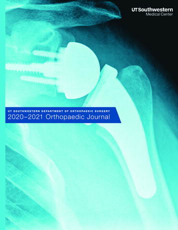
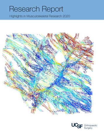
9/29/2015Reflexes and Sensory Exam Deep Tendon Reflexes– Pt. Relaxes– Tendon in slight stretch position– If trouble eliciting response thenhave patient squeeze handstogether Superficial Reflexes– Stroke the skin with a moderatelysharp objectCommon Deep Tendon Reflexes C5C6C7L4S1Grading Deep Tendon Reflexes 0 – Absent1 – Diminished2 – Average (normal)3 – Exaggerated4 – Clonus/Very Brisk10
9/29/2015Common Superficial Reflexes Babinski– Normal Infant – Upgoing Big Toe & fan other toes Usually changes at 1‐2 yrs of age– Normal Child/Adult – Flexion of toesCommon Superficial Reflexes Hoffmann – “Flick” distal phalanx ofindex/middle finger and watch thumb– Normal no movement– Abnormal flexion of distal phalanx of thumbCommon Superficial Reflexes Abdominal – Separate abdomen intoquadrants with vertical and horizontal linethrough umbilicus– Stroke each quadrant away from umbilicus– Normal Moves toward the direction beingstroked– Abnormal Moves away from the direction beingstroked11
9/29/2015Common Superficial Reflexes Abdominal ReflexMotor Neuron Upper Motor Neuron Lesion– Spasticity– Hyperreflexia– Hypertonicitiy Lower Motor Neuron nicityFasciculation/FibrillationsWeakness and atrophySensory Exam Run fingers firmly over the skin to be tested Ask if they can feel it?– Make it a game Is there any difference compared R to L? 2 Point discrimination can also be tested morespecifically in a certain area– Use Paperclip– Test normal side first so child knows what to expect Last resort – needle to see if sensory intact12
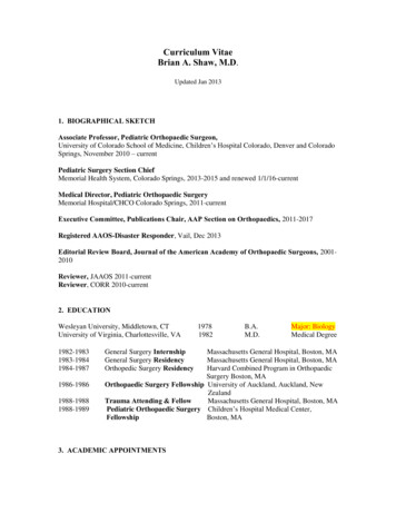
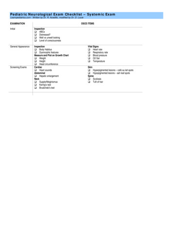
9/29/2015UE Sensory Dermatome LevelsLE Dermatome LevelsPalpation Ensure are a is as relaxed as possible– Make sure area is supported Start with thenoninjured/contralateral/area far awayfrom area of interest Watch the childs face during palpation Child may withdraw and not be astrusting so put this towards the end ofthe exam Start with light pressure and then deeperpressure as tolerated13
9/29/2015Palpation EdemaEffusionSkin tensionAdhesionsBony Prominences/AreasTemperature DifferencePulsesAbnormal Lumps/bumpsEvaluating Diagnostic Imaging Xrays – at least 2 orthogonalviews –– views at 90o to each other– Evaluate the entire xray– When in doubt xray thecontralateral side– Physis and epiphysis makeinterpreting xrays hardThings to Note of Xrays Size and shape of the boneThickness of the cortexGeneral density of the boneMargins of local lesionsSoft Tissue ChangesPeriosteal ChangesJoint spaceBreak in continuity of bone14
9/29/2015Things you can see on Xrays FractureDislocationEffusionTumorsBone QualityOpen/Closed PhysisInfectionsHeterotopic BoneForeign BodyAirLeg Length DiscrepencyLower Extremity alignment (angular deformity)Bone age ( hand xrays )Things you can see on XraysEvaluating Diagnostic Imaging CT scan – gives better bony detail– Shows growth plate closure MRI – givens soft tissue/cartilage details– When concerned for soft tissue injury– Shows bone edema/joint effusion/infection If infection/Tumor – always order with/without contrast Ultrasound – can show cartilaginousstructures easily– Can show an effusion15
9/29/2015Cervical SpineAnatomyPatient History Any restriction of motion? Pain with movement? Numbness or tingling?– Especially down the arms Fine motor movements?– Coloring, Holding utensils/pencils, Buttons How is the child’s vision?Sports Injury? Football?Trouble Walking?Balance difficulty?16
9/29/2015Observation Head and neck posture–––––MidlineHead tiltHead rotationForward headMuscle Prominence (SCM) Shoulders– Level – dominant usually elevated slightly– Rounded– AtrophyExamination of Motion/Movement FlexionExtensionSidebendRotationExamination of Motion/Movement Check shoulder ROM also17
9/29/2015Examination of Motion/Movement Nodding occurs in upper cervical spineFlexion occurs in lower cervical spineOnly use mild overpressure at end rangesIn sidebend ensure not moving shoulder towardear Do resisted isometric movements– Cervical spine– Shouder/UEExamination of Motion/Movement– C1/C2 – Neck Flexion– C3 – Sidebend– C4 – Shoulder Elevation/Flexion– C5 – Shoulder Abduction– C6 – Elbow Flexion– C7 – Elbow Extension– C8 – Thumb Extension– T1 – Finger abductionSpecial Tests Spurling’s Test /Foraminal Compression– Sidebend head & extension /‐ rotation– test if radicular symptoms ( not neck pain)– Indicates cervical nerve root impingement18
9/29/2015Special Tests Romberg Test– Patient stands and Closes eyes– Position held x 20‐30 seconds– excessive sway or lossbalance– Indicates upper motor neuronlesionReflexes and Sensory Exam Deep Tendon Reflexes– C5‐ Bicep– C6‐ Brachioradialis– C7 – Tricep Superficial Reflexes– Hoffman’s ReflexReflexes and Sensory Exam UE Sensory Exam19
9/29/2015Palpation For maximal relaxationhave pt. lie supine Spinous Processes Paraspinal Musculature Sternocleidomastoidmuscles Upper Trapezius muscle Levator Scapulae muscle ScalenesEvaluating Diagnostic Imaging Xrays ‐ AP , Lateral, Open Mouth Odontoid– Cervical Lordosis is normal– Atlanto‐dens intervalThoracic and Lumbar Spine20
9/29/2015AnatomyPatient History Is there pain with inpsiration/expiration?Is there pain with coughing/sneezing/straining?Any numbness or tingling?Any pain shooting down your legs?Any bowel or bladder difficulties?When was deformity noticed?Has the deformity been getting worse?Any family history of scoliosis?When was menarche?Any chest surgery? Heart surgery?Observation Have the patient in a gown(open to the back)– Keep bra on ( sports brapreferred)– Keep shorts/pants on21
9/29/2015Observation Hip heights Shoulder heights Trunk shift– Can use a plumb line Rib humpAsymmetric waistAsymmetric scapulaAsymmetric Trunk RotationExcessive kyphosisSitting postureStanding postureHairy patches/DimplesObservation ATRExamination of Motion/Movement FlexionExtensionSidebendRotation– Any pain with movements?22
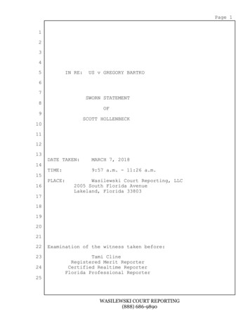
9/29/2015Examination of Motion/Movement Standing Motor Screen– Raise up on toes– Rock back on your heels– Squat down and come back up– Stand on one legExamination of Motion/Movement L2 – Hip flexionL3 – Knee extensionL4 – Ankle DorsiflexionL5 – Great Toe DorsiflexionS1 – Ankle PlantarflexionScheuermann’s Kyphosis23
9/29/2015ScoliosisSpecial Tests Straight Leg RaiseReflexes and Sensory Exam Deep Tendon Reflexes– L4‐ Patellar Tendon– S1‐ Achilles Superficial Reflexes– Abdominal Reflex– Babinski Reflex24
9/29/2015Reflexes and Sensory Exam LE Sensory ExamPalpation Spinous ProcessesParaspinal MusculaturePSISIliac CrestScapulaIschial TuberosityEvaluating Diagnostic Imaging Xrays ‐ PA , Lateral, Entire Spine– PA & lateral thoracic spine– PA, lateral, and bilateral oblique lumbar spine25
9/29/2015Evaluating Diagnostic ImagingEvaluating Diagnostic ImagingEvaluating Diagnostic Imaging 12 thoracic vertebrae 5 lumbar vertebrae Thoracic Kyphosis– Thoracic vertebrae wedgeing Lumbar lordosisPartial/Hemi/Fused 6
9/29/2015Evaluating Diagnostic ImagingEvaluating Diagnostic Imaging CT Scan – gives better bonydefinition– Can give 3D reconstructionsEvaluating Diagnostic Imaging MRI – shows soft tissues– Herniated Nucleus Pulposis– Nerve impingement– Foraminal compression– Central Canal Stenosis27
9/29/2015ShoulderAnatomyPatient HistoryMechanism of injury?Weakness?Sporting Activities?Numbness or Tingling inarm? What hand is dominant? Any muscle wasting/atrophy? 28
9/29/2015Observation Put patient in gown or “tube top”/tank top shirt if girl orhave boys remove shirt AC joint – step deformity Dominant shoulder is usually lower Scapulae of equal size and position Winging of scapula Muscle atrophy– Supra/infraspinatus – suprascapular nerve– Serratus anterior – long thoracic nerve– Upper Trapezius – Spinal Accessory Nerve Clavicles Present/ bump Pectoral Muscles present/atrophy/absentObservationObservation29
9/29/2015Examination of Motion/Movement FlexionAbductionExtensionInternal RotationExternal RotationScaptionAdductionPain with any of these motionsWatch the quality of the movement– From anterior and posterior Can do resisted isometric muscle testing of movements alsoExamination of Motion/Movement Gross Shoulder ROM screen– Field Goal– Jumping Jack– Hands Behind Head– Hands Behind BackSpecial Tests Load and Shift Test– Load the humeral head intoglenoid– Translate anterior andposterior– Normal is 25% or lesstranslation30
9/29/2015Special Tests Apprehension Test– Shoulder abducted 90o and ER– Feeling of apprehension/patient resistence/alarmon the patient face test– Anterior instability/dislocationSpecial Tests Speed’s Test– Resist forward flexion ofstraight arm up to 90o– pain in bicipetalgroove– Indicative of bicipetalTendonitisSpecial Tests Neer Impingement Test– Patients arm is passiveelevated through forwardflexion by examiner– pain– Indicative of shoulderimpingement31
9/29/2015Special Tests Hawkins KennedyImpingement Test– Arm flexed to 90o &internally rotatedmaximally– pain– Indicative ofshoulderimpingementReflexes and Sensory Exam Deep Tendon Reflexes– C5‐ Bicep– C6‐ Brachioradialis– C7 – Tricep Superficial Reflexes– Hoffman’s ReflexReflexes and Sensory Exam UE Sensory Exam32
9/29/2015Common Brachial Plexus Injuries Erb’s Palsy Klumpke’s PalsyPalpationClavicleSternoclavicular jointAcromioclavicular JointCoracoid Process – oftenpainful Humeral Head Scapular Spine Scapula Evaluating Diagnostic Imaging Xrays: Routine AP, True AP (Grashey), Axillary,Scapular Y , Internal Rotation, External Rotationviews Ensure shoulder reduced Evaluate physis to see if wide Ensure humeral head is round33
9/29/2015Evaluating Diagnostic ImagingEvaluating Diagnostic ImagingEvaluating Diagnostic Imaging CT Scan – Gives better bony detail– Can get 3D options MRI – shows soft tissues better– Labrum– Muscles and tendons– Bony edema– Tumors34
9/29/2015ElbowAnatomyPatient History Mechanism of injury?– Fall on outstretched arm– Holding hands and child drops to kneesAny deformity?Does it look similar to other side?What is the child not able to do?What sports/extracurricular activities does thechild do? Any previous injuries/surgeries to arm? 35
9/29/2015Observation Expose the arm Carrying angle – angle of humerusand ulna created when elbowextended and full supination Swelling Joint effusion Resting position of the elbowExamination of Motion/Movement FlexionExtensionSupinationPronation Compare to contralateral side Can place pen/pencil in hand to moreaccurately assess supination/pronationExamination of Motion/Movement36
9/29/2015Special Tests Ligamentous Instability Test– Childs elbow slightly flexed and varus/valgusstress applied– excess motion– Indicates ligamentous instability/laxitySpecial Tests Lateral epicondylitis Test– Pronate the forearm and resist wrist extension orlong finger extension– pain in lateral epicondyle area– Indicative of lateral epicondylitisSpecial Tests Tinel’s at the Elbow– Ulnar nerve is tapped between olecranon &medial epicondyle– tingling sensation in ulnar distribution offorearm37
9/29/2015Reflexes and Sensory Exam Deep Tendon Reflexes– C5‐ Bicep– C6‐ Brachioradialis– C7 – Tricep Superficial Reflexes– Hoffman’s ReflexReflexes and Sensory Exam UE Sensory ExamPalpation Medial epicondyle Lateral epicondyle Supracondylar Humerusarea Olecranon Radial Head Ulnar Nerve in cubitaltunnel Biceps Tendon Triceps Tendon38
9/29/2015Evaluating Diagnostic Imaging Xray – AP, Lateral, InternalOblique– Fat Pad Sign/Sail Sign– Fractures– Internal oblique – lateralcondyle fracture– Dislocations– Medial Epicondyle Fracture– Supracondylar HumerusFracture– Radial neck fractureEvaluating Diagnostic ImagingEvaluating Diagnostic Imaging39
9/29/2015Evaluating Diagnostic Imaging CT Scan– Gives more bone detail and3D possibilities MRI– Evaluates soft tissue,cartilage, joint surface,ligamentous structures,osteochondritis dessicanslesionsWristAnatomy40
9/29/2015Patient History Mechanism of injury? What sports or extracurricular a activities isthe child involved in? Which extremity is dominant? Previous injury or surgery? When was the injury?– Flexor tendons can be repaired if 3 weeks– Flexor tendons require reconstructin if 3 weeksObservation Resting positionForearm alignment/deviation/deformitySoft tissue mass swellingsSwellingExamination of Motion/Movement FlexionExtensionRadial DeviationUlnar DeviationSupinationPronation41
9/29/2015Examination of Motion/MovementUlnar NerveRadial NerveMedian/AINSpecial Tests Finkelstein’s Test– Have patient actively put thumb in palm of fistand ulnar deviate wrist– pain at radial side of wrist– Indicates de Quervain’s tenosynovitis– Have child do both hands as to have a baseline forpain comparisonSpecial Tests Tinel’s at the Wrist– Tap over the carpal tunnel– tingling/paresthesia in to median nervedistribution42
9/29/2015Reflexes and Sensory Exam Deep Tendon Reflexes– C5‐ Bicep– C6‐ Brachioradialis– C7 – Tricep Superficial Reflexes– Hoffman’s ReflexReflexes and Sensory Exam UE Sensory ExamPalpation Anatomic SnuffboxRadial StyloidUlnar StyloidRadial PulseRadial metaphysis43
9/29/2015Evaluating Diagnostic Imaging Xray – AP, Lateral, and Oblique– Scaphoid View – scaphoid fractures– Fractures, bone lesions, massesEvaluating Diagnostic Imaging CT Scan – Shows bone anatomy better– Useful for fractures sometimes MRI – shows soft tissue better– Used to evaluate TFCC tears at tip of ulnar styloidHand44
9/29/2015AnatomyPatient History Mechanism of injury?Do fingers get stuck flexed?Do the fingers pop with attempt at extension?Do fingers overlap/underlap?Are the fingers curved?Have fingers always been fused?Extra digits?Observation Muscle wasting Asymmetry of fingers Rotational or angular deformities of fingers– Check with making fist Mallet Finger Swan Neck Deformity – rupture of terminalextensor tendon (mallet finger) Boutonniere Deformity – rupture of central slip Trigger finger Simean Crease45
9/29/2015Examination of Motion/Movement Finger FlexionFinger ExtensionFinger AbductionFinger AdductionThumb FlexionThumb ExtensionThumb AbductionThumb AdductionExamination of Motion/Movement For flexed digits isolated which joint is flexed– Ex: Trigger ThumbExamination of Motion/MovementUlnar NerveRadial NerveMedian/AIN46
9/29/2015Special Tests Ulnar Collateral Ligament Stress Test– Thumb held in extension & 30 degrees flexion andpassive stress of distal phalanx radially onstabilized proximal phalanx– 10o difference– Indicates Skier’s thumb/Gamekeeper’s thumbSpecial Tests Froment’s Sign– Child grasps a piece of paper between thumb &index finger & examiner tries to pull it away– DIP of thumb flexes to hold the paper– Indicates ulnar nerve paralysis due to weakadductor pollicisReflexes and Sensory Exam Deep Tendon Reflexes– C5‐ Bicep– C6‐ Brachioradialis– C7 – Tricep Superficial Reflexes– Hoffman’s Reflex47
9/29/2015Reflexes and Sensory Exam UE Sensory ExamPalpation Finger Metacarpals Finger Phalanx Radial pulseEvaluating Diagnostic Imaging Xray – AP, Fan Lateral, Oblique of Fingers– MCP joints, DIP joints, PIP joints– Can use AP image to determine skeletal maturity Gruelich and Pyle Atlas– Fractures Rarely need CT scan or MRI48
9/29/2015HipAnatomyPatient History Any sporting or extracurricular activities? Any snapping with hip movement? Infant – Born breech?– First born?– History of hip dysplasia in family? Is patient able to ambulate on leg? Fevers or Chills?49
9/29/2015ObservationEnsure child in shorts or diaperResting position of legStanding postureAssess Weight Bearing amount on eachleg Pelvis heights bilateral Single Leg Stance – Trendelenberg Leg length discrepency Lumbar lordosis – excess if hip flexioncontracture Thigh foldsObservationObservation Gait pattern– Antalgic– Ataxic– Trendelenburg– Foot Progression angle– Patella position– Equal step length– Fat thigh gait50
9/29/2015Examination of Motion/Movement FlexionExtensionAbductionAdductionInternal Rotation – best assessed proneExternal Rotation – best assessed proneExamination of Motion/Movement Active Knee to Chest– Watch for obligatoryexternal rotation SquatSpecial Tests Trendelenburg Sign– Stand on one leg and assess the elevatedhip/gluteal area for dropping51
9/29/2015Special Test Hamstring Contracture Test– Child supine and hip flexed 90degrees & attempt to extend knee Thomas Test– Child supine & flex contralateral hipto chest & look for flexioncontracture in contralateral hipSpecial Tests Barlow TestOrtolani Test Can only be Ortolani OR Barlow positiveSpecial Test Galeazzi Sign– Child supine & hips / kneesflexed 90o– Look for one knee higherthan the other52
9/29/2015Special Test Gower’s SignReflexes and Sensory Exam Deep Tendon Reflexes – L4 & S1 Lower Extremity Dermatome LevelsPalpation Iliac CrestGreater TrochanterASISPSISIschial TuberosityPubic Symphysis53
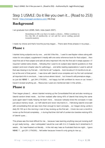
9/29/2015Evaluating Diagnostic Imaging Xray – AP & Frog Lateral of Pelvis–––––––Femoral head seated deep in acetabulumNeck‐shaft angleShenton’s LineKlein’s LineFemoral Head AVNRisser SignAcetabular Index Arthrogram can outline cartilaginous femoralheadEvaluating Diagnostic Imaging Hip Ultrasound – evaluates acartilaginous femoral head orfor hip effusion CT Scan – Gives good bonydetail MRI – shows soft tissue–––––Labral TearHip effusionAbscessDetails AVNCan add arthrogram for moredetails54
9/29/2015Case Study 4 yo male with abmormal gaitCase StudyPhysical Exam What do you see? What do you want to do next? What is going on?55
9/29/2015XraysKnee56
9/29/2015AnatomyPatient History Popping/Clicking?Giving Way?Locking?Mechanism of Injury?Swelling?Sports or Extracurricular Activities?Limp?Knock kneed or bow legged?Observation Lower Extremity alignment– Genu Valgum – intermalleolardistance– Genu Varum – intercondylar distance– Genu Recurvatum Ensure child in shorts or diaperResting position of legStanding postureAssess Weight Bearing amount oneach leg57
9/29/2015ObservationObservation Joint effusionGeneralized SwellingStation of the kneePatella positionScarsRotational DeformityTibial Tubercle ProminenceTibial TorsionObservation Sitting Posture– W position– “Criss cross applesauce”– On knees58
9/29/2015Observation Gait pattern– Antalgic– Ataxic– Trendelenburg– Foot Progression angle– Patella position– Equal step length– Fat thigh gaitExamination of Motion/Movement FlexionExtensionPatellar MobilityPatellar TiltExtensor LagFlexion ContractureVMO activationSpecial Tests Always test as compared to contralateralnormal side MCL & LCL Stress Test59
9/29/2015Special Test Lachman TestSpecial Test Anterior DrawerSpecial Test Posterior Drawer60
9/29/2015Special Test Pivot Shift – ITB changes from an extensor to aflexorSpecial Test McMurray’s TestSpecial Test Patellar Apprehension Test61
9/29/2015Reflexes and Sensory Exam Deep Tendon Reflexes – L4 & S1 Lower Extremity Dermatome LevelsPalpation PatellaTibial TubercleMedial & Lateral Joint LineDistal Femoral CondyleProximal Tibial PlateauMCLLCLPalpation Hamstring TendonsPes TendonsSuprapatellar Pouch (Effusion)VMO62
9/29/2015Evaluating Diagnostic Imaging Xray – AP, Lateral , Tunnel/Notch, ctureDislocationAVNPatella Position – Alta/BajaJoint Effusion – on lateralPhysisTrochlear Dysplasia/AplasiaOsgood Schlatter DiseaseSinding Larson Johanson DiseaseEvaluating Diagnostic Imaging63
9/29/2015Evaluating Diagnostic Imaging CT Scan – Gives better bony detail MRI – evalaute soft tissue– Meniscus– Ligaments – ACL/PCL/MCL/LCL– Bone edema– AVN– Articular CartilageAnkleAnatomy64
9/29/2015Patient History Ankle sprains? Recurrent?Which way did it roll?Have you been able to walk on it since injury?Swelling?Sports or extracurricular activitiesLocation of the painWhat types of shoes does the child wear?Previous injury?Observation Standing and Non weight bearing–––––––––Hindfoot positionForefoot positionPatella position vs. foot positionSwellingBony prominences or soft tissue massesMalleoli positionFoot archShoe WearPelvis Heights – Leg length discrepencyObservation Gait pattern– Antalgic– Ataxic– Trendelenburg– Foot Progression angle– Patella position– Equal step length– Fat thigh gait65
9/29/2015Examination of Motion/Movement tion of Motion/Movement Raise up on toesRock back on heelsSquat DownSingle Leg StancePassive ankle dorsiflexion– With knee extended – Gastroc Tightness– With knee flexed – Soleus TightnessSpecial Tests Transmalleolar Axis– Normal external 40o66
9/29/2015Special Tests Thigh Foot AxisSpecial Test Anterior Drawer– Compare to contralateral sideReflexes and Sensory Exam Deep Tendon Reflexes – L4 & S1 Lower Extremity Sensory Exam67
9/29/2015Palpation TibiaMedial & Lateral MalleoliATFL & CFL ligamentsAchilles TendonCalcaneousPeroneal TendonsAnkle Joint effusionEvaluating Diagnostic Imaging Xray – AP, Mortise, Lateral– Stress Test Gravity Physician Assisted– AVN– Fx– Ligament Instability68
9/29/2015Evaluating Diagnostic Imaging CT Scan – More bony detail– Physeal arrest size MRI– Soft tissue – ligament/tendons– Articular cartilage– AVN– Infection– EffusionFoot69
9/29/2015AnatomyPatient History Location of foot pain?Recurrent ankle sprains?Family history of disorders?Fixed or Flexible deformity of the foot?Shoe wear?Describe injury and position of foot?Previous Treatments (casts or surgeries)?Observation Standing & Supine Foot Position– Hindfoot– Midfoot– Forefoot Resting PositionBony ProminencesSyndactylyPolydactyly70
9/29/2015Observation Gait elenburgFoot Progression anglePatella positionEqual step lengthFat thigh gaitFoot Pronation/Supination Dynamic SupinationObseration Common Foot Deformities– Clubfoot– Congenital Vertical Talus– Metatarsus Adductus– Flatfoot– Hallux Valgus (Bunion)– Hammer Toe/Curly Toe71
9/29/2015Examination of Motion/Movement Ankle – Dorsiflexion/Plantarflexion– Inversion/Eversion Foot– Toe Flexion/Extension– Subtalar Motion – PassiveExamination of Motion/Movement Raise up on toesRock back on heelsSquat DownSingle Leg StancePassive ankle dorsiflexion– With knee extended –Gastroc Tightness– With knee flexed – SoleusTightnessSpecial Tests Heel Elevation Great Toe Passive Extension72
9/29/2015Special Test Heel Squeeze– Medial/LateralReflexes and Sensory Exam Deep Tendon Reflexes – L4 & S1 Lower Extremity Sensory ExamPalpation Bone ProminencesBase 5th metatarsalNavicularMetatarsalPhalanxTalar Head ProminenceMedial archAchilles TendonCalcaneous– Medial/Lateral vs. Plantar vs. Achilles73
9/29/2015Evaluating Diagnostic Imaging Xray – AP/Internal Oblique/Lateral– Single Toe Views – AP/Lateral– Os Calcis View – Hindfoot/LateralCalcaneonavicular CoalitionTalocalcaneal is(Kohler’s, Frieberg’s)– Flatfoot– Cavovarus Foot–––––74
9/29/2015Evaluating Diagnostic Imaging CT Scan – More bony detail– Calcaneonavicular coalition– Talocalcaneal coalition MRI – gives soft tissue details– Soft Tissue masses– AVN– Articular cartilageQuestions75
9/29/2015 1 Pediatric Orthopaedic Physical Exam S. Matt Hollenbeck, M.D., MPT Pediatric Orthopaedic Surgeon Kansas Orthopaedic Center Wichita, KS