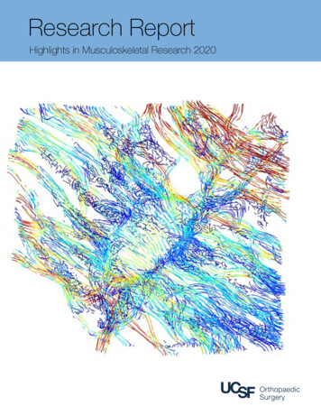
Transcription
Research ReportHighlights in Musculoskeletal Research 2020
Contents3Message from the Chair of theDepartment of Orthopaedic Surgery4NIH Ranking5Our Vision6Research Programs and Activities7 Orthopaedic Translational ResearchOrthopaedic Edge Innovations Laboratory8 Stem Cell LaboratorySkeletal Mechanobiology9 Developmental and Evolutionary Skeletal BiologyOrthopaedic Tissue Engineering and Regeneration10 Orthopaedic Biomechanics and Biotransport11 Skeletal Regeneration / Molecular andCellular Biology / Musculoskeletal Regeneration12 OTI Biomedical Engineering Lab26UCSF Musculoskeletal Research Consortium (METRiCS)27Human Performance Center28Core Center for Musculoskeletal Biology & Medicine29 Core Center for Disruptive Musculoskeletal Innovations(CDMI) Center for Dental, Oral, & Craniofacial Tissue &Organ Regeneration (C-DOCTOR), Industry Research Center30New Faculty32Residency Highlights34Awards, Grants, & Presentations36Trainee Highlights46News and Media62Grants and Fellowships71Research Publications96Philanthropy13 Laboratory for Evolutionary Anatomy14 International Research at IGOT16 OTI Clinical Research Center18 Pediatric Clinical Research20 Sports Medicine Patient-Centered Clinical Outcomes,Sports Medicine Clinical Trials in Knee and ShoulderSurgery, and Digital Health22 Youth Sports Injury Assessment and PreventionCenter, Hip Preservation Center23 Orthopaedic Clinical Research at the Spine Center24 Translational Quantitative Imaging Center25 Orthopaedic Oncology/OsseointegrationProduced by Maryam Farshad, Kathleen Jay, and Erin Simon.May 14, 2021Cover image: Visualization of flow lines from dynamic finite elementfluid modeling representing fluid shear within the pericellular spaceof a single osteocyte lacunae and its associated canaliculardendrites from mouse cortical bone. A special thanks to CharlieShurman (Graduate student, Alliston Lab) Stefaan Verbruggen,and Tamara Alliston, PhD.UCSF Department of Orthopaedic Surgery500 Parnassus Avenue, 3rd Floor Room MU-320WSan Francisco, CA 94117Phone: (415) 476-1166Fax: 476-1304orthosurgery.ucsf.edu
Message from the Chair of theUCSF Department of Orthopaedic SurgeryDear colleagues and friends,The year 2020 will be remembered as aremarkable year of change, challenge, and lossas well as growth, resilience, achievements andsuccess by our researchers.Our day-to-day lives may have been disruptedby the pandemic, but as a Department welearned to adjust and continued to focus on ourcore objectives: looking to the future,answering fundamental questions, andproviding the best available evidence-baseddata from all disciplines of musculoskeletalresearch. We continue to fuel curiosity byexchanging ideas and encouragingcollaborations in orthopaedic research -- basicscience, clinical and translational. Even if it'sover a Zoom.Building upon our sustainable model, we lookforward to continuing success, innovativestudies and evidence-based breakthroughs.Plans are underway to expand and remodellabs in 11 HSE and 95 Kirkham, and our facultyare actively engaged in the process of reenvisioning the entire research community onthe Parnassus campus.We are very proud of all of our researchers forriding out these challenges and slowly,carefully and safely returning to a new normal. Ilook forward to our research enterprise growththroughout 2021 and well beyond!In 2019, we took great pride in sharing thatUCSF was the top public recipient of fundingfrom the National Institutes of Health (NIH), andas a Department we received 39,103,498 inpeer-reviewed NIH research grants. AlthoughThomas Parker Vail, MD2020 posed challenges -- -- including safe waysJames L. Young Professorto re-open labs and conduct clinical researchChair, Department of Orthopaedic Surgerywith our patients-- nonetheless, our Departmentreceived an additional 5,127,654 in NIHFunding to continue our innovative research.3
04 1,192,058 1,050,344 964,495 1,545,182 1,430,868 026216651072011167101314612842231216Fiscal Year 10M 15M 40M 39,103,498 45M 5,127,654 8,406,993 7,852,165 4,825,089 4,440,346 4,440,346 3,695,186 2,217,717 1,864,351 2,536,542 1,085,135 1,256,412 1,720,695 1,638,764 732,376 1,409,230 184,0351997Rank for all institutions 77,032 5M1996Total Award DollarsNIH RankingNIH Research Grants for UCSF Department of Orthopaedic Surgery 35M 30M 25M 20M
Our VisionPioneering musculoskeletal discovery and innovative care to transform lives.Devante Horne, a graduate student, performs musculoskeletal reseach in the Lotz Laboratory forOrthopaedic Tissue Engineering and Regeneration on UCSF’s Parnassus campus.5
Research Programs and ActivitiesResearchers in the Department of Orthopaedic Surgery conduct innovative clinical, basic science, and translational research in musculoskeletalbiology to improve the delivery and outcomes of orthopaedic care. Neha Dole, PhD,above, performs musculoskeletal research in the AllistonLaboratory for Skeletal Mechanobiology at UCSF’s Parnassus Campus.Basic, Translational and Clinical ResearchThe UCSF Department of Orthopaedic Surgery has a diverseand broad basic and translational research program inmusculoskeletal biology. This is in addition to our clinical researchprogram, which spans all orthopaedic subspecialties. Each of ourvarious research programs are aimed at bringing new insights toour understanding of the musculoskeletal system. A major goal isto develop novel treatments for defects, diseases, conditions, andinjuries that affect musculoskeletal function. We are driven by thedesire to improve the delivery and outcomes of orthopaedic care.Additionally, the Department has a strong tradition in clinicalresearch across all subspecialties. Over the past decade, clinicalresearchers have established a large collaborative network bothwithin UCSF as well as with national and international clinicalresearchers. This has improved the impact and depth of ourclinical research.Over the past year, clinical research has been published in allmajor orthopaedic surgery journals including the Journal of Bone& Joint Surgery (JBJS), Journal of Shoulder and Elbow Surgery(JSES), Journal of Orthopaedic Trauma (JOT), Spine journal,6Journal of Pediatric Orthopaedics (JPO), Clinical Orthopaedicsand Related Research (CORR), and the American Journalof Sports Medicine (AJSM). Faculty, fellows, and residentspresented at American Academy of Orthpaedic Surgeons (AAOS),Orthopaedic Research Society (ORS), the American OrthopaedicSociety in Sports Medicine (AOSSM), International Society ofArthroscopy, Knee Surgery and Orthopaedic Sports Medicine(ISAKOS), the Hip and Knee Society, and the OrthopaedicTrauma Association (OTA), among other national and internationalmeetings. For a full list of our departmental contributions tothe 2020 AAOS and ORS conferences, please visit hile the individual projects are too numerous to list in detail,there have been several highlights of collaborative researchacross spine surgery, osseointegration, 3D printing for improvingsurgical outcomes, shoulder arthroplasty and instability, imaginganalysis using high resolution MRI and CT, global health throughUCSF’s Institute for Global Orthopaedics and Traumatology(IGOT), pediatrics and pediatric sports medicine.
Orthopaedic Translational ResearchUCSF VA Health Center, Research Facility at Mission BayThe Laboratory for Orthopaedic Translational Research is directedby Hubert Kim, MD, PhD and Alfred Kuo, MD, PhD at the UCSFVA Research Facility at Mission Bay.The focus of the team’s research effort is to examine themolecular and cellular mechanisms responsible for secondaryinjury cascades that are set in motion after trauma. There isparticular interest in tissues that have an extremely limitedcapacity for healing and regeneration, where preservation ofexisting cells and tissue may be of great clinical significance. Theintention is to apply lessons learned in the laboratory to designbetter treatments for patients.Additionally, Brian Feeley, MD directs the Laboratory for Stem CellRegeneration and Translational Research, located on the UCSF/VA Mission Bay campus focusing on muscle injury problems.Brian Feeley, MD collaborates with Xuhui Liu, MD and researchersat UCSF on developing models to study the molecularmechanisms and cellular mechanisms that are responsible for thedevelopment of muscle atrophy and fatty infiltration after rotatorcuff tears.The focus of the research is to understand the cellular andmolecular changes that occur within the muscle after differentinjuries, but particularly rotator cuff tears. They have developednovel injury and repair models to study the acute and chronicStem cells found within the rotator cuff musclecan be stimulated into fibrotic tissue (red) or fattissue (green) depending on the stimulus (FeeleyLiu Laboratory for Stem Cell Regeneration andTranslational Research)effects of rotator cuff injury on the important signal transductionpathways that govern muscle cell size and stem cell fate withinthe muscle. They also focus on understanding how muscleinjury patterns affect the stem cell populations within the muscle(satellite cells, FAP cells) in an effort to determine treatmentstrategies that would improve muscle function after orthopedicinjuries.Within the UCSF VA Health Center, the Orthopaedic RapidIntelligent Fabrication Group led by Alan Dang, MD and AlexisDang, MD focus on translating orthopaedic ideas into orthopaedicproducts. They maintain a 3-axis CNC mill as well as a small fleetof 3D printers with customized extruders, firmware, and othersoftware optimizations. Active projects include the developmentof advanced surgical lighting technology as well as surgicalinstrumentation and implants.Orthopaedic Edge Innovations LaboratoryMulti-Campus LaboratoryThe Edge Innovations Lab is led by Aenor Sawyer, MD, MS,Alexis Dang, MD and Alan Dang, MD and is focused onEngineering, Designing, and Growth Enabling digital (EDGE) andmanufacturing technologies.This group is responsible for clinical 3D printing across themany campuses of the Department including UCSF ParnassusHeights, The Orthopaedic Institute at Mission Bay, ZSFGH, SFVAHC, UCSF Benioff Children’s Hospital Mission Bay, and UCSFBenioff Children’s Hospital Oakland. Currently, the focus is on 3Dprinting of Precision Anatomic Models for surgical pre-operativeplanning and conducting the research to assess the efficacy andeconomics of the technology.As a result of their work in the 3D imaging arena, Alexis Dang,MD and Alan Dang, MD won the San Francisco Federal ExecutiveBoard “Federal Employee of the Year” award in Science &Technology related to 3D printing in orthopaedics. public-servicerecognition/. The Board represents approximately 70,000 federal,postal and military employees throughout the nine bay areacounties (Alameda, Contra Costa, Marin, Napa, San Francisco,San Mateo, Santa Clara, Solano, Sonoma), as well as agencies inthe Sacramento area.Edge Innovation Laboratory is led by Aenor Sawyer MD, MD,Alexis Dang, MD, and Alan Dang, MD.Additionally, Dr. Aenor Sawyer, Dr. Alexis Dang and Dr. AlanDang spearheaded a multidisciplinary initiative, together with thePediatric Heart group and Radiology, to develop 3D printingtechnologies at UCSF. The “ ” includes augmented reality, virtualreality, and 4D imaging (3D-imaging with a time component). Thishas received 1.4 million in funding.7
Human muscle stem cells and regeneration (Brack Laboratory for SkeletalMuscle Regeneration and Aging).Image by Annarita Scaramozza, PhDStem Cell LaboratoryEli and Edythe Broad Center of Regeneration Medicine andStem Cell Research on Parnassus HeightsThe Brack Laboratory for Skeletal Muscle Regeneration and Aging is directed by Andrew Brack, PhD, and focuseson the development of strategies to accelerate skeletal muscle repair.During aging or in response to radiotherapy, the capacity for muscle repair is diminished, leading to reduced mobility and strength.The Brack Lab uses state of the art machine learning and molecular biology to determine the causes of muscle dysfunctionand identify strategies to rejuvenate the regenerative potential ofskeletal muscle.In the future, the Brack Lab hopes that current projects will leadto strategies that reverse aging and improve recoveryafter radiotherapy.Andrew Brack, PhD, has developed collaborations with clinical faculty, including sports medicine and oncology. Active studies includestudies on muscle aging and muscle recovery after radiotherapy.Skeletal MechanobiologyUCSF Parnassus HeightsThe Laboratory for Skeletal Mechanobiology is directed by Tamara Alliston, PhD.The Alliston Laboratory investigates the molecular pathwayscontrolling skeletal cell behavior, how these pathways coordinatewith physical cues to influence mechanical integrity of healthybone and cartilage, and how they can be harnessed to repairtissue damaged in degenerative skeletal diseases like osteoporosisand osteoarthritis. To answer these questions they combinemolecular, cellular, physiologic, and materials science approaches.In particular, they seek to define the function of TGF insynergistically coordinating physical and biochemical cues inbone and cartilage cells. Since TGFβ is a powerful regulatorof homeostasis throughout the skeleton, understanding thissignaling pathway has helped their team uncover fundamentalnew cellular mechanisms that participate in skeletal health anddisease.This research has provided important new insight on factorsthat cause common musculoskeletal problems, like joint injuries,osteoarthritis, and bone fragility in aging men and women. Nowthe research team is building on what they have learned in thelaboratory to discover new therapeutic strategies to preventskeletal disease and to improve skeletal repair.Osteocyte canalicular networks visualized in silver stained bone.Image by Charlie Schurman 2018 (Alliston Laboratory for SkeletalMechanobiology)8
(A) Skeletal muscle from a duck embryo showing muscles of the head and neck (stained pink with an anti-myosin antibody). (B) At highermagnification (dashed inset box), striated fibers of the jaw closing muscles and their insertions points within skeletal precursor cells can beobserved (Schneider Laboratory for Developmental and Evolutionary Skeletal Biology, confocal images by Dr. Jessye Aggleton).Development and Evolutionary Skeletal BiologyUCSF Parnassus HeightsThe Schneider Laboratory for Developmental and Evolutionary Skeletal Biology is directed by Richard A. Schneider, PhD.Research is broadly aimed at understanding how thedeveloping musculoskeletal system achieves its structural andfunctional integration.To address this question, the lab has created a unique surgicaltransplantation system that involves embryos from two distincttypes of birds (quail and duck), which differ considerably in theirfunctional anatomy and growth rates.Transplanting skeletal and other progenitor cells between themchallenges the resulting chimeric “quck” and “duail” embryos tointegrate two different species-specific developmental programs.By focusing on donor- versus host-controlled changes toembryonic patterning and growth, this strategy has illuminatedmolecular and cellular mechanisms that regulate themusculoskeletal system and enable bones, cartilages, tendons,muscles, and other tissues to achieve their proper size, shape,orientation, and integration.A goal is to devise novel molecular- and cell-based therapies forrepairing and regenerating musculoskeletal tissues affected bybirth defects, disease, and injury. Work from the Schneider Labhas also helped elucidate the role of development in evolution.Orthopaedic Tissue Engineering and RegenerationUCSF Parnassus HeightsThe Orthopaedic Tissue Engineering and Regeneration Laboratory isdirected by Jeffrey C. Lotz, PhD.The Lotz Laboratory is devoted to conducting basic researchin several areas of orthopaedics including biomechanics of thespine, knee, and hand. Biomechanical studies serve to investigatethe physical properties of musculoskeletal (MSK) tissues, as wellas functional performance of MSK patients.The Lotz Laboratory is collaborating with UC Berkeley engineersto design and validate in-clinic tools and sensors that quantifypatient movement and augment traditional physical tests andpatient-reported data. Similar studies are being conductedwith NASA astronauts to understand the adverse effects ofmicrogravity, and to develop countermeasures to maintainastronaut health and safety on long-duration space flight,The Lotz Laboratory has pioneeredbiomechanical, anatomic, and imagingstudies of the human disc/vertebra interface(Lotz Laboratory for Orthopaedic TissueEngineering and Regeneration).such as the planned Mars missions. Additionally, they havefocused on understanding the etiology of different diseases(e.g., disc degeneration, osteonecrosis) and comorbidities (discdegeneration and diabetes).In the area of regenerative medicine, the Lotz laboratory isexploring various uses of mesenchymal stem cells for newtherapies for disc, cartilage, and bone regeneration.The diverse research team includes bioengineers, biologists,biochemists, histologists, and orthopaedic surgeons.9
FieldsLaboratoryBiomechanicsand (FieldsBiotransportNutrienttransport forandOrthopaedicintervertebraldisc cell survivalLaboratory for Orthopaedic Biomechanics and Biotransport)Orthopaedic Biomechanics and BiotransportUCSF Parnassus HeightsThe Orthopaedic Biomechanics and Biotransport Laboratory is directed by Aaron Fields, PhD.The broad research interests of the Fields Lab are related tostructure-function relationships in musculoskeletal tissues, with aparticular focus on the mechanisms of nutrient transport in boneand cartilage, and harnessing nutrient transport for tissue repairand regeneration.The lab combines engineering and biology approaches for: (1)understanding the effects of aging and disease on structuretransport relationships, and (2) developing translatable diagnosticand therapeutic strategies. An overall theme of this researchis the use of advanced experimental and computationaltools to measure how tissue constituents at the nano- and10microscales impact whole-organ behavior. The research involvesclose collaborations with clinicians including spine surgeons,physiatrists, and radiologists.Active projects include: (1) translational studies aimed atharnessing nutrient transport for disc repair and regeneration;(2) clinical studies testing new diagnostic MRI tools for selectingpatients that would most benefit from disc regenerativetherapies; and (3) basic science studies comparing healthyvs. pathologicdisc cell phenotype. These studies are fundedby grants from the National Institutes of Health and the NorthAmerican Spine Society.
Skeletal Regeneration/Molecular and Cellular BiologyZuckerberg San Francisco General Hospital (ZSFGH)The Molecular and Cellular Biology Laboratory is directed by Ralph Marcucio, PhD, and Ted Miclau, MD.The major focus of the work performed is to examine theprocesses that occur during bone regeneration after traumaticinjury. Understanding the events that occur during fracture repairis essential for developing therapies to help people that exhibitdifficulties in bone healing. For example, delayed or non-unionafflict approximately 10% of all people undergoing fracturerepair. By understanding how the body normally respondsto orthopaedic trauma, they are laying the foundation for thedevelopment of new therapeutic regimens to treat a wide varietyof skeletal pathologies.research community. Current areas of study include, the role ofmuscle in bone healing, the role of inflammation in bone healing,the role of angiogenesis in bone healing, genotype-phenotypecorrelations during skeletal development, and the role ofcontinuous phenotypic variation in disease production.Image is from the Molecular and CellularBiology Laboratory.The research utilizes a murine tibia fracture model that wasdeveloped by members of the laboratory and is used in otherlaboratories throughout the national and international orthopaedicMusculoskeletal RegenerationZuckerberg San Francisco General Hospital(ZSFGH)The Laboratory for Musculoskeletal Regeneration isdirected by Chelsea S. Bahney, PhD.The Bahney Laboratory utilizes a developmental engineeringapproach to discover novel therapeutic targets for regenerativemedicine by first studying the normal mechanisms of repair, thenutilizing engineered biomaterials to deliver bioactive signals topromote improved regenerative outcomes.Visualization of the chondro-osseous transition zone in a fracturecallus. (A-C) Low magnification of a murine fracture callus, outlined withblack dashed line, stained with (A) Safranin-O/Fast Green (SO/FG), (B)Modified Milligan’s Trichrome (TC) or (C) Hall and Brunt Quadruple Stain(HBQ). (D-F) A magnified region of cartilage and bone from the fracturecallus, outlined with a red box (A-C), with the TZ indicated by blackbrackets. (G-I) High magnification images of the TZ show the invadingvasculature and the chondro-osseous junction. (Bahney Laboratory forMusculoskeletal RegenerationMusculoskeletal Regeneration).Currently, the focus of the Bahney Lab is primarily on the processof cartilage turning into bone, either naturally during fracturerepair, or in disease processes such as osteoarthritis.A long-term research goal is to translate new biologics thatchange healthcare options in fracture healing and post-traumaticosteoarthritis.11
Laboratory for Evolutionary AnatomyZuckerberg San Francisco General Hospital (ZSFGH)The Laboratory for Evolutionary Anatomy is directed by Nathan Young, PhD.The Young Laboratory addresses biomedical basic researchthrough the lens of evolution, utilizing functional compromise andhistorical constraint as fundamental explanatory principles. Whencombined with mechanistic insights from experimental systems,this approach yields significant insights into the generation ofindividual phenotypes, both normal and abnormal.The lab research program combines classical embryology, modernexperimental and genetic tools, and advanced methods forquantifying and comparing phenotypes at a range of scales.This approach has significance for understanding not only theprocesses that contribute to and constrain evolutionary diversity,but also the individual phenotypic differences found within speciesand among individuals, including dysmorphologies associatedwith human disease states. Research includes the study of normalmechanisms of development as well as the etiology of congenitaldevelopmental defects, and is strongly relevant to longstandinggoals of providing personalized and predictive medicine.Comparison of facial development from embryos to adults in mouse, human, alligator, and chicken (YoungLaboratory for Evolutionary Anatomy).12
Testing facility, Safa Herfat PhD, Transtibial prosthetic socket being 3D printed for a patient at the OTI O&P clinic(OTI Biomedical Engineering Lab).Orthopaedic Trauma Institute (OTI) Biomedical Engineering LabZuckerberg San Francisco General Hospital (ZSFGH)Directed by Safa Herfat, PhD, the OTI Biomedical Engineering Lab specializes in experimental biomechanical testingand finite element analysis of orthopaedic fracture fixation strategies and implants.The lab also collaborates with the UCSF Orthotics & Prostheticsclinics on prosthetic innovation projects incorporating 3Dscanning and printing to design and manufacture patient-specificprosthetic solutions. The lab houses its own 3D lab, with four 3Dprinters, a high accuracy white light 3D scanner, and a highend design workstation. A Hearts Grant from the San FranciscoGeneral Hospital Foundation has also generously funded a large3D printer capable of printing large lower limb sockets, as well asfunding the development of a custom pressure sensor system toobjectively monitor prosthesis fit in the clinic.The lab is collaborating with other UCSF and UC Berkeley labs onan NSF grant-funded project to develop an implantable sensor tomonitor fracture healing.13
International Research at the Institute for Global Orthopaedics andTraumatology (IGOT)Zuckerberg San Francisco General Hospital (ZSFGH)IGOT Research Projects Led by Saam Morshed, MD,MPH, PhD and David Shearer, MD, MPH,:Tanzania- Intramedullary Nailing Versus External Fixation forOpen Tibia Fractures Randomized Controlled Trial Long-TermFollow-UpOpen tibia fractures are among the most common and debilitatinginjuries faced in low-income countries due to high rates of infectionand nonunion. This study aims to address the question of whetherinternal or external fixation is better as definitive treatment for opentibia fractures in Tanzania. The study has enrolled and randomized240 patients and achieved greater than 90% 1 year follow up. Thestudy is currently conducting final data analysis, and we anticipatepublication in the near future.Tanzania- Cost-effectiveness of Prosthetics forAbove Knee AmputeesAlthough the need for greater access to prosthetic services in lowand middle-income countries is well-established, literature on thelong-term sustainability and cost-effectiveness of these prosthesesin low-resource settings is lacking. This study will providelongitudinal follow-up to a previously conducted cost analysis ofabove knee prostheses in a low-resource setting, examining theoriginal target study population to assess the long-term durabilityand impact of the prosthesis on quality of life, functional outcomes,and cost-effectiveness. The study is currently awaiting Tanzanianethical approval, and we anticipate beginning patient enrollmentand data collection in the near future.Tanzania- Cost-effectiveness of Prosthetics forBelow Knee AmputeesLimb loss has profound economic, social, and psychologicaleffects on both the individual and the community, and theseeffects are especially pronounced in low- and middle-incomecountries. However, countries with already strained financialresources rarely prioritize rehabilitation services, and thus there is aneed for economic analyses evaluating cost-effective, sustainable,and appropriate prosthetic technology in LMICs. This multisite,prospective cohort study aims to measure the impact of belowknee prostheses on quality of life, functional outcomes, andcost-effectiveness in a low-resource setting. The study is currentlyawaiting Tanzanian ethical approval, and we anticipate beginningpatient enrollment and data collection in the near future.Tanzania - Low-cost Intramedullary K-wires forPediatric Femur FracturesFemoral shaft fractures in children are commonly treated withsurgery using flexible nails to avoid damage to growth plates.However, titanium flexible nails that are commonly used inhigh-income countries are cost-prohibitive for many familiesin low-income countries where governments do not subsidizeimplant costs. Substituting titanium flexible nails with stainlesssteel “Kirschner wires” could reduce the cost of these implantsnearly 40-fold, thereby markedly increasing access to surgery for14children globally. IGOT is supporting a randomized controlled trialin Tanzania comparing these low-cost implants to the high-costtitanium nails for children with femoral shaft fractures.Tanzania - A Pilot Masked, Randomized Controlled TrialEvaluating Locally-applied Gentamicin versus Saline inOpen Tibia Fractures (pGO-Tibia)Tibial shaft fractures are the most common long-bone fracture.Deep infection remains a common, devastating complication ofopen injuries leading to lifelong impairment that disproportionatelyaffects low- and middle-income countries (LMICs). Thoroughsurgical debridement, followed by fracture stabilization usinginternal or external fixation, is the mainstay of treatment. Oneproposed adjunctive measure is prophylactic local antibioticdelivery, which can achieve much higher antibiotic concentrationsat the surgical site than can be achieved safely with systemicadministration. There is a growing body of literature evaluatinglocal antibiotic administration in both aqueous and powder format the time of wound closure. While demonstrating potentiallypromising results, these studies are heterogeneous, of poorgeneral methodologic quality, and none originate from LMICswhere this technique would have the greatest potential benefits.Local gentamicin is particularly promising given the broadspectrum of activity against common pathogens in osteomyelitis(staphylococcus, gram-negative rods), wide availability, and lowcost ( 1 per 80mg vial). IGOT propose a prospective, blinded,randomized controlled trial enrolling adult open tibial shaft fracturesat the Muhimbili Orthopaedic Institute (MOI) in Dar es Salaam,Tanzania. At the time of initial debridement, participants will berandomly assigned to receive aqueous gentamicin after closure orplacebo saline injection. The primary outcome will be deep surgicalsite infection at 1 year. Secondary outcomes include health-relatedquality-of-life (HRQOL), modified Radiographic Union Scale forTibial Fractures (mRUST), FIX-IT score for clinical healing, and costof treatment using time-driven activity-based costing (TDABC) andsurvey methods.Tanzania – Total Joint Replacement RegistryOur partners at Muhimbili Orthopaedic Institute in Tanzaniacreated an excel sheet registry ten years ago with 900 patients.This project will use this excel sheet to create and implementa formal registry prospectively using REDCap for total jointreplacement procedures.Africa– Open tibial shaft fracture treatment practices in AfricaOpen tibial shaft fractures are the most common open long bonefracture and a significant source of morbidity and mortality in lowand middle-income countries (LMICs). Appropriate managementof open tibia fractures includes timely antibiotic administration,surgical debridement, and definitive stabilization, which havebeen
UCSF's Institute for Global Orthopaedics and Traumatology (IGOT), pediatrics and pediatric sports medicine. Researchers in the Department of Orthopaedic Surgery conduct innovative clinical, basic science, and translational research in musculoskeletal biology to improve the delivery and outcomes of orthopaedic care.