Transcription
Ni et al. Stem Cell Research & Therapy(2021) SEARCHOpen AccessMiR-375 reduces the stemness of gastriccancer cells through triggering ferroptosisHaiwei Ni1, Hai Qin1, Cheng Sun2, Yichen Liu1, Guojing Ruan1, Qianqian Guo3, Tao Xi1*, Yingying Xing1* andLufeng Zheng1*AbstractBackground: Gastric cancer stem cells (CSCs) are the main causes of metastasis and drug resistance. We previouslyindicated that miR-375 can inhibit Helicobacter pylori-induced gastric carcinogenesis; here, we aim to explore theeffects and mechanisms of miR-375 on gastric cancer (GC) cell stemness.Methods: Lentivirus infection was used to construct GC cells with ectopic expression of miR-375. In vitro andin vivo experiments, including analysis of tumor spheroid formation, CD44 sub-population with stemness,stemness marker expression, and tumor-initiating ability, were performed to evaluate the effects of miR-375 on thestemness of GC cells. Furthermore, microarray and bioinformatics analysis were performed to search the potentialtargets of miR-375 in GC cells. Luciferase reporter, RNA immunoprecipitation, and RNA-FISH assays were carried outto verify the targeting of miR-375. Subsequently, combined with tissue microarray analysis, erastin-resistant GC cells,transmission electron microscopy, a series of agonists and oxidative stress markers, the underlying mechanismscontributing to miR-375-mediated effects were explored.Results: MiR-375 reduced the stemness of GC cells in vitro and in vivo. Mechanistically, SLC7A11 was identified as adirect target of miR-375 and miR-375 attenuated the stemness of GC cells mainly through triggering SLC7A11dependent ferroptosis.Conclusion: MiR-375 can trigger the ferroptosis through targeting SLC7A11, which is essential for miR-375mediated inhibition on GC cell stemness. These results suggest that the miR-375/SLC7A11 regulatory axis couldserve as a potential target to provoke the ferroptosis and thus attenuate the stemness of GC cells.Keywords: MiR-375, SLC7A11, Ferroptosis, Stemness, Gastric cancerIntroductionGastric cancer (GC) ranks fifth in incidence among allmalignant tumors worldwide and the mortality rate isthird (powered by GLOBOCAN 2018); this is closelydue to tumor relapse, metastasis, and drug resistance.Cancer stem cells (CSCs) have been regarded as the rootof tumor relapse, metastasis, and drug resistance [1, 2].* Correspondence: Xitao18@hotmail.com; cpuskyxyy@126.com;zhlf@cpu.edu.cn1School of Life Science and Technology, Jiangsu Key Laboratory ofCarcinogenesis and Intervention, China Pharmaceutical University, 639Longmian Road, Nanjing 211198, People’s Republic of ChinaFull list of author information is available at the end of the articleAs a consequence, it may facilitate GC treatment to elucidate the underlying mechanisms contributing to theprogression gastric CSCs.Ferroptosis, a newly established form of cell death, isdifferent from conventional programmed death such asapoptosis, necrosis, and autophagy [3]. Biochemically,ferroptosis is initiated by oxidative perturbations ofintracellular microenvironment which involves lipid peroxidation, lethal reactive oxygen species (ROS), and freeferrous iron (Fe2 ) [4]. Although cancer cells usuallyhave a higher level of ROS, which is essential for tumorprogression, such as migration, proliferation, and The Author(s). 2021 Open Access This article is licensed under a Creative Commons Attribution 4.0 International License,which permits use, sharing, adaptation, distribution and reproduction in any medium or format, as long as you giveappropriate credit to the original author(s) and the source, provide a link to the Creative Commons licence, and indicate ifchanges were made. The images or other third party material in this article are included in the article's Creative Commonslicence, unless indicated otherwise in a credit line to the material. If material is not included in the article's Creative Commonslicence and your intended use is not permitted by statutory regulation or exceeds the permitted use, you will need to obtainpermission directly from the copyright holder. To view a copy of this licence, visit http://creativecommons.org/licenses/by/4.0/.The Creative Commons Public Domain Dedication waiver ) applies to thedata made available in this article, unless otherwise stated in a credit line to the data.
Ni et al. Stem Cell Research & Therapy(2021) 12:325survival [5, 6], while CSCs are believed to possess alower level of ROS [7]. Thus, it might be effective toeliminate CSCs by stimulating ROS generation [8]. Simultaneously, cancer cells can be eliminated via elevatingROS with inhibiting antioxidant defense [9]. Therefore,it encourages us to focus on a ROS-dependent cell deathmechanism for gastric CSCs. Notably, iron metabolism[10, 11] and ferroptosis have recently been shown as apromising therapeutic strategy to eliminate CSCs [12–14]. Meanwhile, ferroptosis is associated with chemoresistance and the anti-tumor efficacy of immunotherapy[15, 16]. However, the involvement of ferroptosis in gastric CSC progression was still confusing.A number of genes required for ferroptosis have beenidentified, including those involved in lipid peroxidationand amino acid metabolism, in which system xc-, asodium-independent antiporter of cystine and glutamate,takes part in amino acid metabolism [3]. Cystineglutamate antiporter xCT (SLC7A11), which is the subunit of system xc-, together with SLC3A2 (CD98), is selective for uptaking cystine into cells in exchange forglutamate [17]. This system synthesizes glutathione(GSH) by taking extracellular cystine into cells and thusrepresses oxidative stress response during ferroptosis.Many studies have found that xCT is abnormallyexpressed due to the overloaded oxidative stress and increased nutrient metabolism requirements in variouscarcinomas [18]. CD44, which is the cell adhesion molecule, has been identified as a marker of gastric CSCs[19, 20]. The previous studies have shown that a CD44variant (CD44v) interacts with xCT [21, 22] andSLC7A11 overexpression is associated with increasedCSC-like properties [23]. However, the relationship between SLC7A11 and the stemness in GC is stillundefined.MicroRNAs are short endogenous and evolutionarilyconserved single-strand RNA molecules, approximately19 to 25 nucleotides in size [24] and have been widelyshown to be associated with CSC-like traits [25]. MiR375 is discovered as a pluripotent miRNA involved inpancreatic islet development, glucose homeostasis, andcell differentiation [26]. Additionally, miR-375 has beenidentified as a tumor suppressor in various tumors [27,28]. Notably, miR-375 is found to be lowly expressedand considered as a possible biomarker in GC [29, 30].We previously showed that miR-375 can inhibit Helicobacter pylori-induced gastric carcinogenesis [31]. Furthermore, ours and other studies have indicated thatmiR-375 can suppress the stemness of breast cancercells [32]. Thus, we assume that miR-375 may play critical roles in GC cell stemness.Here, we found that miR-375 suppressed the stemnessof GC cells in vitro and in vivo. Further mechanisticstudies revealed that miR-375 could induce ferroptosisPage 2 of 17by targeting SLC7A11 and thus attenuate the stemnessof GC cells. This study identified a novel miR-375/SLC7A11 regulatory axis responsible for GC cell stemness by mediating ferroptosis. Since conventional chemotherapeutic drugs which induce cancer cell apoptosishave been confirmed to induce cancer cell stemness anddrug resistance, this miR-375/SLC7A11 regulatory axismight serve as a potential target to suppress the stemness of GC cells via triggering ferroptosis and provideclues for the development of ferroptosis inducer.Materials and methodsCell culture and chemical agentsHuman GC cell lines SGC-7901 and BGC-823 were obtained from China Academia Sinica Cell Repository(Shanghai, China). Human gastric mucosal epithelial cellline GES-1 was a gift from the Shanghai Institute of Digestive Disease. Cells were cultured in RPMI-1640medium (KeyGEN BioTECH, Nanjing, China) supplemented with 10% fetal bovine serum (FBS, Gibco, GrandIsland, NY, USA), 100 μg/mL streptomycin, and 100units/mL penicillin [33, 34]. HEK-293 T cells preservedin our laboratory were cultured in high glucose DMEM(KeyGEN BioTECH, China). All cell lines were incubated at 37 C in 5% CO2. Ferroptosis agonists erastin,sorafenib, and ferroptosis inhibitor Ferrostatin-1 (Fer-1)were purchased from MCE (MedChemexpress, USA).Another ferroptosis agonist sulfasalazine (SAS), pancaspase inhibitor Z-VAD-FMK, and necrosis inhibitorNecrostatin-1 (Nec-1) were purchased from TargetMol(Target Molecule Corp, USA).Plasmids, miR-375 mimics, and siRNA transfectionWhen cell growth reached 40–50% in density, cells wereused for transfection using jetPRIME transfection reagent (Polyplus Transfection, France) following the manufacturer’s protocol. MiR-375 mimics and miRNA NC(negative control) were synthesized by GenePharma(Shanghai, China). Small interfering RNA (siRNA) ofSLC7A11 (5′-GGAGTTATGCAGCTAATTA-3′; 5′GGGTGGAACTCCTCATAAT-3′; 5′-GGGAACAACTATAAAGAAA-3′) or si-NC (5′-TTCTCCGAACGTGTCACGT-3′) were synthesized by Biomics Biotechnology(Nantong, China). SLC7A11 overexpression vectorpcDNA3.4-SLC7A11 and empty plasmid (NC) were purchased from MiaolingBio (Wuhan, China). For luciferasereporter assay, the sequences of SLC7A11 3′UTR (WT)containing miR-375 binding site and SLC7A11 3′UTR(MUT) containing mutant miR-375 binding site werecloned into pMIR-Report, respectively. The detailed procedure for plasmid construction was referred in our previous work [35]. All plasmids were verified by DNAsequencing.
Ni et al. Stem Cell Research & Therapy(2021) 12:325Page 3 of 17Quantitative real-time PCR (qRT-PCR)Microarray analysisTotal RNA was extracted from GC cell lines using TRIzol(TransGen Biotech, China) following the standard recommendation. The cDNA was reversely transcribed with MMLV (Vazyme, Nanjing, China). MiRNA and U6 snRNART primer mix and miRNA qRT-PCR primer set (GenePharma, China) were used to detect and quantify miRNAexpression. SYBR Green master mix (Vazyme) was usedfor qRT-PCR assay on an ABI Prism 7500 Sequence Detector (Applied Biosystems, USA). The primer sequencesused were listed as below: SLC7A11 forward, 5′-TCTCCAAAGGAGGTTACCTGC-3′; SLC7A11 reverse, 5′AGACTCCCCTCAGTAAAGTGAC-3′; GAPDH forward, 5′-TGTGGGCATCAATGGATTTGG-3′; GAPDHreverse, 5′-ACACCATGTATTCCGGGTCAAT-3′. ThemRNA and miRNA levels were normalized to an internalcontrol (GAPDH or U6 snRNA), respectively. The relativeexpression level was calculated using 2 ΔΔCt method [36].Total RNA was extracted in SCG7901 cells with or without miR-375 stable overexpression using Trizol reagent(Life Technologies, Carlsbad, CA, USA) and purifiedwith an RNeasy mini kit (Qiagen, Valencia, CA, USA).Biotinylated cDNA was prepared according to the standard Affymetrix protocol from 250 ng total RNA usingAmbion WT Expression Kit. Following labeling, fragmented cDNA was hybridized for 16 h at 45 C on Clariom D Assay (Human, Affymetrix). GeneChips werewashed and stained in the Affymetrix Fluidics Station450. All arrays were scanned by using Affymetrix GeneChip Command Console (AGCC) which was installed inGeneChip Scanner 3000 7G [40]. The operation ofmicroarray analysis was outsourced by Beijing CnkingbioBiotechnology Corporation (Cnkingbio, China). All datawas deposited in Gene Expression Omnibus (GEO) repository (GSE147698).Construction of erastin-resistant GC cellsWestern blottingErastin-resistant cells were constructed as a ferroptosisresistant model [16]. To generate erastin-resistant GCcells, BGC-823 or SGC-7901 cells were seeded in 24well culture plates and treated with erastin (10 μM) fortwo weeks. Fresh culture medium containing erastin waschanged every 2 days. Then, surviving clones were collected and further amplified. The resistance of erastinwas confirmed by cell viability assay [37].The detailed procedure was described in our previousstudy [41]. Firstly, cells were scraped from the 6-wellculture plates for centrifugal collection at 300 g for 5min at 4 C. Cell-derived protein lysate was harvested byRIPA lysate (Beyotime, China). Ten micrograms of protein was fractionated by 10% SDS/PAGE, transferred topolyvinylidene difluoride membranes, and then incubated with the primary antibody at 4 C overnight. Afterincubating with a secondary antibody for 45 min, theECL system was used to detect the intensity of proteinexpression [42, 43]. Primary antibodies were SLC7A11/xCT (1:1000, proteintech, China), ALDH1A1 (1:1000,proteintech, China) and Oct3/4 (1:1000, Wanlei, China),CD44 (1:1000, Wanlei, China), and GAPDH (1:5000, YIFEI XUE BIOTECHNOLOGY, Nanjing, China) was usedas an internal reference.Stable cell line construction with lentivirus infectionBriefly, miR-375, SLC7A11 coding, and SLC7A11shRNA sequences were sub-cloned into pGLV3/H1/GFP Puro Vector (LV3 lentiviral vector) (GenePharma),respectively. The lentivirus package was performed byGenePharma. The final viral titer was 1 108 TU/mLand 20 μl lentivirus was added into 1 mL medium to infect GC cells. Stable cell lines were selected with puromycin (Sigma, 2 μg/mL) for 2 weeks. Fluorescenceobservation and qRT-PCR were used to verify infectionefficiency [38].Tissue microarray analysisThe tissue microarray was purchased from OutdoBiotech (Shanghai, China), which contains 17 primary GCtissues, 12 normal adjacent tissues, 16 metastatic lesions,and 5 normal gastric mucosa tissues [39]. Further immunohistochemistry assay was performed by Servicebio(Wuhan, China) to detect SLC7A11 expression whichwas quantified using Quantity-one Software. Additionally, RNA-fluorescence in situ hybridization assay wasperformed by Boster Biological Technology to detectmiR-375 and SLC7A11 levels in this tissue microarray.RNA immunoprecipitation (RIP) assayRIP assay was performed using the Protein A/G AgaroseResin (YEASEN, China) following the manufacturer’sprotocol. For RIP assay, SLC7A11 3′UTR sequencescontaining miR-375 binding sites or mutated bindingsites were inserted into pcDNA3.1 ( ) plasmid, namedas SLC7A11 3′UTR (WT) or SLC7A11 3′UTR (MUT),respectively. SGC-7901 and BGC-823 cells were transfected with SLC7A11 3′UTR (WT) and SLC7A11 3′UTR (MUT). After 24 h, cells were extracted and lysedby NP-40 lysis buffer (Beyotime, China). After that,100 μL lysate was incubated with NP-40 buffer containing Protein A/G Agarose Resin bound with human antiAgo2 antibody (Cell Signaling Technology, Danvers,MA, USA) at 4 C overnight. Agarose resin was isolatedby centrifugation and digested with protease K (YEAS
Ni et al. Stem Cell Research & Therapy(2021) 12:325EN) to isolate Ago2-RNA complex from the beads. qRTPCR was performed to detect miR-375 level [44].RNA-fluorescence in situ hybridization (RNA-FISH)RNA-FISH for miR-375 and SLC7A11 was performedon GC cells. Cell slides fixed with 4% paraformaldehydewere treated with several buffers in RNA-FISH Kit (GenePharma). After denaturing at 73 C for 5 min, probemixture was hybridized in cell slide overnight in darkness at 37 C. 5′CY3-labeled Locked Nucleic Acid probestargeting miR-375 and 5′FAM labeled Locked NucleicAcid probes targeting SLC7A11 (GenePharma). Afterwashing, the signal was observed under confocal microscope. Detailed procedures were followed by the standard protocol (GenePharma) [44].Dual-luciferase reporter assayMiR-375 mimics or miR-375 NC and SLC7A11 3′UTR(WT), SLC7A11 3′UTR (MUT), or pMIR-Report wereco-transfected into HEK293T cells using JetPRIME.After 48 h, cell lysate was collected following the instructions’ protocol of Dual Luciferase Reporter Gene AssayKit (Yeason, China). The luciferase activity was quantified by POLARstar Omega multimode microplatereader. The relative luciferase activity was calculated byfirefly luciferase activity/Renilla luciferase activity [44].Flow cytometry analysisBGC-823 and SGC-7901 cells were seeded into 6-wellplates and transfected with miR-375 mimics, si-SLC7A11,pcDNA3.4-SLC7A11(SLC7A11), or pcDNA3.4 (NC). Flowcytometry analysis was performed to detect CD44 subpopulation [19]. Briefly, cells were harvested at a densityof 106 cells/mL by centrifugation at 300 g and 4 C for 5min. Cells were stained with anti-CD44-APC (BD Biosciences, USA) or an isotype control at 4 C in darkness. Thestained cells were washed using PBS and measured byflow cytometer (BD, USA) [43].Cell viability assay (Cell Counting Kit-8)Cell viability was assessed using CCK-8 (Cell CountingKit-8) assay (Target Mol, USA). Cells were seeded in a96-well plate at a density of 3000 cells/well in 100 μL ofculture medium in a 5% CO2 incubator at 37 C for 24 h.Then, different concentrations of erastin, sorafenib, orsulfasalazine were added to the culture plate, and DMSOwas used as a NC group. After incubating for 24 h, 10 μLCCK-8 was added to each well for 1 h incubation. Theabsorbance was measured at 450 nm with a microplatereader [45].Spheroid formation assayGC cells were cultured in serum-free DMEM/F12medium supplemented with B27 supplement, 20 ng/mLPage 4 of 17EGF, and 20 ng/mL bFGF (ThermoFisher Scientific,USA) in 96-well ultra-low attachment dishes (Corning,USA) [46]. Two hundred cells were seeded into 200 μLserum-free medium per well for at least 7 days [47].Spheres with the size bigger than 70 μm were countedand obtained. All images were recorded with a LeicaDMI microscope (DE).Nude mouse xenograft models and metastasisexperimentAll in vivo experiments with mice xenotransplant modelwere performed in accordance with the approval of theEthics Committee for Animal Experimentation of ChinaPharmaceutical University. And the experiments werecomplied with the ARRIVE guidelines and should becarried out in accordance with the UK Animals (Scientific Procedures) Act, 1986. Four- to eight-week-old female BALB/c nude mice were purchased from theModel Animal Research Center of Nanjing University(Nanjing, China). For the tumor-limiting dilution assay,1 107, 5 106, and 2.5 106 of LV3-miR-375 cells,LV3-SLC7A11 cells, LV3-shSLC7A11 cells, LV3-miR375-SLC7A11 cells, and LV3-NC cells (BGC-823 andSGC-7901) were subcutaneously implanted into theunderarm of mice. Fifteen days later, all mice were euthanized and tumor tissues were collected and weighed.The stem cell frequencies were calculated using anELDA (http://bioinf.wehi.edu.au/software/elda/) [48].For metastasis experiment, mouse models were established by intravenous injection of cells. Three mice pergroup were injected with 2 106 cells in 200 μL RPMI1640 serum-free media. Six weeks after injection, micewere sacrificed for collecting lung tissues. Tissue samples were then sectioned and stained with hematoxylineosin. All procedures and image scanning were conducted by Servicebio (Wuhan, China).Glutathione (GSH), intracellular cysteine, and lipidperoxidation assaysTotal GSH levels in six-well cell plates were determinedusing a Glutathione Assay Kit (Beyotime, China) following the standard protocol. The intracellular cysteine testkits were purchased from Nanjing Jiancheng Bioengineering Institute (Nanjing, China). Cysteine level wasmeasured using Cysteine (Cys) content test kit (A126-11) (Nanjing Jiancheng Bioengeering Institute, Nanjing,China) in the six-well cell plates according to the manufacturer’s protocol. C11-BODIPY581/591 (Thermo FisherScientific, Waltham, MA, USA) was used to detect thelevel of lipid peroxidation. Cells were incubated inRPMI-1640 medium with C11-BODIPY581/591(1 μM) for30 min at 37 C. Flow cytometry was carried out at theabsorbance of 484/510 nm [3].
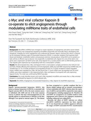
Ni et al. Stem Cell Research & Therapy(2021) 12:325Transmission electron microscopyCells were added with electron microscopy fixative at4 C for 24 h and centrifuged at low speed, then wrappedin 1% agarose and rinsed 3 times with 0.1M PBS (PH 7.4) for 15 min each time. Then, cells were fixed with1% osmic acid-0.1M PBS (PH 7.4) at roomtemperature (20 C) for 2 h. Subsequently, cells wererinsed with 0.1 M PBS (pH 7.4) 3 times for 15 mineach time. In the period of dehydration, the sampleswere dehydrated by placing 50%, 70%, 80%, 90%, 95%,100%, and 100% alcohol and 100% acetone in order for15 min each time, respectively. After being embed with812 embedding agent for 5–8 h, the samples wereinserted into the embedding plate for 37 C over night.Next, the samples were polymerized in an oven at 60 Cfor 48 h. Samples were cut into ultra-thin slices (60–80nm) with ultra-thin microtome. After uranium-leaddouble staining, the sections were dried at roomtemperature overnight and observed under the transmission electron microscope [49].Prussian blue stainingPrussian blue staining kit was purchased from Servicebio(Wuhan, China). The paraffin embedded slices of GCtumor tissues were continuously transferred into xyleneI for 20 min, xylene II for 20 min, anhydrous ethanol Ifor 5 min, anhydrous ethanol II for 5 min, and 75% alcohol for 5 min, respectively. Then, the slices were washedwith distilled water. After mixing the Prussian blue dyeA and B in equal proportions into the Prussian blue dyesolution, the slices were put into the dye solution for 1 hand washed twice with distilled water. Nucleus stainingwas performed with Prussian blue C for 3 min andrinsed with running water. Subsequently, slices werecontinuously put into anhydrous ethanol I for 5 min, anhydrous ethanol II for 5 min, anhydrous ethanol III for5 min, xylene I for 5 min, and xylene II for 5 min, respectively. Finally, the slides were sealed with neutralgum and observed under microscope [50].Statistical analysisGraphPad Prism 5 (GraphPad Software, Inc., La Jolla,CA, USA) was utilized for the statistical analysis. All results were obtained from three independent experimentsand presented as the mean standard deviation (SD).The statistical significance between different groups wasanalyzed by Student’s t test. P 0.05 or less was considered to be statistically significant.ResultsMiR-375 inhibits the stemness of GC cells in vivo andin vitroTo explore the roles of miR-375 in GC progression, weconstructed two GC cell lines (BGC-823, SGC-7901)Page 5 of 17with or without miR-375 stable overexpression (LV3miR-375 vs LV3-NC) by lentivirus infection. The overexpression efficiency was verified by qRT-PCR and fluorescence observation (Supplementary Figure S1A – C).Since we previously showed that miR-375 can inhibitHelicobacter pylori-induced gastric carcinogenesis [31]and tumor cell stemness is regarded as one of the mainfactors contributing to the recurrence and metastasis oftumors, we further investigated the effects of miR-375on GC cell stemness. Since the non-adherent tumor cellspheres are highly enriched with CSC-like cells compared to the adherent tumor cells [51], we determinedwhether miR-375 has effects on sphere-formation abilityand found that both the size and amount of sphereswere decreased in GC cells with miR-375 overexpression(Fig. 1A, B). Surprisingly, SGC-7901 cells overexpressingmiR-375 could hardly form spheres. Additionally, the expression of pluripotent transcription factors (OCT3/4,ALDH1A1, CD44) was significantly decreased by miR375 overexpression (Fig. 1C). Besides, the CD44 subpopulation of GC cells with stemness was decreased bymiR-375 overexpression (Fig. 1D). Furthermore, wefound that the tumorigenic ability of GC cells with miR375 overexpression was decreased, which was evident bythe decreased tumor formation rate and stem cell frequency using the tumor-limiting dilution assay (Fig. 1E–H). Consistently, GC cells with or without miR-375overexpression were injected into nude mice throughthe tail vein. Six weeks later, the metastatic ability ofmiR-375-overexpressed cells was weaker than that ofNC group, as characterized by the decrease of metastaticnodules (Fig. 1I). Notably, the online dataset analysis(Kaplan Meier-plotter tool: http://kmplot.com) showedthat GC patients with a higher level of miR-375 held alonger lifetime (Fig. 1J). These results suggest that miR375 can reduce the stemness of GC cells.SLC7A11 is identified as the direct target of miR-375Then, we set out to explore the mechanisms by whichmiR-375 inhibits GC cell stemness. We screened out thepossible downstream targets of miR-375 through performing gene-chip sequencing analysis. Due to the expression level of miR-375 in SGC-7901 being relativelylow among several GC cell lines (Supplementary FigureS1D), we chose SGC-7901 LV3-miR-375 and LV3-NC asthe experimental models. It was found that 6723 geneswere increased and 4798 genes were decreased in SGC7901 cells with miR-375 overexpression (Fig. 2A, B),among which SLC7A11, NFIX, CAST, PITPNA, GMFB,SLC16A2, CADM1, and RBPJ were predicted to be thepotential targets of miR-375 through online databaseprediction (Targetscan, miRDB, miRwalk) (Fig. 2C).Since SLC7A11, NFIX, and CAST were decreased bymiR-375 overexpression and targeting SLC7A11 has
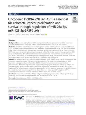
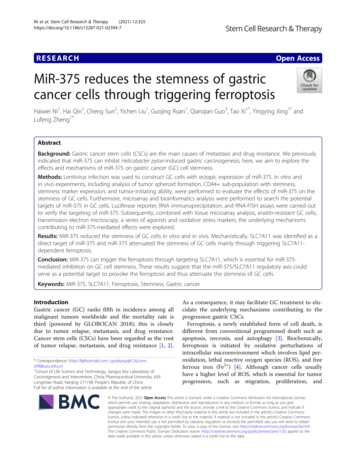
Ni et al. Stem Cell Research & Therapy(2021) 12:325Page 6 of 17Fig. 1 MiR-375 attenuates the stemness of GC cells. A, B The sphere size and number were examined in GC cells with or without miR-375overexpression. C The protein expression of stemness markers was evaluated in GC cells with or without miR-375 overexpression. D CD44 subpopulation was determined in GC cells with or without miR-375 overexpression. E The images of tumors derived from different numbers ofSGC7901 cells with or without miR-375 overexpression. F The tumor formation rate and confidence intervals for 1/(stem cell frequency) werecalculated based on the results shown in E. G The images of tumors derived from different numbers of BGC-823 cells with or without miR-375overexpression. H The tumor formation rate and confidence intervals for 1/(stem cell frequency) were calculated based on the result shown in G.I HE staining images of lung obtained from mice with injection of GC cells with or without miR-375 overexpression. J The correlation betweenmiR-375 expression and the overall survival probability of GC patients was evaluated using the online analysis tool (http://kmplot.com). Data arepresented as the mean SD, n 3, **p 0.01 vs control, ##p 0.01 vs LV3-NCrecently been shown to kill colorectal CSCs [52],SLC7A11 was chosen for further research. As expected,SLC7A11 protein and mRNA levels were reduced in GCcells with miR-375 overexpression (Fig. 2D). To furtherdemonstrate that miR-375 can directly target SLC7A11,we constructed luciferase reporter plasmids containingSLC7A11 3′UTR with miR-375 binding site mutant ornot (Fig. 2E) and found that the luciferase activity ofpMIR-3′UTR-WT was remarkably suppressed by miR375 overexpression, while no significant effect on the
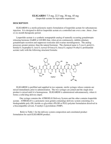
Ni et al. Stem Cell Research & Therapy(2021) 12:325Page 7 of 17Fig. 2 SLC7A11 is a direct target of miR-375 in GC cells. A The volcano map indicating the number of mRNAs in SGC-7901 cells with or withoutmiR-375 overexpression. B The mRNA heatmap showing the change of mRNA expression in SGC-7901 cells with or without miR-375overexpression. C The venn map showing the potential targets of miR-375. D SLC7A11 protein expression is detected in GC cells with or withoutmiR-375 overexpression. Data are presented as the mean SD, n 3, *p 0.05, **p 0.001 vs control. E The diagram indicating the binding ofmiR-375 on SLC7A11. F Luciferase reporter analysis was performed to determine the effect of miR-375 on the activity of SLC7A11 3′UTR WT andSLC7A11 3′UTR MUT. Data are presented as the mean SD, ns means no significance, n 3, *p 0.05 vs NC vector. G RIP assay wasconstructed to measure miR-375 level in RNA pulled down by Anti-Ago2 in GC cells with SLC7A11 3′UTR WT or SLC7A11 3′UTR MUToverexpression. Data are presented as the mean SD, n 3, **p 0.01 vs pMIR-report empty group. H RNA-FISH was performed to evaluate theco-localization of miR-375 and SLC7A11 in GC cellsactivity of pMIR-3′UTR-MUT was observed (Fig. 2F).Additionally, miR-375 was increasingly enriched inRNA complex pulled down by Ago2 in cells withSLC7A11-3′UTR-WT overexpression but decreased incells with SLC7A11-3′UTR-MUT overexpression(Fig. 2G). Furthermore, RNA-FISH assays showed aclear overlap between miR-375 and SLC7A11 in twoGC cell lines, mainly in the cytoplasm (Fig. 2H).These results indicate that miR-375 can directly targetSLC7A11 in GC cells.MiR-375 triggers ferroptosis via targeting SLC7A11 in GCcellsSince SLC7A11 is a critical suppressor of ferroptosisthrough uptaking cystine into the cells, we wonderedwhether miR-375 can trigger ferroptosis via targetingSLC7A11 in GC cells. Indeed, analysis on our gene-chipresults revealed that cysteine metabolism- and ferroptosisrelated signaling pathways (Ferrous iron binding, Cysteinetype endopeptidase activity involved in apoptotic signaling,Cysteine-type endopeptidase activity, Glutamate-cysteine
Ni et al. Stem Cell Research & Therapy(2021) 12:325ligase activity, and Selenocysteine insertion sequence binding) were enriched in GC cells with miR-375 overexpression (Fig. 3A). Consistently, GSEA analysis obtained thesimilar result that KEGG CYSTEINE AND METHIONINEMETABOLISM was positively correlated with miR-375overexpression (Fig. 3B). Then, three independent siRNAsagainst SLC7A11 were used and we chose SLC7A11-2 forthe subsequent experiment as SLC7A11-2 showed thestrongest knockdown efficiency (Supplementary FigureS1E–F). Additionally, transmission electron microscopyPage 8 of 17(TEM) assay showed that the mitochondria becamesmaller and membrane density was increased with vestigial cristae in GC cells with miR-375 overexpression orSLC7A11 knockdown and miR-375-mediated effect wasreversed by SLC7A11 overexpression (Fig. 3C). The efficiency of SLC7A11 overexpression was verified later (Supplementary Figure S1G – I). Erastin, a ferroptosis inducer,served as a positive control. Furthermore, lipid ROS leve
was quantified using Quantity-one Software. Addition-ally, RNA-fluorescence in situ hybridization assay was performed by Boster Biological Technology to detect miR-375 and SLC7A11 levels in this tissue microarray. Microarray analysis Total RNA was extracted in SCG7901 cells with or with-out miR-375 stable overexpression using Trizol reagent