Transcription
Chang et al. BMC Systems Biology 2016, 10(Suppl 1):1DOI 10.1186/s12918-015-0242-3PROCEEDINGSOpen Accessc-Myc and viral cofactor Kaposin Bco-operate to elicit angiogenesis throughmodulating miRNome traits of endothelial cellsHsin-Chuan Chang1, Tsung-Han Hsieh1, Yi-Wei Lee2, Cheng-Fong Tsai2, Ya-Ni Tsai1, Cheng-Chung Cheng3*and Hsei-Wei Wang1,2,4From The Fourteenth Asia Pacific Bioinformatics Conference (APBC 2016)San Francisco, CA, USA. 11 - 13 January 2016AbstractBackground: MicroRNAs (miRNAs) have emerged as master regulators of angiogenesis and other cancer-relatedevents. Discovering new angiogenesis-regulating microRNAs (angiomiRs) will eventually help in developing newtherapeutic strategies for tumor angiogenesis and cardiovascular diseases. Kaposi’s sarcoma (KS), which is inducedby the etiological infectious agent KS-associated herpesvirus (KSHV), is a peculiar neoplasm that expresses bothblood and lymphatic endothelial markers and possesses extensive neovasculature. Using KSHV and its proteins asbaits will be an efficient way to discover new angiomiRs in endothelial cells. Kaposin B is one of the latent viralgenes and is expressed in all KSHV tumor cells. Since Kaposin B is a nuclear protein with no DNA-binding domain, itmay regulate gene expression by incorporating itself into a transcription complex.Results: We demonstrated that c-Myc and Kaposin B form a transcription complex and bind to the miR-221/-222promoter, thereby affecting their expression and anti-angiogenic ability. By small RNA sequencing (smRNA-Seq), werevealed that 72.1 % (173/240) of Kaposin B up-regulated and 46.5 % (113/243) of Kaposin B down-regulatedknown miRNAs were regulated by c-Myc. We also found that 77 novel miRNA were up-regulated and 28 novelmiRNAs were down-regulated in cells expressing both c-Myc and Kaposin B compared with cells expressingKaposin B only. The result was confirmed by RNA-IP-seq data.Conclusions: Our study identifies known and novel c-Myc-regulated microRNAs and reveals that a c-Myc-orientedprogram is coordinated by Kaposin B in KSHV-infected cells.Keywords: MicroRNAs, Angiogenesis, KS-associated herpesvirus, c-Myc, Kaposin B, Small RNA sequencingBackgroundKaposi’s sarcoma-associated herpesvirus (KSHV), alsoknown as human herpesvirus 8 (HHV8), is associated withKaposi’s sarcoma (KS) [1]. KS is the most common cancerin individuals with AIDS and one of the most commoncancers in patients who have immunosuppression [2]. KStypically appears as colored lesions or blotches on the skin,although it can spread to internal organs [3]. This disease* Correspondence: chengcc@mail.ndmctsgh.edu.tw3Division of Cardiology, Department of Internal Medicine, Tri-Service GeneralHospital, National Defense Medical Center, Taipei, TaiwanFull list of author information is available at the end of the articlehas been recognized as a peculiar neoplasm that has adiverse cellular makeup and an extensive neovasculature.In host endothelial cells, KSHV infection alters the expression of mRNAs and microRNAs required for many aspectsof tumorigenesis, including angiogenesis [4]. Therefore, KSand KSHV are recognized as an excellent model to studyangiogenesis and to discover new angiogenic mechanisms,including coding and non-coding RNAs.Recently, the effect of KSHV infection on cellularmicroRNA (miRNAs) expression has been reported[5]. It was found that pre-miRNA signatures delineatethe stages of KSHV-associated HUVEC transformation 2016 Chang et al. Open Access This article is distributed under the terms of the Creative Commons Attribution 4.0International License (http://creativecommons.org/licenses/by/4.0/), which permits unrestricted use, distribution, andreproduction in any medium, provided you give appropriate credit to the original author(s) and the source, provide a link tothe Creative Commons license, and indicate if changes were made. The Creative Commons Public Domain Dedication o/1.0/) applies to the data made available in this article, unless otherwise stated.
Chang et al. BMC Systems Biology 2016, 10(Suppl 1):1Fig. 1 (See legend on next page.)Page 2 of 119
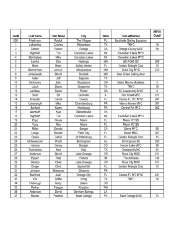
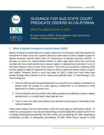
Chang et al. BMC Systems Biology 2016, 10(Suppl 1):1Page 3 of 119(See figure on previous page.)Fig. 1 Kaposin B binds to c-Myc for regulating endothelial cell angiogenic activities. a Nuclear distribution pattern of KSHV Kaposin Bprotein in cells. Immunofluorescence staining for Kaposin B proteins. HMEC1 cells with stable Kaposin B (KapB) expression were fixed, and KaposinB proteins were detected using anti-Flag mAb, followed by anti–mouse IgG secondary antibody conjugated with FITC (green). Cell nuclei werecounterstained with Hoechst 33342, while actin filaments with Texas Red phalloidin (Alexa Fluor 568). b HMEC1 cells stably expressing Kaposin Bwere subjected to the Transwell cell-migration assay (n 3). c Kaposin B increases cell motility in HUVEC. Primary HUVEC stably transduced withKaposin B or the vector control by lentivirus were used for Transwell cell-migration assays (n 3). d Kaposin B enhances microvascular formationof HUVEC in an in vitro MatriGel angiogenesis assay. Pictures were taken after 6 h of incubation (left), and tube length was then measured andcompared (right). e Schematic representation of miR-221/-222 proximal promoter. Three E-boxes (E1 and E2/3, in red) were found. f Coimmunoprecipitation assays show Kaposin B and c-Myc form a protein complex. Cell lysates were prepared from HMEC1 cells stably expressingKaposin B. Five micrograms of anti-FLAG (clone M2; left panel), anti-HA (right panel) or isotype IgG control were incubated with 500 μl of cellextracts and then analyzed by western blotting with indicated mAbs. g The interaction between c-Myc and Kaposin B was independent of promoterDNA. Co-immunoprecipitation assays were performed with or without DNase pre-treatment on cell lysates prepared from HMEC1 cells stably expressingKaposin B. Anti-FLAG (clone M2) or isotype IgG control were incubated with cell extracts, and pull-down products were analyzed by western blottingwith anti-HA mAb for c-Myc (upper panel) or anti-FLAG M2 mAb for Kaposin B (lower panel). h-i Knockdown of endogenous c-Myc levels in Kaposin B( )HUVECs inhibits Kaposin B-induced cell migration (h, left) and microvasculature formation (i, right) (n 3)[6, 7]. MiRNAs are small RNAs of 18–24 nucleotides inlength that have emerged as master regulators of cancerand key modulators of angiogenesis [8–10]. An increasingnumber of studies have shown that some miRNAs, calledangiomiRs (microRNAs that regulate angiogenesis), play acrucial role in regulating angiogenesis [11]. For example,miR-221 and miR-222, two closely related miRNAsencoded in cluster from a genomic region on chromosome X, can modulate the angiogenic properties of humanumbilical vein endothelial cells (HUVECs) by targeting c-Kit [11] and endothelial nitric oxide synthase(eNOS) [12]. MiR-221 and miR-222 also regulatelymphatic endothelial cell (LEC) motility by targetingETS2 and ETS1 transcription factors, respectively [4].Loss of miR-221 expression demarks the transitionfrom merely immortalized to fully tumorigenicHUVECs [6]. KSHV also induces endothelial cell motility by inducing miR-31 expression while suppressingthat of miR-221 and miR-222 [4, 13].Kaposin B, one of the three Kaposin proteinsencoded from the KSHV genome K12 locus, is oneof the viral genes expressed during latency and istherefore expressed in all KSHV tumor cells [4].Kaposin B stabilizes various cytokine (such as IL6and GM-CSF) mRNA containing AU-rich elements(AREs) via the p38/MK2 pathway. In response toLPS, Kaposin B and MK2 were shown to be exportedto the cytoplasm, where mRNA stability is regulated[14]. Kaposin B also enhances the PROX1 mRNAstability during lymphatic reprogramming of vascularendothelial cells [15]. Kaposin B can influence cellular gene expression by regulating promoter activitiesof host genes: both Kaposin B and KSHV virallatency-associated nuclear antigen (LANA) proteinscan down-regulate miR-221 and miR-222 levels byrepressing the activity of miR-221/-222 cluster promoter [4]. Since there is no predictable DNA-bindingdomain on Kaposin B, how this nuclear protein canregulate mRNA and miRNA expression remainsunclear.c-Myc achieves its oncogenic effects by regulatingtranscription of protein-coding genes as well as microRNA genes such as miR-29b-1/miR-29a [16, 17]. c-Mycis also essential for vasculogenesis and angiogenesisduring development and tumor progression [18] via inducing the expression of miR-17 92 angiogenic miRNAcluster [19]. Revealing the angiomiRs regulated by cMyc and the underlying regulatory mechanisms will helpto further understand c-Myc and endothelial cellbiology.Here, we showed that Kaposin B and c-Myc are in thesame transcription complex that directly regulates themiR-221/-222 cluster promoter activity. A c-Mycoriented circuit is therefore formed in the presence ofKaposin B in KSHV-infected endothelial cells. Furthermore, we also provide a global microRNA signaturewhich is regulated by c-Myc and Kaposin B. We hopeour roadmap will help the search and development ofnew therapeutic targets for virus- or cancer-inducedangiogenesis, cancer formation and metastasis.MethodsCell culture and KSHV infectionHuman primary umbilical vein endothelial cells(HUVECs) were purchased from Clonetics Inc.(Walkersville, Md.) and were cultured as described [4].HMEC1, an immortalized human microvascularendothelial cell line, was cultured in endothelial cellgrowth medium MV (C-22020; PromoCell, Heidelberg,Germany). A recombinant virus, rKSHV.219, that expresses the red fluorescent protein (RFP) from theKSHV lytic PAN promoter, the green fluorescent protein(GFP) from the EF-1alpha promoter, and with a gene forpuromycin resistance as a selectable marker, was constructed using JSC-1 cells as described previously [20].
Chang et al. BMC Systems Biology 2016, 10(Suppl 1):1Fig. 2 (See legend on next page.)Page 4 of 119
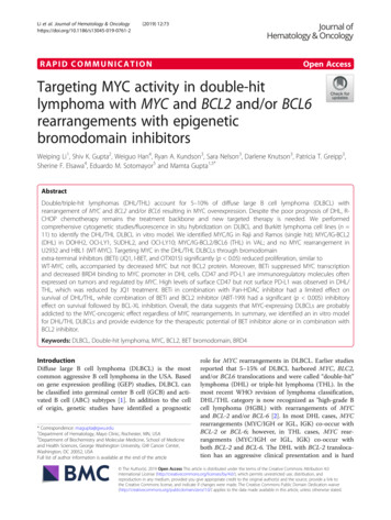
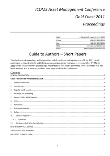
Chang et al. BMC Systems Biology 2016, 10(Suppl 1):1Page 5 of 119(See figure on previous page.)Fig. 2 c-Myc cooperates with Kaposin B to regulate cellular known miRNAs. a smRNA-Seq revealed differentially expressed miRNAs betweenKaposin B( ) and HUVECs ( 1.5 folds, left), and between Kaposin B( )c-Myc( ) and HUVECs ( 1.5 folds, right). b Venn diagrams summarizingsignificant overlap between c-Myc and Kaposin B known miRNAs signature. (upper panel) up-regulated miRNAs; (lower panel) down-regulatedmiRNAs. c Gene Set Enrichment Analysis (GSEA) verified the significant overlap between c-Myc and Kaposin B miRNAs signature. d RT-qPCRdetected various known miRNAs expression. Mean expression levels of the target miRNAs are compared with the U6 control (n 3). *: P .05.e RT-qPCR detected miR-221/miR-222 expression in Kaposin B( ) and Kaposin B( )c-Myc( ) cells. f The biological pathway of genes which weretargeted by the Kaposin B( )c-Myc( ) induced/reduced miRNAs. The P value were marked on the figurePlasmid constructionPlasmids expressing KSHV Kaposin B and miR-221,miR-222, or miR-221/-222 were constructed as described previously [4]. c-Myc expression constructs(pcDNA3-HA-c-Myc and pHR-c-Myc) and knock downconstruct (pLKO.1) was kindly provided by Prof. Kenneth CW Wu [21]. The full-length miR-221/-222 promoter reporter plasmid was constructed as describedpreviously [4], and primers for cloning miR-221/-222promoter mutants are listed in Additional file 1.Immunofluorescence assay (IFA)Cells were fixed with 4 % paraformaldehyde (Sigma-Aldrich), permeabilized with 0.2 % Triton X-100 (SigmaAldrich), and then blocked with PBS containing 1 % bovine serum albumin (Sigma-Aldrich). For Kaposin Bstaining, cells were incubated with the monoclonal antibody anti-FLAG M2 at a 1:500 dilution for 60 to120 min at 25 C followed by incubation with FITCconjugated goat anti-Mouse IgG (1:500, Jackson ImmunoResearch) for 60 to 120 min at 25 C. Rhodaminephalloidin (Molecular Probes, Invitrogen) was used tolabel actin cytoskeleton. Cell nuclei were counterstainedwith Hoechst 33342 (Sigma-Aldrich), and examined byfluorescence confocal microscopy (Olympus FV1000).Transwell cell migration and endothelial cell tubeformation assaysCell migration ability was evaluated using Costar Transwell Polycarbonate Permeable Supports (Corning, NY,USA) as described previously [13]. In brief, 5 104 cellsin 500 μl of culture medium were applied to the upperchamber of the device, and 750 μl of medium containing10 ng/ml human VEGF (R&D Systems, Minneapolis,MN, USA) was added to the lower chamber. A polycarbonate membrane with a pore size of 8 μm was placedin between the two chambers. After 6 h of incubation at37 C for HMEC1 and HUVEC cells, the membrane wasfixed in 4 % paraformaldehyde (Sigma-Aldrich) for20 min at room temperature and then stained withHoechst 33342 solution (Sigma-Aldrich) for 30 min. Onthe upper side of membrane were identified un-migratedcells and removed. The cells under the membrane werecounted under a microscope.In vitro angiogenic activity of vascular endothelial cellswas examined by tube formation assays: primaryHUVECs were placed onto MatriGel Basement Membrane Matrix gel (Becton-Dickinson, Franklin Lakes, NJ,USA) for 6 h at 37 C. The formation of tubule structurewas observed every hour under a microscope.Small RNA sequencing (smRNA-Seq) and data analysisTotal RNA was collected and small RNA fractions weresequenced using Illumina HiSeq2000 sequencer (Illumina, San Diego, CA USA) according to the manufacturer’s instructions. For data analysis, quality Fastqsequences, which were without poly-A, ambiguous nucleotides or a 5′ adapter, yet flanking 6–18 nt of 3′adapter sequence, had the adapter sequences trimmedand the identical sequences were then collapsed tounique sequences. The resulting unique sequences thatdid not align to the mRNA database (UCSC genomebrowsers) but were aligned to known microRNA sequences (miRBase R20; http://www.mirbase.org/) weresubjected to further quantification analysis. Sequencingreads were used to obtain a RPM (reads per millionmapped reads) [22, 23] value calculated as C/(MN) 109, where C is “read numbers aligned to a given miRNAchromosomal region”, M is “multiple mapping numbersacross all miRNA regions” and N is “total read numbersthat map to human genome sequence”.RNA extraction, real-time quantitative polymerase chainreaction (qPCR) and microRNA expressionRNA extraction and reverse transcription were performed as previously described [13]. The expressionof mature human miR-221 and miR-222 was determined by a stem-loop real-time PCR system using theprimer pairs previously published [4]. MicroRNA expression data were normalized to that of U6 snRNA.Details of qPCR primers are in Additional file 1.Coimmunoprecipitation (co-IP), chromatinimmunoprecipitation (ChIP) and ImmunoblottingCo-IP and ChIP assays were performed as previously described [24]. Briefly, Kaposin B or c-Myc expressionplasmid was co-transfected into 293T cells using TurboFect (R0531; Fermentas, Glen Burnie, Maryland, USA).After 48 h, cells were lyzed in NET lysis buffer
Chang et al. BMC Systems Biology 2016, 10(Suppl 1):1Page 6 of 119Table 1 Top 50 miRNAs induced by both c-Myc and Kaposin BNamecMyc KapB/vec (Fold)KapB/vec a-let-7e-5pHU KapB cMyc 139-3p2636.49HU KaposinB 8980982.710852004
Chang et al. BMC Systems Biology 2016, 10(Suppl 1):1Page 7 of 119Table 1 Top 50 miRNAs induced by both c-Myc and Kaposin B 56862.096381478containing protease and phosphatase inhibitors and celllysate (500 μl) was immunoprecipitated with anti-FLAGM2 (for detecting FLAG-tagged Kaposin B protein;Sigma-Aldrich), anti-HA or isotype IgG antibody. Theimmunoprecipitates were probed with anti-FLAG M2(1:2000) and anti-c-Myc Tag (clone 9E10; MILLIPORE,05–419, 1:2000) monoclonal antibodies, and visualizedwith horseradish peroxidase-conjugated secondary antibodies using the ECL reagent (Amersham PharmaciaBiosciences). For chromatin immunoprecipitation assay,chromosomal DNA fragments were prepared as described [25]. Briefly, lysates were incubated with isotypeIgG or anti-FLAG antibody. The approximately 100-basepair products from miR-221/-222 proximal promoter(E1 region: 600 (45,607,128-45,607,214); E2/3 region: 1100 (45,607,635-45,607,764); NC region: 2600(45,609,170-45,609,265)) were detected by real-timePCR.Luciferase reporter assaysFor reporter assays, 293T cells (5 104/well) in 24-wellplates were transfected with either Kaposin B or c-Mycexpression plasmid, along with the firefly luciferasereporter gene construct (500 ng) and 50 ng pRL-TKRenilla luciferase construct (for normalization) usingTurboFect . Cells were harvested 48 h after transfection,and the luciferase activity was measured using theDual-Luciferase Reporter Assay System (E1910; Promega,Charbonnières, France).ResultsKaposin B binds to c-Myc for regulating endothelial cellangiogenic activitiesKaposin B is a nuclear protein which can regulatemiR-221 and miR-222 expressions without any predictable DNA-binding domain [4, 26]. Therefore, wehypothesized that Kaposin B may be a novel transcription cofactor which regulates promoter activitiesby binding to other transcription factor(s). To investigate, we overexpressed Kaposin B in HMEC1 andprimary HUVEC cells and tested their migration andtube formation abilities. We verified expression ofKaposin B protein in lentivirus-transduced HUVECusing immunoblotting (Additional file 2-A), andimmunofluorescence staining showed that localizationof Kaposin B was in nucleus (Fig. 1a). Expression ofKaposin B induced an increase in cell migration ratein HMEC1 and primary HUVEC cells (Fig. 1b-c).Kaposin B also induced more microvascular structures formation by HUVEC in the in vitro tube formation assay (Fig. 1d).To maintain KSHV latency and oncogenesis,KSHV-latency proteins, LANA and vIRF3, stabilizedand activated c-Myc proteins [27–30] and c-Myc wasshown to reduce miR-222 expression in mouse mammary tumors [31]. Therefore, we examined the proximal promoter region of miR-221/-222 and identifiedthree putative c-Myc binding sites (E1, E2 and E3 Eboxes with a motif sequence CANNTG) (Fig. 1e).We hypothesized that Kaposin B may associate withc-Myc in regulating host gene expression. We therefore tested whether Kaposin B and c-Myc are in thesame protein complex by co-transfecting FLAGtagged Kaposin B and HA-tagged c-Myc into 293Tcells. c-Myc was detected in immunoprecipitation(IP) assays using anti-FLAG monoclonal antibody(mAb) against Kaposin B. On the other hand,Kaposin B was detected in IP assay using anti-HAmAb against c-Myc (Fig. 1f ). The interaction between c-Myc and Kaposin B was independent of promoter DNA since c-Myc was still detected in theanti-FLAG mAb pull-down products even when celllysates were pre-treated with DNase digestion forremoval of genomic DNA fragments (Fig. 1g,Additional file 2-B).We further checked whether c-Myc is required forKaposin B functions. Knockdown of endogenous cMyc in Kaposin B-expressing HUVEC cells abolishedKaposin B-induced cell migration and microvasculature formation. (Fig. 1h-i, Additional file 2-C). Wealso knocked down c-Myc in Kaposin B-expressingHMEC1 cells and found that cell migration abilitywas down-regulated. (Additional file 2-D-E).Small RNA sequencing reveals known miRNAs co-regulatedby c-Myc and Kaposin B in primary endothelial cellsPrevious study showed that Kaposin B can downregulate miR-221 and miR-222 levels by repressing the
Chang et al. BMC Systems Biology 2016, 10(Suppl 1):1Page 8 of 119Table 2 Top 50 miRNAs reduced by both c-Myc and Kaposin BNamehsa-miR-3614-5pHU KapB cMyc (RPM)25.7006HU KaposinB (RPM)35.6277cMyc KapB/vec (Fold)KapB/vec 6590.3352904640.654970387hsa-miR-548 51808667
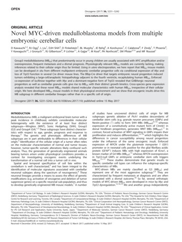
Chang et al. BMC Systems Biology 2016, 10(Suppl 1):1Page 9 of 119Table 2 Top 50 miRNAs reduced by both c-Myc and Kaposin B 909622820.376417558hsa-miR-548 ctivity of miR-221/-222 cluster promoter [4]. However,there are few studies exploring Kaposin B-induced globalmiRNAs alteration. Therefore, we overexpressed Kaposin B in HUVEC to examine its regulated miRNA expression. We have demonstrated that c-Myc binds toKaposin B (Fig. 1f-g) and is required for its functions(Fig. 1h-i), hence we overexpressed c-Myc in KaposinB( ) HUVEC to examine its ability to augment KaposinB-regulated miRNA expression.We analyzed small RNA sequencing (smRNA-Seq)data according to an in-house bioinformatics pipeline[32]. The Illumina next generation sequencing platformgenerated more than 29 million high-quality sequencingreads for HUVEC transduced with empty lentiviral vector alone, Kaposin B alone and Kaposin B with c-Myc.The initial operations included identifying sequencematches to the coding RNA database for eliminatingreads of degraded mRNAs. Non-coding RNA reads thatmatch previously annotated miRNAs deposited in themiRBase database (release 21) were subjected tonormalization and quantitative profiling. Firstly, weexplored known miRNAs expression. When we overexpressed Kaposin B alone (KapB/vec), we found 240 upregulated and 243 down-regulated known miRNAs. Incontrast, 312 up-regulated and 200 down-regulated miRNAs ( 1.5 fold change; Fig. 2a) were identified uponsimultaneous overexpression of Kaposin B and c-Myc(KapB c-Myc/vec). We further interrogated the contribution of c-Myc in Kaposin B-regulated miRNomechanges. Comparison of the c-Myc( )Kaposin B( )miRNA signature with that triggered by Kaposin B alonein HUVEC indicated that c-Myc helped to further induced the expression of 72.1 % (173/240) of Kapsoin Bup-regulated miRNAs (Fig. 2b, upper panel, fold change 1.5). Similarly, the miRNAs down-regulated by KaposinB and c-Myc were 46.5 % (113/243); Fig. 2b, lower panel,fold change 1.5). c-Myc and Kaposin B-regulated miRNAs are enumerated in Tables 1 and 2 (only top 50 of cMyc( )Kaposin B( ) miRNA are listed). Gene Set Enrichment Analysis (GSEA) revealed that the expressionof up- or down-regulated miRNAs by c-Myc correlatedwith those induced or repressed by Kaposin B inHUVEC (Fig. 2d). We also performed qPCR to detectmiRNAs expression in Kaposin B( ) and Kaposin B( )cMyc( ) cells. We identified that miR-193b-5p, miR-1975p, miR-210, and miR-1246 were up-regulated in Kaposin B( ) and Kaposin B( )c-Myc( ) cells. In contrast,miR-100, miR-146a-3p and miR-1271-5p were downregulated (Fig. 2d). RT-qPCR also demonstrated thatmiR-221 and miR-222 were down-regulated in KaposinB( ) and Kaposin B( )c-Myc( ) cells (Fig. 2e). ThemiRNAs detected in RT-qPCR, except miR-1246 andmiR-1271-5p, were similar to the ones detected inKSHV-infected HUVECs (Additional file 3). Moreover,we had used up to 3 different kind of tools (TargetScan(http://www.targetscan.org/vert 70/), mirDB (http://mirdb.org/miRDB/index.html) and TargetMiner (http://www.isical.ac.in/ bioinfo miu/targetminer20.htm)) to analyzethe targeted genes of c-Myc and Kaposin B induced/reduced top 5 miRNAs (Table 1). Based on our results, 806genes were targeted by the up-regulated miRNAs(Additional file 4-A) and 818 genes were targeted by thedown-regulated miRNAs (Additional file 4-B), respectively. The biological pathways were further analyzed,genes targeted by the up-regulated miRNAs contribute toaxonal guidance, cardiac β-adrenergic signaling and αadrenergic signaling; while the genes targeted by thedown-regulated miRNAs contribute to Wnt/β signaling,reelin signaling and molecular mechanisms of cancer(Fig. 2f). This indicates Kaposin B c-myc regulated miRNAs may trigger cells to more tumorigenic stage.Small RNA sequencing identifies novel miRNAsco-regulated by c-Myc and Kaposin B in primaryendothelial cellsTo identify novel miRNAs, we first removed mRNAcontamination and known miRNA loci. Using threeindependent bioinformatics algorithms (miRDeep2,mireap and miRanalyzer), 20–26 unique genomic lociyielded at the same time in Kaposin B( )-, KaposinB( )c-Myc( )-expressing cells and HUVEC controlcells (Fig. 3a), and we listed all novel miRNA candidates in (Additional file 5). We found 325 and 374novel miRNA candidates in Kaposin B-expressingcells and Kaposin B( )c-Myc( ) cells (KapB c-Myc/vec) that were up-regulated, respectively. In contrast,
Chang et al. BMC Systems Biology 2016, 10(Suppl 1):1Fig. 3 (See legend on next page.)Page 10 of 119
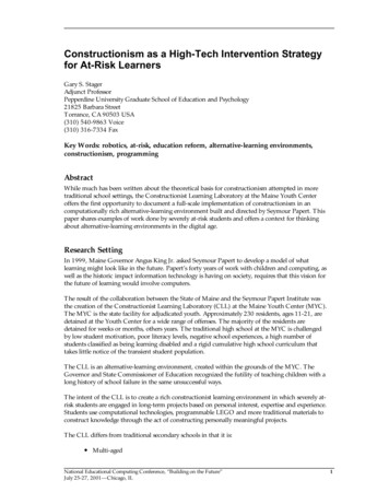
Chang et al. BMC Systems Biology 2016, 10(Suppl 1):1Page 11 of 119(See figure on previous page.)Fig. 3 c-Myc cooperates with Kaposin B to regulate cellular novel miRNAs. a The analysis pipeline for identification of known and novel miRNAsfrom smRNA-seq data. Reads or sequences pass each filtration process are indicated. b Venn diagrams summarizing significant overlap betweenc-Myc and Kaposin B signature novel miRNAs. (upper panel) up-regulated miRNAs; (lower panel) down-regulated miRNAs. c Venn diagramssummarizing significant overlap between up-regulated or down-regulated novel miRNA candidates and Ago1/2( ) RNA-IP-s
Hospital, National Defense Medical Center, Taipei, Taiwan . (eNOS) [12]. MiR-221 and miR-222 also regulate . transcription of protein-coding genes as well as micro-RNA genes such as miR-29b-1/miR-29a [16, 17]. c-Myc is also essential for vasculogenesis and angiogenesis