Transcription
Application NoteMarch 15, 2021Keywords or phrases:Octet, Bio-Layer Interferometry, epitope binning,antibody, mAb, cross competition assays, hybridoma,phage, ELISA, in-tandem, classical sandwich, premix,binChart, matrix generation, binding, kinetics, affinityCross-Competition or Epitope Binning Assayson the Octet RH96 SystemRashi Takkar 1, Amrita Yadav 1, Sriram Kumaraswamy 1, Sindy Liao-Chan2, Jan-Willem Theunissen21. Sartorius, Fremont, CA2. Igenica, Inc., Burlingame, CACorrespondenceEmail: octet@sartorius.comAbstractEarly identification of antibody candidates with desired affinity and dissociation kinetics, binding epitopes, and critical qualityattributes such as glycosylation profiles can be vital for avoiding later-phase failures caused by selecting non-ideal leads. Theepitope binning process enables antibody candidates that bind similar epitope regions in the antigen into bins. Octet Bio‑Layer Interferometry platforms enable epitope binning analysis using cross-competition assays where competitivebinding of antibody pairs are assessed. These assays can be easily extended to characterizing large antibody libraries withhigh-throughput Octet platforms and specifically developed epitope binning software analysis tools to evaluate largeantibody data sets. Octet epitope binning assays can also accommodate multiple assay orientations that enable analysis ofmultivalent antigens and antibodies in crude or hybridoma supernatants. This application note describes Octet systemcapabilities and guides into developing epitope binning assays and data analysis.Find out more: www.sartorius.com
Table of ContentsIntroduction. . . . . . . . . . . . . . . . . . . . . . . . . . . . . . . . . . . . . . . . . . . . . . . . 2Octet RH96 Instrument . . . . . . . . . . . . . . . . . . . . . . . . . . . . . . . . . . 2Advantages Over ELISA and SPR. . . . . . . . . . . . . . . . . . . . . . . . . 3Developing Cross-Competition/Binning Assays . . . . . . . . . 4Choosing the appropriate assay format. . . . . . . . . . . . . . . . . 4In-tandem assay . . . . . . . . . . . . . . . . . . . . . . . . . . . . . . . . . . . . . . . . . 4Choosing the right biosensor . . . . . . . . . . . . . . . . . . . . . . . . . 5Capture using affinity tags-based approach. . . . . . . . . . 5Streptavidin-based approach . . . . . . . . . . . . . . . . . . . . . . . . . 5Amine-reactive coupling (AR2G)-based approach. . . 6Antigen loading or antigen immobilization . . . . . . . . . . . 6Hydration of biosensors . . . . . . . . . . . . . . . . . . . . . . . . . . . . . . . 6Baselines. . . . . . . . . . . . . . . . . . . . . . . . . . . . . . . . . . . . . . . . . . . . . . . 7Ensuring the antigen is active . . . . . . . . . . . . . . . . . . . . . . . . . 7Association . . . . . . . . . . . . . . . . . . . . . . . . . . . . . . . . . . . . . . . . . . . . 8Ensuring complete self-blocking. . . . . . . . . . . . . . . . . . . . . . 8Classical sandwich and premix assays. . . . . . . . . . . . . . . . . . . 9Choosing the right biosensor . . . . . . . . . . . . . . . . . . . . . . . . . 9Amine-reactive coupling (AR2G)-based approach. . . 9Capture-based approach . . . . . . . . . . . . . . . . . . . . . . . . . . . . 10Streptavidin-based approach . . . . . . . . . . . . . . . . . . . . . . . . 10Antibody loading or antibody immobilization . . . . . . . 10Hydration of biosensors . . . . . . . . . . . . . . . . . . . . . . . . . . . . . . 10Baseline . . . . . . . . . . . . . . . . . . . . . . . . . . . . . . . . . . . . . . . . . . . . . . 10Ensuring mAbs are active . . . . . . . . . . . . . . . . . . . . . . . . . . . . . 11Association . . . . . . . . . . . . . . . . . . . . . . . . . . . . . . . . . . . . . . . . . . . . 11Classical sandwich. . . . . . . . . . . . . . . . . . . . . . . . . . . . . . . . . . . . . 11Ensuring complete self-blocking. . . . . . . . . . . . . . . . . . . . . . 11Premix. . . . . . . . . . . . . . . . . . . . . . . . . . . . . . . . . . . . . . . . . . . . . . . . . . 11Cross reactivity between antibodies . . . . . . . . . . . . . . . . . 12Biosensor Regeneration. . . . . . . . . . . . . . . . . . . . . . . . . . . . . . . . . . 12Running Cross-Competition/Epitope Binning Assays. . . 13In-tandem. . . . . . . . . . . . . . . . . . . . . . . . . . . . . . . . . . . . . . . . . . . . . . . 15Classical sandwich. . . . . . . . . . . . . . . . . . . . . . . . . . . . . . . . . . . . . . 16Premix. . . . . . . . . . . . . . . . . . . . . . . . . . . . . . . . . . . . . . . . . . . . . . . . . . . 16Data Analysis (Version 8.0 or Higher) . . . . . . . . . . . . . . . . . . . . 16Analysis post-data export. . . . . . . . . . . . . . . . . . . . . . . . . . . . . . . 18Conclusion. . . . . . . . . . . . . . . . . . . . . . . . . . . . . . . . . . . . . . . . . . . . . . . . 22References. . . . . . . . . . . . . . . . . . . . . . . . . . . . . . . . . . . . . . . . . . . . . . . . 232IntroductionEpitope binning is a term used to describe segmentationof a panel of monoclonal antibodies (mAbs) into binsbased upon the antigen region, or epitope, bound by eachantibody. This grouping is performed using cross competition assays, in which the competitive binding of antibodypairs to a specific antigen is characterized. If the antigenbinding of one mAb prevents the binding of another, thenthese mAbs are considered to bind to similar or overlapping epitopes. Conversely, if binding of a mAb to the antigen does not interfere with the binding of another, thenthey are considered to bind to distinct, non-overlappingepitopes. Two criteria must be fulfilled in order to assignmAbs into the same bin. First, all mAbs in the same binshould block each other’s ability to bind the antigen. Second, all mAbs in the same bin should have similar blockingprofiles when paired with other mAbs in the panel.In early drug development, cross-competition assays areused to characterize hundreds of antibody clones and canbe performed with hybridoma supernatants, phage lysatesor purified samples. Because mAbs in different bins bindto distinct epitopes and display diverse functional characteristics, epitope binning studies can increase the likelihood of choosing a lead antibody with the desiredbiological activity. Cross-competition assays also are performed to identify mAbs that bind similar epitopes to apreviously characterized mAb as in the generation of biosimilars or biobetters. These assays may also be useful inselecting reagents for sandwich or ELISA-type assays,such as those used for biomarker testing or pharmacodynamic assays, to identify good antibody pairs that bind tothe antigen simultaneously.Octet RH96 InstrumentOctet systems are ideally suited to run cross-competitionassays, with their combination of assay speed, versatility ofassay design and parallel, independent biosensor format. Theworking principle is based on Bio-Layer Interferometry (BLI),a label-free technology that measures molecular interactionsin real time for the purpose of quantitation and kinetic analysis. Binding analyses can be performed in standard 96-well,half area 96-well, standard 384-well or tilted-bottom 384well microplates depending on throughput requirementsand available sample volume. 384-well tilted-bottom microplates (Sartorius, Part No. 18-5076) only require 40 µL ofsample or reagent per well.
The Octet RH96 system has the highest throughput andversatility of any instrument in the Octet family, and isideally suited for cross-blocking assays and epitope binningthroughput needs. The instrument monitors up to 96interactions simultaneously, enabling label-free analysis atan unprecedented speed of 2 minutes for a full 96-wellmicroplate. The system’s ability to read 8, 16, 32, 48 or96 wells in parallel enables tailoring of assay design tomaximize analytical throughput or sensitivity. The 8 and16 biosensor modes provide high sensitivity for measuringsmall molecule or peptide binding interactions and proteinquantitation down to ng/mL for 1-step or pg/mL for multistep assays. The Octet RH96 instrument also features twosample plate locations which doubles sample capacity, andit is also amenable to automation. The Dip and Readbiosensors used in Octet assays can be regenerated andre-racked for maximum flexibility and operational costsavings. Furthermore, specialized software tools areavailable for epitope binning analysis, including matrixgeneration, BinChart, and normalization of the binningtraces. More information on the Octet RH96 and otherOctet systems can be found on the Sartorius website.Advantages Over ELISA and SPRThe Octet RH96 System provides several compellingadvantages over ELISA and SPR as summarized in Table 1.The Octet RH96 system is significantly faster than competing methods, compatible with crude matrices, allowsdiscrete, independent, parallel interactions to be monitoredsimultaneously and is easy to operate.Octet RH96 systemELISASPRLabeled detection reagentsNoYesNoMonitoring of all stepsYesNoYesReal-time monitoringYesNoYesRun Time (32x32)8 hours24 hours14 hours*Crude lysate compatibility(hybridoma supernatants, lysates)YesNoNo; potential clogging issues whenusing crude, unpurified samplesDetection of low-affinity mAbsYesNoYesParallel interactionsDiscrete, independent, parallelinteractions monitoredsimultaneouslyDiscrete, independent interactionMAY be monitored simultaneouslybut throughput is compromised;for higher throughput, restrictednumber of multiple, independentinteractions analyzedCombined flow paths restrict abilityto monitor multiple, independentinteractions simultaneouslySamples are recoverable andcan be re-usedYesNoNoBiosensor choicesMany biosensor chemistriesavailableN/ALimited choice of chip biosensorchemistriesRegenerationBiosensors are inexpensive andmay be regeneratedCan tailor specific regenerationbuffers for each mAbCannot regenerate and re-usesamplesChips are expensive and have to beregeneratedCannot tailor regeneration for eachmAb (universal regenerationsolution needed)VersatilityCompatible with allbinning formatsMore amenable to sandwich andpremix binning as capturingantigen on plate surface forin-tandem binning may distort itsconformationCompatible with all binning formats;rarely used for in-tandem assaybecause throughput is significantlycompromisedOther considerationsEasy experiment set-upMinimal user-training requiredExtensive analyst time neededMinimal user training requiredAutomation required for highthroughput runsNeed experienced usersRequires optimization of surfaceregenerationTable 1: Octet RH96 system advantages over ELISA and SPR.*20-channel SPR system.3
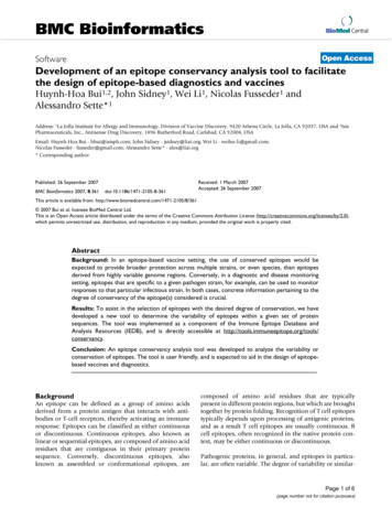
Developing Cross-Competition/Binning AssaysTo confirm binning results, it is imperative to perform bidirectional binning, regardless of the assay format, i.e. testingthe mAb pairs in both orientations. Furthermore, multiplebinning assay formats may be performed to corroboratebinning results.There are three primary binning assay formats: in-tandem,classical sandwich and premix.11.In-tandem format (Figure 1A): the antigen immobilizedonto a biosensor is presented to the two competingantibodies in consecutive steps. Binding to distinctnon-overlapping epitopes is indicated if saturation withthe first antibody does not block binding of the secondantibody. However, immobilization of the antigen maydistort its structural conformation, masking some epitopes and/or exposing some non-native epitopes. It istherefore recommended to immobilize the antigenusing mild methods such as capture using tags.For all binning formats, there are two ways of performingthe assay on a panel of mAbs.1.OR2. Antigen-loaded (in-tandem), or bare (classical sandwich or premix) biosensors are saturated with a fixedmAb, and then an array of mAbs is presented as competing mAbs.2. Classical sandwich format (Figure 1B): involves immobilizing the first antibody onto the biosensor, followedby incubation with the antigen, and then the secondsandwiching antibody. Classical sandwich binning isamenable only to monomeric and homogenous antigens; multimeric or heterogeneous antigens wouldresult in the second antibody binding to multiple subunits or different subpopulations of the antigen, respectively, resulting in a false binning profile.2Choosing the appropriate assay formatThe flow chart in Figure 2 outlines various factors in deciding the appropriate assay format for a particular antigenand panel of antibodies. Figure 2 also highlights someconsiderations and recommended early optimizations foreach assay format.3. Premix format (Figure 1C): antigen is incubated with alarge molar excess of the second antibody to form apremix sample. The premix is then presented to the biosensor loaded with the first antibody. The premix assayformat is often used to clarify ambiguous responsesseen with the other binning methods. One drawback ofthe premix method is that it requires large amounts ofreagents and approximate knowledge of KD values forthe interaction between each antibody in the panel andthe antigen.AIn-tandem assayAgCompetingmAb (Ab2)In-tandem binning involves first immobilizing a purifiedantigen on a biosensor, and then presenting a saturatingmAb (Ab1) followed by a competing mAb (Ab2). This formatcan be applied for binning crude mAb supernatants as wellas purified mAbs.CPremix assayBiosensorBiosensor12SaturatingmAb (Ab1)3In-tandem assayB Classical sandwich assayBiosensor1Antigen-loaded (in-tandem), or bare (classical sandwich or premix) biosensors are saturated with an array ofmAbs from the panel, and a fixed mAb is introduced asthe competing mAb on all biosensors.21CoupledmAb (Ab1)AgSandwichingmAb (Ab2)32Coupled mAb (Ab1)AgAgAgPremixed mAb Ag (Ab2 Ag)Figure 1: Three formats of epitope binning assays. (A) In-tandem assay: Antigen is immobilized on the biosensor, followed by the binding of thesaturating mAb (Ab1) and competing mAb (Ab2), respectively. (B) Classical sandwich assay: One mAb is immobilized on the biosensor (Ab1), antigen iscaptured using this mAb and then a second sandwiching mAb is tested (Ab2). (C) Premix assay: One mAb is immobilized on the biosensor (Ab1), thebiosensor is then exposed to a premix solution containing the antigen and a large molar excess of the second mAb (Ab2).4
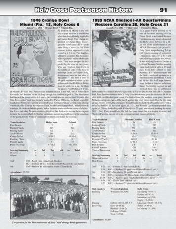
Choosing the right biosensorFor in-tandem binning studies, it is critical to choose abiosensor that maintains the structure and activity of theimmobilized antigen. For a complete list of biosensors,please refer to the Biosensor Selection Guide.Fc Capture (AHC), Anti-Mouse Fc Capture (AMC) andNickel-NTA (NTA). Alternatively, custom biosensors canbe made by immobilizing an antibody against a specificmotif or tag on the antigen, onto the biosensor. Once anoptimized regeneration protocol has been developed,these biosensors may be regenerated and reused toreduce assay costs.Capture using affinity tags-based approachCapture biosensors are pre-immobilized with a high affinitycapture antibody or protein that binds to the antigen via aknown motif or tag. Since the tag or the motif has beenengineered at a specific antigen location, the capture-based approach allows the most favorable orientationwith the least structural distortion of the antigen. Therefore, the capture-based approach is the preferredmethod for antigen immobilization for in-tandem binningstudies. The interaction between the antigen and the biosensor should be stable i.e., with low or minimal dissociation.Some ready-to-use biosensors available for tagged antigens include Anti-GST, HIS2, Anti-Penta His, Anti-HumanStreptavidin-based approachIn vivo site-specific biotinylation methods that introduceone streptavidin binding site at a specific location on theantigen are recommended. However, in-vitro biotinylationof the antigen can be performed if necessary. For detailedbiotinylation protocols for protein ligands with StreptavidinBiosensors, refer to Technical Note 28, Biotinylation of Protein for Immobilization onto Streptavidin Biosensors.Streptavidin Biosensors (SA) are regenerable to the level ofthe immobilized formatPremixClassicalsandwichBinning formatconsiderationsRecommendedearly optimizationexperimentsCrude(Hybridoma supes,phage lassical Sandwich Ag concentration must be ator greater than KD Premixed mAbs must be inlarge molar excess over Ag Block unsaturatedbiosensor surface whenAHC/AMC Biosensors used Ag should not dissociaterapidly from biosensorcoupled mAbs Block unsaturated biosensorsurface when AHC/AMCBiosensors used Weak affinity/High off-ratemAbs coupled to biosensormay lead to ambiguous data Approx. KD required fromkinetic screen Ensure no cross-reactivitybetween mAbs Ensure mAbs still active postcoupling to SA or AR2G Ensure complete self-block(Hybridoma supes,phage lysates) Ensure no cross-reactivitybetween mAbs Ensure mAbs still active postcoupling to SA or AR2G Ensure complete self-blockBinningformatIn-tandemIn-tandemIn-tandem Ag may be loaded onbiosensor using tags ordirectly (AR2G) First mAb must saturate andremain bound to Ag toprevent “free Ag” frombinding to competing Ab Weak affinity/high off-ratemAbs presented assaturating Abs may lead toambiguous data Direct coupling of Ag tobiosensor with AR2G maylead to Ag inactivation —ensure Ag still active Ensure complete self-blockFigure 2: Assay format flow chart.5
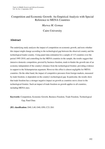
Antigen loading or antigen immobilizationOptimal antigen loading of the biosensor is critical for goodquality binning data. Oversaturation of the biosensor withthe antigen to maximize signal is not recommended andmay lead to data artifacts due to steric hindrance, possibleaggregation on the surface, or non-specific interactionsbetween the antibody and antigen.When performing the antigen loading step, slow loading foran extended period of time is preferable to rapid ligandimmobilization. The loading curve should show a gradualincrease in signal to 0.5–1 nm, but may be higher or lowerdepending on the antigen size. 50–200 nM of antigen is atypical concentration range for biosensor loading. Importantly, ensure low antigen levels are captured on the biosensor to minimize crowding artifacts and the interactionbetween the antigen and the biosensor is stable with lessthan 15% dissociation of the antigen in 10 minutes. It isimportant to establish a stable baseline with minimal signaldrift prior to proceeding to the association steps becausethe antigen needs to remain bound to the biosensor whendipped into the saturating and competing mAbs.Biosensor selection, antigen concentration and loadingduration can all be optimized in one assay on Octet systemsby loading a few antigen concentrations — 50, 100 and200 nM — for 10 minutes followed by dissociation in bufferfor 10 minutes. Figure 4 shows 10–minute loading and dissociation steps of a 100 and 200 nM 50 kDa histidine‑taggedantigen on Anti-Penta His (Figure 3A) and Ni-NTA (Figure 3B) Biosensors. The loading curve characteristics andsignal (nm shift) of the antigen at both concentrations ontoboth biosensors are acceptable; however, greater than 15% ofantigen appears to dissociate from the Anti-Penta His Biosensor (Figure 3A). Antigen loading at 100 nM onto Ni-NTABiosensors shows fairly slow initial binding and an optimalloading signal of 0.7 nm after 10 minutes, and the interaction remains stable with minimal dissociation ( light bluetrace in Figure 3B); this was therefore selected as the optimalloading condition for further experiments.Hydration of biosensors Anti-Penta HIS BiosensorBiosensors should go through hydration in media or buffer0.5that matches the media or buffer in the corresponding0.4samplesfor at least 10 minutes prior to running the assay.0.3During in-tandem binning, hydration of biosensors shouldbe0.2performed in the Octet Kinetics Buffer, PBS or bufferthat0.1 has been used to dilute the antigen.Binding (nm)Amine-reactive coupling (AR2G)-based approachDirect immobilization of the antigen is established by forming a covalent bond to free lysine residues via an AmineReactive Biosensor (AR2G). AR2G Biosensors should onlybe considered if the capture-based approach or biotinylation of the antigen is not feasible. Covalent linking of theantigen to the biosensors could potentially distort its structure, and may also lead to steric hindrance if the biosensorsurface is overly saturated. Diluting the antigen in 10 mMacetate buffer at pH 4–6 for immobilization onto the AR2GBiosensor works well. This method is typically regenerableto the level of the immobilized ligand. For a detailed protocol on AR2G Biosensor immobilization, refer to TechnicalNote 26, Dip and Read Amine Reactive Second-Generation(AR2G) kel-NTA BiosensorAnti-Penta HIS Biosensor1.10.50.40.90.30.7Binding (nm)Binding (nm)600Time (sec)0.20.10.50.300.1-0.10200400600Time (sec)80010001200-0.10200400600Time (sec)Nickel-NTA BiosensorFigure 3: Antigen loading and dissociationof a 100 and 200 nM 50 kDa His-tagged antigen onto (A) Anti-Penta His and (B) Ni-NTA Biosensors.The1.1Anti-Penta His Biosensors show significant ( 15%) dissociation of the antigen from the surface within 10 minutes, whereas the Ni-NTABiosensors exhibit minimal dissociation. Therefore, Ni-NTA Biosensors were selected for further experiments with this antigen.Binding (nm)0.960.70.50.30.1
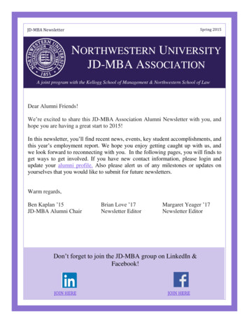
BaselinesIn all binning assays, there are usually three baseline steps.The three baseline steps for in-tandem binning are: 1) thestep prior to antigen loading (sensor check), 2) the stepbefore the association of the saturating mAb (Ab1), and 3)the step preceding the association of the competing mAb(Ab2) (Figure 4). A baseline should be established usingbuffer or media matched to the one used in the subsequent loading and the two association steps to mitigateany non-specific binding and drift from buffer effects.Baseline steps are performed to remove any unboundantigen and Ab1 from the biosensor.If mAbs are present in cell culture supernatants, then thebaseline steps preceding the mAb binding steps shouldbe performed in mock-transfected supernatants, spentmedia or low IgG FBS supplemented in growth media.Some biosensors, such as Streptavidin and Ni-NTA, maybe more prone to non-specific binding by free biotin orirrelevant histidine-containing proteins present in supernatants. In these cases, performing 2–5 minute baselinesin spent or FBS supplemented media becomes vital.Ensuring the antigen is activeCoupling of the antigen to the biosensor surface occasionally can alter the antigen’s structural conformation, revealing non-native epitopes and/or masking epitopes. Beforeproceeding to binning studies, it is important to ensure thatthe antigen remains active and able to bind the mAb panelfollowing immobilization. Antigen activity can be confirmedvia a kinetic screening assay performed in the same formatas the binning assay.For this kinetic screen, antigen loading is followed by association with 200–350 nM mAbs for 10–15 minutes, and thena 10–15 minute dissociation step. This screening assay canalso be used to flag mAbs with weak affinities to the antigen. Weak affinity mAbs are unreliable when presented assaturating mAbs (Ab1); they will dissociate quickly andexpose free antigen to competing mAbs (Ab2). The binningdata for these mAbs will be more reliable when they are presented as competing mAbs (Ab2). Figure 5 shows a kineticscreen of a panel of six mAbs. In this panel, antibodies 100,102 and 103 exhibit high off-rates and may produce ambiguous binning profiles when used as saturating mAbs (Ab1)in an in-tandem assay format.If binning is performed with purified mAbs, then the baseline steps preceding the association of mAbs should bedone in the same buffer that the mAbs are diluted in,either Octet Kinetics Buffer or PBS. When binning purified mAbs, non-specific binding typically is minimal. How-Baselineever, if some non-specific binding is observed, thenblocking reagents such as BSA (up to 1–2%) and/or nonionic detergents such as Tween-20 (up to 0.05%) may beadded to the buffer.BaselineAntigenBaselineSaturating mAb (Ab1)Competing mAb (Ab2)4.0Binding (nm)3.02.01.002000220024002600Time (sec)280030003200Figure 4: Representative data from an in-tandem binning assay. The arrows indicate the three baselines for this assay format: before antigen loading(sensor check), after antigen loading before binding of the saturating mAb (Ab1) and before binding of competing mAb (Ab2). The baseline stepsremove any unbound antigen/ antibody from the biosensor, and the baseline after Ab1 can also be used to flag for mAbs with fast off-rates.7
Ab 100AB 101Ab 1020.20.10250500750Time (sec)Binding (nm)0.2Binding (nm)Binding (nm)0.20.110000250500750Time (sec)Ab 10310000500750Time (sec)1000Binding (nm)Binding (nm)Binding (nm)00.10250500750Time (sec)500750Time (sec)1000Ab 1050.2250250Ab 1040.100.110000.20.10250500750Time (sec)1000Figure 5: Binding kinetics of a panel of 6 mAbs interacting with an immobilized antigen on Ni-NTA Biosensors. The binding of all mAbs confirms thatthe antigen retains its activity upon immobilization. Antibodies 100, 102 and 103 have high off-rates, and may result in ambiguous binning profileswhen used as saturating mAbs (Ab1) in an in-tandem binning format.AssociationThe in-tandem binning assay utilizes two association steps –binding of the saturating mAb (Ab1) to the antigen coupled-biosensor and binding of the competing mAb (Ab2) tothe antigen in the presence of Ab1 (Figure 1A). The firstassociation step should be run long enough to see somecurvature in the data trace and until the binding curve hasflattened for an extended period of time. The flattening ofthe curve indicates that the Ab1 has saturated the antigen-loaded biosensor, which is necessary for this assay format. In general, a 10–15 minute Ab1 association step isrecommended. The competing antibody (Ab2) associationshould also be run long enough to see some curvature inthe data traces; however, the Ab2 binding curve does notneed to reach equilibrium. For the Ab2 association step, a5–10 minute association should be sufficient.Ensuring complete self-blockingPrior to running the binning assay, Ab1 and Ab2 concentrations should be optimized in order to ensure completeself-blocking. Self-blocking is when the same antibody isused as both the saturating and competing antibody. TheAb2 binding signal should be zero in this case, given thatthe antibody completely saturates the antigen on the biosensor as Ab1. In general, Ab1 and Ab2 are tested at thesame concentration during optimization, in the range of100–400 nM, to ensure complete self-blocking. During theactual binning assay, however, Ab2 concentrations may betested at one-half to one-third the concentration of Ab1 toconserve reagents.8Figure 6A shows the self-blocking curves for 14 purifiedmAbs in a panel. The antibody concentrations were keptconstant at 250 nM when presented as saturating andcompeting mAbs. The association step durations for saturating and competing mAbs were 15 and 10 minutes,respectively. All Ab2 signals were minimal, indicating no significant self-binding. To get a clearer picture, aligned Ab2binding traces for all mAbs are shown in Figure 6B.Self-binding signals of 0.1 nm were observed for red, yellow, green, dark blue, black and light blue traces (Figure 6B).The small self-binding signals likely result from lack of fullsaturation of the antigen-loaded biosensor by Ab1, leavingsome free epitopes available for Ab2 binding. A higher concentration of saturating mAb may be necessary to fully saturate the antigen on the biosensor and allow for completeself-blocking. Therefore, these six antibodies were re-testedby using a higher concentration of saturating mAb(333.3 nM) while maintaining the competing mAbs at250 nM (Figure 6C). As seen in Figure 6C, use of a higherconcentration of saturating mAb diminished self-bindingfor all six mAbs. Consequently, association of 333.3 nM ofsaturating mAb (Ab1) for 15 minutes, and association of250 nM of competing mAb (Ab2) for 10 minutes were chosen as the final conditions to perform the in-tandem binning experiments (as shown in Figure 4).
AAntigen3.5CompetingmAb (Ab2)Saturating mAb (Ab1)Because several aspects of these binning formats are similar, assay development discussions for classical sandwichand premix binning assays have been combined here. Anydiscrepancies between the two formats will be highlightedand discussed in detail.3.0Binding (nm)2.52.01.51.00.50340036003800B4000 4200Time (sec)4400460048005000Competing mAb (Ab2)0.250.200.15Binding (nm)0.10Classical sandwich assays involve immobilization of Ab1onto the biosensor, followed by stepwise binding of theantigen and the sandwiching Ab2. The Ab2 molecule canbind the captured antigen only if Ab2s binding epitopedoes not overlap that of immobilized Ab1. The classicalsandwich binning format is compatible with mAb supernatants and purified mAbs. One limitation of the classicalsandwich format is that it requires monovalent antigens andhomogenous mixtures of antigen. A false positive result canarise if the antigen forms multimers or aggregates. In thiscase, the immobilized mAb will not saturate the antigen,and free antigen epitopes will be available for sandwichingmAb (Ab2) 05000Time (sec)50505100Competing mAb (Ab2)0.25In the premix assay, Ab1 is immobilized onto the biosensor.Following immobilization, Ab1 binds pre-complexed antigen-Ab2, which have been pre-incubated for 4–5 hours.Premix binning assays usually are performed on purifiedsamples and not on supernatants because of the largeamount of Ab2 needed to perform the initial premix. Thisassay format is amenable to monovalent as well as multivalent antigens and is often the format of choice for confirmation of ambiguous binning profiles or “intermediate”responses from other f
ping epitopes. Conversely, if binding of a mAb to the anti-gen does not interfere with the binding of another, then they are considered to bind to distinct, non-overlapping epitopes. Two criteria must be fulfilled in order to assign mAbs into the same bin. First, all mAbs in the same bin should block each other's ability to bind the antigen. Sec-