Transcription
Yu et al. Journal of Animal Science and 496-5(2020) 11:87RESEARCHOpen AccessTannic acid prevents post-weaning diarrheaby improving intestinal barrier integrity andfunction in weaned pigletsJie Yu1,2†, Yanyan Song1,2†, Bing Yu1,2, Jun He1,2, Ping Zheng1,2, Xiangbing Mao1,2, Zhiqing Huang1,2,Yuheng Luo1,2, Junqiu Luo1,2, Hui Yan1,2, Quyuan Wang1,2, Huifen Wang1,2 and Daiwen Chen1,2*AbstractBackground: Tannic acid (TA) is potential to reduce diarrhea in weaning pigs, but knowledge about the influenceof TA on intestinal barrier integrity and function is still scarce. This experiment was conducted to investigate theeffects of dietary TA supplementation on growth performance, diarrhea rate, intestinal barrier integrity and functionof weaned pigs.Methods: A total of 108 crossbred (Duroc Landrace Yorkshire) piglets, with an initial average body weight of6.60 0.27 kg, were allotted to 3 groups (6 pigs/pen and 6 replicates/group) in a randomized complete blockdesign according to their gender and body weight. Piglets were fed the basal diet with 0 (control, CON), 0.2% and1.0% TA, respectively. The trial lasted for 28 d.Results: Compared with the CON group, dietary 0.2% and 1.0% TA supplementation didn’t affect ADFI, ADG and F:G (P 0.05), but reduced diarrhea rate, diarrhea index and diarrhea score of piglets (P 0.05), reduced diamineoxidase (DAO) activity and D-lactic acid concentration in serum (P 0.01). The higher occludin expression andlocalization were observed in the duodenum, jejunum and ileum after supplementation with 0.2% or 1.0% TA(P 0.05). Adding 0.2% TA to diet significantly decreased crypt depth, increased villus height/crypt depth ratioin the duodenum (P 0.05), and dietary 1.0% TA tended to decrease crypt depth (P 0.10) and significantlydecreased villus height (P 0.05) of the ileum. Moreover, lower malondialdehyde content in the ileum wasdetected in the pigs fed 1.0% TA (P 0.05). In the duodenum, both 0.2% and 1.0% TA groups had higheroccludin (OCLN) mRNA and 0.2% TA group had higher zonula occludens-2 (ZO-2) level (P 0.05). Meanwhile,dietary 1.0% TA supplementation tended to up-regulate OCLN mRNA levels in the jejunum (P 0.10) and 0.2%TA supplementation tended to up-regulate zonula occludens-1 (ZO-1) mRNA levels in the ileum (P 0.10).Conclusion: In conclusion, dietary supplementation of 0.2% or 1.0% TA could effectively alleviate postweaning diarrhea without altering growth performance in weaned piglets, which might be achieved byimproving intestinal barrier integrity and function.Keywords: Intestine barrier, Post-weaning diarrhea, Tannic acid, Weaned piglets* Correspondence: dwchen@sicau.edu.cn†Jie Yu and Yanyan Song contributed equally to this work.1Animal Nutrition Institute, Sichuan Agricultural University, Chengdu 611130,People’s Republic of China2Key Laboratory for Animal Disease-Resistance Nutrition, Ministry ofEducation of China, Chengdu 611130, People’s Republic of China The Author(s). 2020 Open Access This article is licensed under a Creative Commons Attribution 4.0 International License,which permits use, sharing, adaptation, distribution and reproduction in any medium or format, as long as you giveappropriate credit to the original author(s) and the source, provide a link to the Creative Commons licence, and indicate ifchanges were made. The images or other third party material in this article are included in the article's Creative Commonslicence, unless indicated otherwise in a credit line to the material. If material is not included in the article's Creative Commonslicence and your intended use is not permitted by statutory regulation or exceeds the permitted use, you will need to obtainpermission directly from the copyright holder. To view a copy of this licence, visit http://creativecommons.org/licenses/by/4.0/.The Creative Commons Public Domain Dedication waiver ) applies to thedata made available in this article, unless otherwise stated in a credit line to the data.
(2020) 11:87Page 2 of 11BackgroundPost-weaning diarrhea usually results in high morbidityand mortality of piglets, which is a serious issue in pigproduction. The subsequent decline in growth rate hasbrought enormous economic losses in swine industry.Diarrhea in weaning piglet is mainly caused by weaningstress, lactogenic immunity deprivation, diet and environmental changes. Stress associated with early weaningleads to continuous impairment of intestinal barrierfunction in pigs [1]. The intestinal barrier consists of asingle layer of columnar epithelium and intercellulartight junctions of enterocytes [2], which serves as an important defense mechanism against hostile environmentin the intestinal lumen. The complete intestinal barriernot only allows passage of select solutes that are beneficial to the host, but also effectively prevents the passageof antigens, bacterial toxins, and pathogens [3]. Damagein the intestinal barrier, characterized by increased intestinal permeability, usually augments the risk of entericinfection, promotes the translocation of luminal bacteria,toxins, and antigens into subepithelial tissues, and finallyresults in inflammatory reactions and gastrointestinaldiseases [2]. These are the most common pathogenicfactors of severe diarrhea in piglets. Therefore, the integrity and function of intestinal barrier play an importantrole in alleviating diarrhea of weaned piglets.For the sake of animal food safety and environmentalprotection, the era of banning antibiotics has come and thepolicy of limiting high zinc and high copper has beenimplemented. Therefore, it is extremely urgent to seekeffective and natural pollution-free alternatives like plantextracts as substitutes for antibiotics, zinc oxide, highcopper, etc. Tannic acid (TA) is one of the typical representatives of high molecular weight polyphenol compoundsthat mainly exist in a wide variety of plants, such as plantderived feeds, grains, and fruits [4]. TA was usually considered as an anti-nutritional factor (ANF) in the past becauseit forms complexes with proteins, polysaccharides, digestiveenzymes and metal ions, which are not conducive to thedigestion and absorption of nutrients by animals and evenconsidered to be toxic [5, 6]. Interestingly, some recentstudies have shown that dietary TA has no effect or hassome beneficial effect on the nutrient digestibility andgrowth performance of animals, which is contrary to theprevious public cognition of TA [7, 8]. In addition, manymedicinal plants with effect on mitigating diarrhea have revealed the presence of TA, like Gallnut contains massiveTA, which even can reach 50–70% of its weight [9]. Subsequently, the anti-diarrhea, anti-oxidation, microbial homeostasis regulation and other effects of TA have been foundone after another, which have attracted extensive attentionof animal nutritionists [4, 10].TA has a strong convergence effect in the gastrointestinaltract of pigs and poultry, which slows down intestinalperistalsis, strengthens colonic water and electrolytereabsorption, and further alleviates the occurrence ofdiarrhea [9, 11]. Moreover, TA has been proved to improveintestinal damage, increase villus height [12] and reducecrypt depth [13], thus improving the integrity of intestinalmorphology and structure. However, knowledge about theinfluence of dietary supplementation of TA in piglets onintestinal barrier integrity and function is still scarce. Therefore, this study was aimed to investigate the effects of dietaryTA supplementation on growth performance, diarrhea rate,intestinal barrier integrity and function of weaned piglets. Inthis way, we try to explain the effects and the underlyingmechanism of TA for alleviating post-weaning diarrhea.Yu et al. Journal of Animal Science and BiotechnologyMethodsThe experimental procedures used in this study wereapproved by the Institutional Animal Care and UseCommittee of Sichuan Agricultural University.Experimental animals, diet and designA total of 108 crossbred piglets (Duroc Landrace Yorkshire, weaned at 21 1 d of age), with an initialaverage body weight of 6.6 0.27 kg, were allocated to 3groups (6 pigs/pen and 6 replicates/group) on the basisof their gender and body weights. Piglets were fed thebasal diet supplemented with 0 (control, CON), 0.2%and 1.0% TA, respectively. These TA levels were obtained by adding tannalbin containing 51% TA and40.17% protein. Namely, tannalbin was added to the dietfor 0, 0.4% and 2.0% at the expense of the soybean mealsin the equal amount. Tannalbin, a compound of tannicacid with protein was provided by Guangzhou InsighterBiotechnology Co., Ltd. (GuangZhou, China) and inwhich tannic acid is extracted from Gallnut. Accordingto pharmacopoeia, tannalbin decomposes into TA afterentering the small intestine. The feeding experimentlasted for 28 d. The basal diet was formulated according tothe National Research Council (NRC 2012) recommendations to meet or exceed the nutritional requirements(Table 1). The analyzed levels of TA in experimental dietsfor 0.2% and 1.0% TA groups were 1973.09 mg/kg,12,004.84 mg/kg, respectively. All pigs had free accessto feed and water in this 28-d experimental period. Theambient temperature was maintained at 26 2 C, andrelative humidity was controlled at 60% 5%.Growth performance and diarrheaAll piglets were individually weighed at the beginning (0d), middle (14 d) and end (28 d) of the experiment after12 h of fasting, and average daily gain (ADG) was calculated. The feed intake of each pen was recorded everyday to calculate the average daily feed intake (ADFI),and feed to gain ratio (F:G) was calculated using feed intake and body weight gain. The incidence of diarrhea for
Yu et al. Journal of Animal Science and Biotechnology(2020) 11:87Table 1 Ingredients composition and nutrient levels of basaldiets (as-fed basis)Item%Calculated compositiona%Corn31.37DE, MJ/kg14.69Extruded corn29.15CP18.57Soybean meal8.00Ca0.74Fermented soybean meal5.00Total P0.59Extruded soybean4.00Available P0.42Soybean protein concentrate5.00D-Lysine1.31Soybean oil1.50D-Methionine0.44Sucrose3.00D-Methionine Cystine0.69Whey powder6.70D-Threonine0.79Fish meal3.50D-Tryptophan0.22Salt0.40L-Lysine .02Choline chloride0.10Limestone0.75Dicalcium phosphate0.58Vitamin premixb0.05Mineral premixc0.20Page 3 of 11based on average body weight. Blood samples werecollected from anterior cava vein into vacuum tubeswithout anticoagulant, and were then centrifuged at 3,500 g for 10 min at 4 C. The serum was separated intocentrifuge tubes and stored at 20 C for further analysis. After the blood collection was completed, the same18 pigs were euthanized with intravenous injection ofchlorpromazine hydrochloride (3 mg/kg body weight) reported by Chen et al. [18]. The abdominal cavity of thepiglets was then opened, and duodenum, jejunum andileum were quickly separated on the basis of anatomicalstructures. Intestinal tissue segments of about 2 cm wereimmediately separated from the proximal sections of theduodenum, jejunum and ileum with carefulness to avoidsqueezing, and fixed in 4% paraformaldehyde solutionfor intestinal morphology and immunofluorescenceanalysis. The remaining duodenum and ileum segmentsand the jejunum segment of about 10 cm (each pig wasselected at the same site) were cut longitudinally, andgently washed by 0.9% of pre-cooled saline. Then, theintestinal mucosa was gently scraped by a sterile microscope glass slide into a sterile frozen storage tube (a newglass slide was required for each intestinal segment andthe whole process was operated on ice), and then storedto 80 C so as to facilitate the determination of geneexpression and MDA content.aValues are calculatedbThe premix provides following per kilogram of diet: vitamin A, 15,000 IU;vitamin D3, 5,000 IU; vitamin E, 40 mg; vitamin K, 5 mg; vitamin B1, 5 mg;vitamin B2, 12.5 mg; vitamin B6, 6 mg; vitamin B12, 0.06 mg; folic acid, 2.5 mg;nicotinic acid, 50 mg; D-pantothenic acid, 25 mg; D-biotin, 0.25 mgcThe premix provides following per kilogram of diet: Fe (as ferrous sulfate),100 mg; Cu (as copper sulfate), 6 mg; Mn (as manganese sulfate), 4 mg; Zn(zinc sulfate), 100 mg; I (potassium iodide), 0.14 mg; Se (as sodiumselenite), 0.35 mgeach pen was observed and recorded at 09:00 and 20:00h each day during the experimental period. The incidence and severity of piglet diarrhea were assessed byscoring fecal consistency: Scores were 0 normal, firmfeces; 1 soft feces, possible slight diarrhea; 2 definitelyunformed, moderately fluid feces; 3 very watery andfrothy diarrhea. Piglets were considered to be diarrheicwhen the diarrhea score was 2 or above, and data werereported as a cumulative score for all pigs on each day[14, 15]. Diarrhea rate was calculated according to theformula [16]: diarrhea rate (%) Σ (the number of pigswith diarrhea per pen days of diarrhea)/(total numberof piglets 28 d) 100. The diarrhea index was calculated in accordance to our previous report [17]: diarrheaindex sum of diarrhea scores of pigs per pen/(numberof piglets per pen total days).Sample collectionIn the morning of the 29th day after fasting for 12 h, sixpigs from each group (one pig per pen) were selectedSerum parameter measurementsThe D-lactic acid concentration and diamine oxidase(DAO) activity in serum were measured spectrophotometrically by using the corresponding ELISA kits (JiangsuMeimian Industrial, Inc., Jiangsu, China) according to themanufacturer’s instructions.MDA content in serum and intestinal mucosaThe MDA content in serum was directly detected by usingthe commercial reagent kits (Nanjing Jiancheng Bioengineering Institute, Jiangsu, China) with UV-VIS Spectrophotometer (UV1100, MAPADA, Shanghai, China) accordingto the manufacturer’s instructions. The frozen small intestinal mucosal samples were weighed (approximately 0.5 g)and homogenized in pre-cooled physiological saline solution (1:9, weight/volume). The supernatant solution wascollected into centrifuge tubes after homogenate mixturewas centrifuged at 3,500 g for 10 min at 4 C to determineintestinal MDA content. MDA content in small intestinalmucosa was detected using commercial kits (NanjingJiancheng Bioengineering Institute, Jiangsu, China) with aUV-VIS Spectrophotometer (UV1100, MAPADA, Shanghai, China) according to the manufacturer’s instructions.Total protein concentration of supernatant solution wasdetermined as the protein standard using the Brafordbrilliant blue method by the commercial kits (Nanjing
(2020) 11:87Page 4 of 11Jiancheng Bioengineering Institute, Jiangsu, China). Allsamples were measured in triplicate.a confocal scanning microscope (NIKON ECLIPSE TI)and NIKON DS-U3 software. The localization and expression of occludin in the small intestine was analyzed byfluorescence evaluation. DAPI stain in the images indicates live cells.Yu et al. Journal of Animal Science and BiotechnologyIntestinal morphologyThe morphology including villus height, crypt depth,intestinal wall thickness and mucosal thickness of duodenum, jejunum and ileum were measured after sampleswere fixed in 4% paraformaldehyde solution for 24 haccording to the procedure described in the study of Heet al. [17]. Simply speaking, the fixed intestinal sampleswere dehydrated in ethanol, cleared in xylene, and embedded in paraffin wax. Then, the samples were transverse sectioned at a 5-μm thickness and installed onglass slides. Paraffin sections were dewaxed to waterwith xylene, ethanol and distilled water, and stained withhematoxylin and easin. Finally, the slices were sealedwith neutral gum after being dehydrated again for a lightmicroscopy examination. A minimum of 10 welloriented, intact villi and crypt was measured withImage-pro plus 6.0 (Media Cybernetics, Inc., Rockville,MD, USA) for each intestinal sample. In the same way,the thickness of mucosal layer and intestinal wall wasmeasured at 10 sites of each sample.Immunofluorescence analysisImmunofluorescence assay was used to determine thelocalization and expression of occludin in duodenal,jejunal and ileal tissue. The preparation steps of paraffinsections were the same as intestinal morphologyanalysis. After dewaxed to water with xylene, ethanoland distilled water, the tissue sections were placed inethylene diamine tetraacetic acid (EDTA) buffer (pH 8.0,Servicebio Technology Co., Ltd., Wuhan, China) forantigen retrieval. And then, the slides were washed 3times in phosphate buffer saline (PBS, pH 7.4). A histochemical pen (Gene Technology Co., Ltd., Shanghai,China) was used to draw a circle around the tissue toprevent the antibody from flowing away, autofluoquencher were added for 5 min, and then incubated with3% BSA. Subsequently, the tissue sections were incubated overnight at 4 C with rabbit anti-occludin polyclonal antibody (1:200; Abcam plc., Cambridge, UK).After slides were washed 3 times with PBS, the goatanti-rabbit IgG-Cy3 secondary antibody (ServicebioTechnology Co., Ltd., Wuhan, China) was added to thecircle to cover the tissues and then incubated at roomtemperature in the dark for 50 min. Slides were washedwith PBS and the 4′,6-diamidino-2-phenylindole (DAPI,Servicebio Technology Co., Ltd., Wuhan, China) wasdripped into the circle and incubated at room temperaturein the dark for 10 min. Finally, slides were washed withPBS and sealed with anti-fluorescence quenching sealer(Servicebio Technology Co., Ltd., Wuhan, China). Allslides were observed, and the images were collected usingRelative quantitative real-time PCRThe frozen small intestinal mucosa sample (approximately 0.1 to 0.2 g) was ground into powder (liquidnitrogen was continuously added during grinding tomaintain a lower temperature) and added into a sterilecentrifuge tube containing 1 mL RNAiso Plus reagent(TaKaRa, Dalian, China). Then, the total RNA wasextracted from duodenal, jejunal and ileal mucosa following the manufacturer’s instructions. For each sample,the concentration and quality of total RNA were verifiedby using a spectrophotometer (NanoDrop Technologies,Inc., Wilmington, DE, USA) at 260 and 280 nm. Theoptical density (OD) ratio (260 nm/280 nm) was between1.8 and 2.0. Moreover, the integrity of RNA was determined by formaldehyde agarose gel electrophoresis. RNAof each sample was reverse-transcribed using the PrimeScript RT reagent Kit (TaKaRa Biotechnology Inc., Dalian, China) according to the manufacturer’s directions.Quantitative real-time PCR (qRT-PCR) was performedto determine the mRNA expression levels of zonulaoccludens-1 (ZO-1), zonula occludens-2 (ZO-2), occludin (OCLN), claudin-1 (CLDN-1) and claudin-2 (CLDN2) using an CFX96 Real-Time PCR Detection System(Bio-Rad Laboratories, Inc., Hercules, CA) and SYBRGreen reagents (TakaRa, Dalian, China) according to themanufacturer’s protocol. The specific primers for themwere commercially synthesized and purchased fromSangon Biotech Co., Ltd. (Shanghai, China) and werelisted in Table 2. The 10 μL qRT-PCR system consistedof 5 μL SYBR Green (TaKaRa, Dalian, China), 0.5 μL forward primer, 0.5 μL reverse primer, 3 μL nuclease-freeH2O and 1 μL cDNA template. The reactions were performed at 95 C for 30 s, 40 cycles of denaturization at95 C for 5 s, and annealing at annealing temperature for30 s with a final extension at 72 C for 5 min. And GAPDHwas used as the reference gene transcript. The specificity ofPCR amplification was confirmed by melting curveanalysis. All samples were repeated in triplicate and resultsof the relative expression ratio of target genes relative tothe reference gene were calculated using the 2–ΔΔCtmethod [19].Statistical analysisData were analyzed by a one-way ANOVA analysis usingthe GLM procedure of SAS 9.2 (SAS Institute Inc., Cary,NC, USA) in a completely randomized design. Each penformed the experimental unit for pig growth performance and diarrhea indicators, and the selected pig served
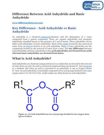
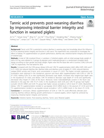
Yu et al. Journal of Animal Science and Biotechnology(2020) 11:87Table 2 Primers used for real-time quantitative PCRGeneAccession No.ZO-1XM 005659811.1 F: CAGCCCCCGTACATGGAGAPrimer sequences (5′ 3′)Size, bp114Page 5 of 11Table 3 Effects of tannic acid (TA) on growth performance inweaned pigletsItemNM 001206404.1 F: ATTCGGACCCATAGCAGACATAG90R: GCGTCTCTTGGTTCTGTTTTAGCOCLNNM 001163647.2 F: CTACTCGTCCAACGGGAAAG158R: ACGCCTCCAAGTTACCACTGCLDN-1 NM 001258386.1 F: GCCACAGCAAGGTATGGTAAC140R: AGTAGGGCACCTCCCAGAAGCLDN-2 NM 001161638.1 F: GCATCATTTCCTCCCTGTT156R: TCTTGGCTTTGGGTGGTTGAPDH NM 001206359.1 F: TGAAGGTCGGAGTGAACGGAT114R: CACTTTGCCAGAGTTAAAAGCAZO-1 Zonula occluden 1, ZO-2 Zonula occluden 2, OCLN Occludin, CLDN-1Claudin 1, CLDN-2 Claudin 2, GAPDH Glyceraldehyde-3-phosphatedehydrogenase, F Forward, R Reverseas the experimental unit for other traits. All data fordiarrhea evaluation were converted by arcsine squareroot transformation for statistics. Results were presentedas means and standard error of means (SEM). ADuncan’s multiple comparison was applied to analyzethe differences among groups. Linear and Quadraticcontrasts were used to determine the dose effect of tannic acid in weaned piglets. A P-value of P 0.05 wasconsidered statistically significant, and 0.05 P 0.10were accepted as representing tendencies to differences.ResultsGrowth performance and diarrheaAs shown in Table 3, there were no differences in ADFI,ADG and F:G between the 3 groups of pigs (P 0.05).As shown in Table 4, the addition of 0.2 and 1.0% TA tothe diet linearly reduced the diarrhea rate, diarrheaindex and diarrhea score of weaned piglets compared tothe CON group (P 0.05).Intestinal permeability and the localization of occludinThe effects of dietary TA supplementation on serumparameters are presented in Table 5. Compared with theCON group, 0.2% and 1.0% TA in the diet significantlyreduced DAO activity and D-lactic acid concentration inserum (P 0.01). The representative image of the occludin stained using immunofluorescence in the small intestinal epithelium is shown in Fig. 1. In CON group,occludin staining was diffused with less staining in theepithelium membrane, indicating possible disruption ofthe tight junction. Meanwhile, occludin was localized tothe cell membrane region in the duodenal epithelium ofpiglets fed 0.2% or 1.0% TA diet. The expressions ofP-value0ANOVA Linear Quadratic0.21.0Initial BW, kg 6.66.66.60.271.0000.9960.999Final BW, kg12.713.412.70.690.7270.7580.469ADFI, g265.6281.8271.924.44 0.8950.9760.644ADG, g154.3165.1151.616.20 0.8260.7600.598F:G ratio1.761.711.800.060.5870.4420.487ADFI, g528.7560.6540.224.86 0.6610.9750.372ADG, g281.8318.4280.820.58 0.3640.6140.188F:G ratio1.881.791.940.070.2940.2640.271ADFI, g397.1421.2406.123.08 0.7610.9740.467ADG, g218.1241.7216.216.44 0.4900.6410.277F:G ratio1.831.761.890.050.1480.171R: GCGCAGACGGTGTTCATAGTTZO-2Added tannic acid, % SEM0 to 14 d15 to 28 d0 to 28 d0.152CON, piglets receiving a basal diet; CON 0.2% TA, piglets receiving a basaldiet supplemented with 0.2% TA; CON 1.0% TA, piglets receiving a basal dietsupplemented with 1.0% TA; ADFI, average daily feed intake; ADG, averagedaily gain; F:G, feed:gain ratiooccludin in the duodenum, jejunum and ileum were remarkably increased by 0.2% and 1.0% TA diet.Intestinal morphologyThe intestinal morphology is given in Table 6. In theduodenum, dietary TA supplementation had no effecton villus height and the thickness of mucosa and duodenal wall (P 0.05). Notably, the 0.2% TA group significantly reduced crypt depth and increased villus height/crypt depth ratio compared to the CON group and the1% TA treatment (P 0.05). In the jejunum, dietary TAsupplementation had no effect on jejunal morphology,including villus height, crypt depth, mucosal and intestinal wall thickness (P 0.05). In the ileum, no significant effect of dietary TA supplementation was observedon the thickness of mucosa and ileal wall (P 0.05).Strangely, although the 1.0% TA group tended to reducethe crypt depth (P 0.063), it significantly reduced thevillus height compared with the CON group (P 0.05).MDA content in serum and small intestineTable 7 presents the MDA content in serum and smallintestine of weaned pigs. Dietary TA supplementationhad no effect on MDA content in serum, duodenum andjejunum (P 0.05), but 1.0% TA group significantly reduced the MDA content in the ileum compared with theCON group (P 0.05).
Yu et al. Journal of Animal Science and Biotechnology(2020) 11:87Page 6 of 11Table 4 Effects of tannic acid (TA) on diarrhea rate, diarrhea index and diarrhea score in weaned pigletsItemAdded tannic acid, %SEM00.21.0Diarrhea rate, %19.4a13.0ab8.7bDiarrhea index0.50.30.2Diarrhea .0010.00020.6280 to 14 d15 to 28 dDiarrhea rate, %16.8aa10.2aaDiarrhea index0.40.2Diarrhea .050.0090.0030.354abb0.310.0080.0030.4520 to 28 dDiarrhea rate, %aDiarrhea index0.4Diarrhea scorea130.3105CON, piglets receiving a basal diet; CON 0.2% TA, piglets receiving a basal diet supplemented with 0.2% TA; CON 1.0% TA, piglets receiving a basal dietsupplemented with 1.0% TAa,bMean values with unlike superscript letters were significantly different (P 0.05)Gene expression of tight junction proteinThe expression levels of ZO-1, ZO-2, OCLN, CLDN-1and CLDN-2 mRNA in the small intestinal mucosa areshown in Fig. 2. In the duodenum, compared with theCON group, the higher ZO-2 mRNA expression levelsof supplemented with 0.2% TA and OCLN mRNA levelsof supplemented with 0.2% and 1.0% TA were determined (P 0.05). In addition, supplementation with 1.0%TA tended to up-regulate OCLN mRNA expressionlevels in the jejunum (P 0.054), and meanwhile, similartrend was shown for ZO-1 mRNA levels of dietary 0.2%TA supplementation in the ileum compared with theCON group (P 0.055).DiscussionGenerally, TA is unfavorable to animal growth in nonruminant animals [20–22], but the experiment resultsare varied. Dietary TA supplementation at 125, 250, 500and 1000 mg/kg levels have shown to linearly reduceADG and feed efficiency in weanling pigs [6]. Studies inbroilers showed that adding 0.07% and 0.2% TA (sweetchestnut wood extract) to the diet had no effect onADFI, ADG and feed conversion ratio [23]. In agreementwith this, the present work demonstrated that adding0.2% or 1.0% TA to the diet had no significant effect onADFI, ADG and F:G in weaned piglets. Moreover,dietary supplemented with 0.5% or 1% TA indicated noadverse effect on growth performance, but the higherdosage, 1.5% TA, decreased ADFI in pigs [24], suggesting the biological effects of TA are dose-dependent. Onthe contrary, other studies have shown that tanninscould improve the growth performance of pigs andbroiler chicks [13, 25, 26], or improve feed utilizationand final body weight of broilers [7]. It’s well known thatTA has positive effects on gastrointestinal tract of animals, such as anti-oxidation and antibacteria, howevertoxic and anti-nutritional effects also exist [27]. Theseeffects seem to be closely related to the source, concentration and chemical structure of TA and animal species[5]. Thus, we hypothesized that TA concentration in ourexperimental diet might be not high enough to impairgrowth performance of weaned piglets. On the otherhand, dietary tannalbin was applied to introduce TA tothe pigs in our experiment. Tannalbin has a less adverseeffect on the palatability of the diet, because the proteinneutralizes the astringency of TA in oral cavity, whichmay also explain why TA has no obvious effect on thegrowth performance, especially the feed intake.Table 5 Effects of tannic acid (TA) on serum parameters in weaned pigletsItemDAO, IU/LD-Lactate, μg/mLAdded tannic acid, eANOVALinearQuadratic3.65 0.001 0.0010.0090.030.0060.0040.074CON, piglets receiving a basal diet; CON 0.2% TA, piglets receiving a basal diet supplemented with 0.2% TA; CON 1.0% TA, piglets receiving a basal dietsupplemented with 1.0% TA; DAO, diamine oxidasea,bMean values with unlike superscript letters were significantly different (P 0.05)
(2020) 11:87Yu et al. Journal of Animal Science and BiotechnologyPage 7 of 11Fig. 1 Effect of tannic acid (TA) on expression and localization of occludin protein in small intestine of weaned piglets (scale bar: 100 μm). Thelocalization of tight junction protein occludin in duodenum (a), jejunum (b) and ileum (c) of weaned piglets was visualized usingimmunofluorescence technique. The localization of occludin (red), DAPI (blue), as well as merged occludin and DAPI are shown. DAPI stainindicates live cells. CON, piglets receiving a basal diet; CON 0.2% TA, piglets receiving a basal diet supplemented with 0.2% TA; CON 1.0% TA,piglets receiving a basal diet supplemented with 1.0% TATable 6 Effects of tannic acid (TA) on intestinal morphology in weaned pigletsItemAdded tannic acid, %SEM00.21.0Villus height, μm354.4337.6357.2Crypt depth, llus height: Crypt depth1.962.302.040.080.0300.7670.010Mucosal thickness, μm794.6747.7847.940.960.2550.1970.296Intestinal wall thickness, mm1.401.381.560.060.1240.0530.545Villus height, μm357.8386.4399.915.090.1670.1050.319Crypt depth, μm161.3163.9169.79.880.8270.5480.945JejunumVillus height: Crypt depth2.242.382.390.130.6740.5360.533Mucosal thickness, μm683.9657.2646.231.770.6950.4790.648Intestinal wall thickness, mm1.031.001.040.040.7380.6670.523Villus height, μm353.6a385.3a288.9b21.110.0170.0120.124Crypt depth, μm176.7166.1140.910.050.0630.0210.794IleumVillus height: Crypt depth2.012.382.040.150.1950.6260.085Mucosal thickness, μm542.0533.2593.063.850.7770.5080.821I
Post-weaning diarrhea usually results in high morbidity and mortality of piglets, which is a serious issue in pig production. The subsequent decline in growth rate has brought enormous economic losses in swine industry. Diarrhea in weaning piglet is mainly caused by weaning stress, lactogenic immunity deprivation, diet and envir-onmental changes.