Transcription
ARTICLE IN PRESSCytotherapy 000 (2020) 1 8Contents lists available at ScienceDirectCYTOTHERAPYjournal homepage: www.isct-cytotherapy.orgFull-length articleCryopreserved bone marrow aspirate concentrate as a cell source for thecolony-forming unit fibroblast assayDustin R. Berger1, Ellis T. Aune1, Christopher J. Centeno1,2, Neven J. Steinmetz1,*12Regenexx, LLC, Broomfield, Colorado, USACenteno-Schultz Clinic, Broomfield Colorado, USAA R T I C L EI N F OArticle History:Received 23 September 2019Accepted 16 April 2020Available online xxxKeywords:AgingBone marrow aspirate concentrate (BMAC)Cell cultureColony-forming unit fibroblast (CFU-F)CryopreservationA B S T R A C TPurpose: The prevalence of connective tissue progenitor cells within a cell-based therapy is often quantifiedusing the colony-forming unit fibroblast (CFU-F) assay. The present study investigates the feasibility of usingcryopreserved bone marrow aspirate concentrate (BMAC) as an alternative cell source to fresh BMAC forCFU-F quantification.Methods: Freshly prepared and corresponding cryopreserved BMAC samples from patients receiving autologous cell therapy (n 98) were analyzed using the CFU-F assay for comparison. Cultures were established bydirectly plating BMAC at low cell densities and maintained for a 2-week growth period. Colonies were enumerated to determine CFU-F frequency, and a subset of cultures was imaged and analyzed to quantify colonyarea and density.Results: A nonlinear relationship was observed between plating density and CFU-F frequency over a widerange in plating densities ( 30-fold). Cryopreserved BMAC yielded recoverable (77 § 23%) and viable (73 §9%) nucleated cells upon thawing. After cryopreservation, CFU-F frequencies were found to be significantlylower (56.6 § 34.8 vs. 50.3 § 31.7 colonies per million nucleated cells). Yet the number of CFU-F in fresh andcryopreserved BMAC were strongly correlated (r 0.87) and had similar area and densities. Further, moderate correlations were observed between the number of CFU-F and nucleated cells, and both the mean colonyarea and density were negatively correlated with patient age. Notably, no relationship was found betweenCFU-F frequency and age, regardless of whether fresh or cryopreserved BMAC was used.Conclusions: Freshly prepared and cryopreserved BMAC yielded correlated results when analyzed using theCFU-F assay. Our findings support the cryogenic storage of patient BMAC samples for retrospective CFU-Fanalyses, offering a potential alternative for characterizing BMAC and furthering our understanding of progenitor cells in relation to clinical outcome. 2020 International Society for Cell and Gene Therapy. Published by Elsevier Inc. This is an open access article under the CC BY-NC-ND license. )IntroductionOrthopedic regenerative medicine is a rapidly growing medicalspecialty that uses both autologous and allogeneic orthobiologics toassist in tissue healing [1]. Autologous bone marrow stands as a common source of progenitor cells, and one predominant orthobiologic,bone marrow aspirate concentrate (BMAC), has been safely used totreat a variety of orthopedic conditions [2 4]. The existence of connective tissue progenitor cells within bone marrow was firstdescribed by Friedenstein and colleagues, when bone formation wasobserved within diffusion chambers populated with bone marrowcell suspensions and implanted intraperitoneally [5]. Later studiesrevealed these progenitor cells to adhere in culture when plated at* Correspondence: Neven J. Steinmetz, PhD, Chief Scientific Officer, Regenexx, LLC403 Summit Boulevard, #201 Broomfield, CO 80021, USA.E-mail address: nsteinmetz@regenexx.com (N.J. Steinmetz).low density and proliferate to form discrete colonies containing fibroblast-like cells [6,7], capable of differentiating into cells of differentmesenchymal lineages, including bone, cartilage and fat, in vitro [8].However, the in vivo multipotency of connective tissue progenitorcells has been disputed [9], and paracrine secretion of trophic andimmunomodulatory factors is currently thought to be the primarymode of action [10]. Notwithstanding, several studies have nowreported improved patient outcomes when autologous BMAC treatments with greater numbers of progenitor cells are used [11,12].Unfortunately, point-of-care assays are not currently available toassess the number of progenitor cells within a BMAC therapy, hindering efforts to correlate their prevalence with patient outcomes.A colony-forming unit fibroblast (CFU-F) assay adopts traditionalcell culture techniques to determine the frequency of plastic adherent, connective tissue progenitor cells contained within a biologicalsample, such as BMAC. One important premise of the CFU-F assay 249/ 2020 International Society for Cell and Gene Therapy. Published by Elsevier Inc. This is an open access article under the CC BY-NC-ND -nd/4.0/)
ARTICLE IN PRESS2D.R. Berger et al. / Cytotherapy 00 (2020) 1 8that each colony is generated from a single progenitor cell, and therefore, the number of colonies observed is representative of the number of progenitor cells as a fraction of the total cells plated [13].Unfortunately, owing to the inherent nature of the CFU-F assay,results cannot be obtained a priori because patients are treated withautologous BMAC long before a clinician has knowledge of the progenitor cell composition. Moreover, fresh samples of BMAC must beassayed within a timely manner for every patient investigated, anuntenable option for most clinicians because large studies would beencumbered with persistent periods of cell culture. As the field oforthopedic regenerative medicine advances, there is a growing needto improve upon the quantitative characterization of BMAC therapies.Cryopreserved BMAC, if similar to fresh BMAC with respect to cultureperformance, could serve as an alternative option to conduct theCFU-F assay once a retrospective cohort of patients is identified. Thisproposed methodology would minimize the overall assay timerequired (batch vs. continuous), minimize the number of samples tobe analyzed (retrospective samples of interest vs. all treated patients)and additionally minimize sources of laboratory variation potentiallyinherent upon culturing samples at many time points.Strong correlations have been observed between pre-freeze andpost-thaw bone marrow across several cell populations, includingmononuclear cells, committed myeloid precursors and hematopoieticstem cells [14]. Additionally, connective tissue progenitor cells havebeen recovered from cryopreserved bone marrow and successfullyculture expanded with clinical applications in mind [15,16]. Althoughseveral groups have performed the CFU-F assay with both fresh andcryopreserved bone marrow and reported no differences betweenthe two, data are limited [15] or not shown [17]. In the present study,patient-matched fresh and cryopreserved BMAC samples were compared using the CFU-F assay to determine whether the prospectivecryopreservation of patient samples is a feasible storage strategy toemploy when collecting samples to be used retrospectively for comparing the prevalence of connective tissue progenitor cells withpatient outcomes.MethodsBMACPatients undergoing orthopedic procedures with autologousBMAC for musculoskeletal injuries elected to have a small portion oftheir injectate retained for cellular analysis. Informed consent wasobtained from all individual participants included in the study. Usingsterile technique, bone marrow was harvested under ultrasound orfluoroscopic guidance and processed into BMAC, as previouslydescribed [4]. In brief, 5 10 mL of whole bone marrow aspirate(BMA) was harvested from each of 3 6 locations per side within theposterior superior iliac spine into syringes containing heparin. TheBMA was serially centrifuged at 200g for 6 min, and the resultantnucleated cell rich buffy coat was manually isolated using aseptictechnique. Before clinical use, a small representative volume ofBMAC ( 0.2 mL) was collected for cell counting, cryopreservationand CFU-F cell culture. Patient age, gender, BMA volume and cell concentration, and BMAC volume and cell concentration are summarizedin Table I.Nucleated cell countComplete blood counts were performed on all BMA and BMACsamples at the time of laboratory processing using an automatedhematology analyzer (ABX Micros 60, Horiba Medical, Montpellier,France). Before counting, the BMAC samples were diluted tenfold insterile saline to remain within the linear range of the instrument. Theresults from the white blood cell parameter were used to report totalnucleated cell counts.Table IPatient and sample data presented as mean § SD,median, and (minimum-maximum).PatientsN 98Age (years)Gender (male, female)BMA (mL)BMA (106 cells/mL)BMAC (mL)BMAC (106 cells/mL)48 § 17, 53, (13 78)58 (59%), 40 (41%)81 § 20, 88, (22 120)25 § 10, 23, (11 50)3.4 § 1.7, 3.0, (0.9-8.7)408 § 113, 406, (113 672)Cryopreservation of BMACTen million nucleated cells from each BMAC sample (10 85 mL)were directly transferred into cryogenic vials and resuspended in chilledcryopreservation medium (1 mL) consisting of 63% minimal essentialmedium alpha (Corning, Corning, NY, USA), 1% L-alanyl-L-glutamine(Corning), 1% penicillin-streptomycin (GE Healthcare, Chicago, IL, USA),30% fetal bovine serum (Access Biologicals, Vista, CA, USA) and 5%dimethyl sulfoxide (Corning). The cryogenic vials were immediatelyadded to an isopropyl alcohol based controlled rate freezing containerand placed in a 80 C freezer. After 24 h, the BMAC samples wereremoved from the controlled rate freezing container and remained at80 C for up to 1 week before being transferred to liquid nitrogen forcryogenic storage.Cryorecovery of BMACFrozen BMAC samples were removed from cryogenic storage andgently swirled within a 37 C water bath for 2 min to permit rapidthawing. The contents of the thawed vials were subsequently dilutedtenfold in warmed complete culture medium consisting of 88% minimal essential medium alpha, 1% L-alanyl-L-glutamine, 1% penicillinstreptomycin, 10% fetal bovine serum and 1 ng/mL human basicfibroblast growth factor (Gold Biotechnology, St. Louis, MO, USA).Nucleated cell count and viability measurements were obtained fromeach BMAC sample post-thaw using 0.4% Trypan Blue stain and anautomated cell counter (TC20, BioRad, Hercules, CA, USA) before further dilution in complete culture medium for cell culture.CFU-F assayInitially, freshly prepared BMAC (n 30) was serially diluted in complete culture medium and directly plated at densities of 90, 30, 10 and3.3 103 nucleated cells per cm2 within standard tissue culture six nster, Austria). In subsequentwell plates (Greiner Bio-One, Kremsmuexperiments, fresh and cryopreserved BMAC samples (n 98) wereplated at 30 and 10 103 nucleated cells per cm2. Viability was takeninto account when plating the thawed BMAC to ensure equivalent numbers of living nucleated cells in both the fresh and cryorecovered conditions. Following a 48 h attachment period, non-adherent cells wereremoved by washing with phosphate buffered saline (PBS), and theremaining cells were maintained at 37 C and 5% CO2 with biweeklyreplacement of complete culture medium. After 2 weeks of culture, thesix-well plates were washed with PBS and subsequently stained with3% (w/v) crystal violet (Sigma Aldrich, St. Louis, MO, USA) in methanol(EMD Millipore, Burlington, MA, USA). Macroscopic colonies, greaterthan 1 mm in diameter and containing a minimum of 100 spindleshaped cells (supplementary Figure 1), were identified and counted bythree independent observers.Image analysis of colony area and intensityHigh-resolution images (1200 dpi) of a subset of six-well platescontaining CFU-F from fresh and cryopreserved BMAC (n 52) were
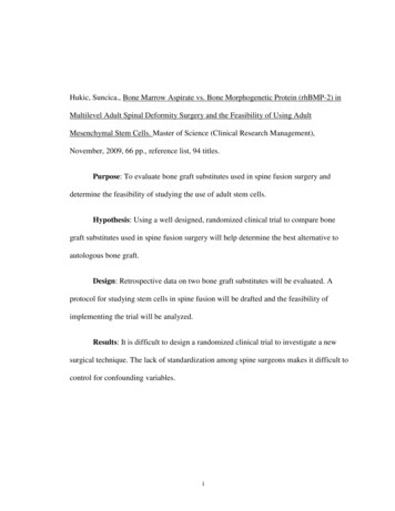
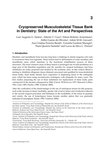
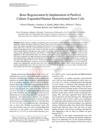
ARTICLE IN PRESSD.R. Berger et al. / Cytotherapy 00 (2020) 1 83Figure 1. The relationship between plating density and colony formation in fresh BMAC samples is non-linear. (A) The average number of counted CFU-F per well plated withinstandard six-well plates at densities of 3.3, 10, 30 and 90 103 nucleated cells per cm2; a quadratic least square regression illustrates the non-linear relationship; (n 30 4). (B)Colony frequencies obtained by normalizing the average CFU-F per well to the input number of nucleated cells; lines represent the median; Friedman test with Dunn’s multiplecomparisons: **P 0.001, *P 0.01 versus 90 103 nucleated cells per cm2. (C) Representative high-resolution well scans from a single donor; circles demarcate individual CFU-Fas counted by one of three independent observers.acquired using a commercial flatbed scanner (CanoScan LiDE220,Cannon, Tokyo, Japan). A java-based plugin (ColonyArea) for ImageJ(National Institutes of Health, Bethesda, MD, USA) was used to automatically crop the images to individual wells, convert them to grayscale and apply a background threshold for colony detection [18].The mean colony area for each well was subsequently calculated bydividing the number of pixels having an intensity greater than zeroby the total number of manually counted colonies and scaling theresult to square millimeters. Similarly, the mean colony density foreach well was calculated by dividing the raw integrated density(pixel area * mean gray value) of the thresholded regions by the number of manually counted colonies and scaling the result by a factor of10 5.normalizing to the total number of input cells, the calculated CFU-Ffrequency (CFU-F per million nucleated cells) trended downwardwith plating density and was significantly lower at the highest density (Figure 1B). Representative high-resolution well scans show differences in colony formation across the investigated plating densities(Figure 1C). Overcrowding caused difficulty in discerning individualcolonies at the highest plating density, whereas the majority ofBMAC cultures (22 of 30) contained wells with one or no CFU-F at thelowest plating density. All BMAC samples consistently produced atleast three CFU-F per well at one or both intermediate plating densities. Thus, 30 and 10 103 nucleated cells per cm2 were selected asideal plating densities to evaluate cryopreserved BMAC as a cellsource for the CFU-F assay.Statistical analysisAll colony data are presented as averages of the individual wellsacross a six-well plate, and all other data are presented as averages(mean § SD) among patient BMAC samples. Normality was assessedusing the D’Agostino-Pearson test, and the data were not normallydistributed. Therefore, non-parametric statistical tests were performed (GraphPad Prism, GraphPad Software, La Jolla, CA, USA). Differences between multiple pairwise groups were evaluated using theFriedman test with Dunn’s post hoc multiple comparison testing, differences between two pairwise groups were evaluated using the Wilcoxon signed rank test and strengths of association were evaluatedusing Spearman’s rank-order correlation. Comparisons and associations were considered statistically significant at P 0.05.Cryopreserved BMAC yields viable nucleated cellsSmall sample volumes of BMAC, placed within dimethyl sulfoxide containing cryopreservation medium, subjected to a controlledrate freezing process, and cryopreserved for an average duration of16 § 14 days, produced viable nucleated cells upon thawing(Figure 2). An average of 77 § 23% of cells, ranging from 34% to 160%,were recovered from a target number of 10 million cryopreservednucleated cells (Figure 2A). Cell viability ranged from 49% to 88%with an average of 73% § 9% (Figure 2B).ResultsOptimizing the plating density of BMAC for the CFU-F assayA limited number of freshly prepared BMAC samples were platedover a 3-fold dilution series to determine the appropriate platingdensity, or densities, to evaluate CFU-F formation (Figure 1). Whenplated at 90, 30, 10 and 3.3 103 nucleated cells per cm2 and cultured for a period of 2 weeks, a non-linear relationship between plating density and CFU-F count was observed (Figure 1A). AfterFigure 2. Viable nucleated cells are obtained from cryopreserved BMAC. (A) Cell recovery and (B) viability measurements following cryorecovery; lines represent the median(n 98).
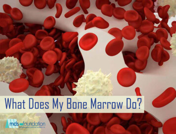
ARTICLE IN PRESS4D.R. Berger et al. / Cytotherapy 00 (2020) 1 8Cryopreserved BMAC as a cell source for the CFU-F assayFresh and corresponding cryopreserved BMAC samples were cultured at two plating densities to obtain CFU-F counts, and a subset ofculture plates were scanned to quantify colony area and density(Figure 3). The lower plating density of 10 103 nucleated cells percm2 was used to calculate the CFU-F frequency of a BMAC sampleonly if there was an average of three or more CFU-F per well in bothexperimental conditions, otherwise the higher plating density wasused (supplementary Figure 2). Approximately 40% (38 of 98) ofBMAC samples required plating at 30 103 nucleated cells per cm2to calculate the CFU-F frequency using this criterion. Overall, compared with fresh BMAC, the CFU-F frequencies were lower withinsamples that underwent cryopreservation (Figure 3A). Average CFUF frequencies of 50.3 § 31.7 (0.0050% § 0.0032%) and 56.6 § 34.9(0.0057% § 0.0035%) were quantified in cryopreserved and freshBMAC, respectively, representing a small, yet significant, pairwise difference (fold change 0.95 § 0.38, P 0.001).A strong positive correlation (r 0.87, P 0.001) was observedbetween fresh and cryopreserved BMAC with respect to CFU-F frequency (Figure 3B). No significant differences in mean colony area ordensity were found following cryopreservation (Figure 3C). Moreover, fresh and cryopreserved BMAC samples were moderately correlated (r 0.49, P 0.001) with respect to colony area (Figure 3D) anddensity (data not shown). Representative images of CFU-F assayplates from a single donor illustrate the comparable nature of CFU-Fquantified from fresh (Figure 3E) and cryopreserved (Figure 3F)BMAC samples.Cryopreserved BMAC samples from a limited number of patientswere thawed a second time, approximately 10 months (330 § 30days) after having been initially thawed, to investigate the effectslonger periods of cryopreservation have on cell viability and CFU-Ffrequency. Viability was not significantly affected (75% § 7% vs. 78%§ 7%), whereas CFU-F frequency remained strongly correlated(r 0.79, P 0.005) between the first and second instance of cryorecovery (supplementary Figure 3).CFU-F, Nucleated cells and patient ageNucleated cell and CFU-F counts within fresh BMAC samples werecompared to determine their strength of association (Figure 4). Theaverage concentrations of nucleated cells and CFU-F were 4.1 §1.1 108 per mL and 2.3 § 1.6 104 per mL, respectively, and a moderate correlation (r 0.37, P 0.001) was observed between the two(Figure 4A). When accounting for the total volume of BMAC prepared, 3.4 § 1.7 mL (Figure 4B), the strength of the positive correlation (r 0.64, P 0.001) between the overall number of CFU-F andtotal nucleated cells increased (Figure 4C). Similar trends wereobserved when using the CFU-F data acquired from cryopreservedBMAC samples (supplementary Figure 4).Colony formation and CFU-F frequency were compared withpatient age to determine whether any relationships exist (Figure 5).Patient age ranged from 13 to 78 years, with an average age of 48 §17 years. When the CFU-F frequency (Figure 5A) was compared withage, no correlation was present (P 0.05). However, when normalized to the volume of bone marrow aspirated, a weak negative trend(r 0.19, P 0.068) emerged (Figure 5B). Moreover, moderate negative correlations (r 0.3, P 0.05) with age were observed forboth mean colony area (Figure 5C) and density (Figure 5D). The CFUF data obtained from cryopreserved BMAC samples yielded similarresults (supplementary Figure 5).DiscussionIn the present study, both freshly prepared and cryopreservedBMAC from the same group of patients were evaluated by the CFU-Fassay and compared to determine the feasibility of using cryopreserved samples to estimate the connective tissue progenitor cell content of bone marrow based treatments. A direct plating method wasused to quantify CFU-F because others have reported commonly useddensity gradient separation techniques reduce the recovery of progenitor cell populations [19 21]. Our results demonstrate a non-linearrelationship between plating density and colony formation, challenging a seminal report describing the relationship as linear [7]. However,culture duration is a contributing factor, and our longer period (14 vs.8 days) could allow adjacent colonies to fuse to a greater extent, resulting in the underestimation of CFU-F at higher plating densities. In support of this, a decrease in CFU-F counts between days 7 and 10 ofculture with higher plating densities has been reported [22], and afternormalizing to the input number of cells from directly plated bonemarrow, the relationship between plating density and CFU-F frequency has been described as negatively correlated [20,22].Every BMAC sample was plated at two densities, one three timesgreater than the other, to quantify colonies in donors having a widerange of CFU-F frequencies, as recommended in the literature [22].The lower plating density was selected based on both the resultsfrom our initial dilution series and previous work identifying10 103 cells per cm2 as the most advantageous density for platingwhole bone marrow to obtain representative CFU-F counts [19]. Atypical well within a six-well tissue culture plate has an available surface area of approximately 10 cm2. In requiring an average of three ormore CFU-F per well, the lower limit of detection of CFU-F frequencywas established at roughly 30 or 10 CFU-F per million nucleated cells(0.003% or 0.001%), depending on the plating density used. Almost allpatient samples (95 of 98) yielded an average of three or more CFU-Fper well at one or both plating densities, suggesting the currentapproach adequately captures the prevalence of progenitor cellsacross our patient population. Although a small portion of patientsamples (3 of 98) did not yield an average of three or more CFU-F perwell at the higher plating density of 30 103 nucleated cells per cm2,sporadic colonies were observed within all culture plates.Previous studies have documented success in recovering viablenucleated cells from cryopreserved bone marrow [15,16,23,24]. However, the primary focus of these earlier studies was on the cultureexpansion of progenitor cells for clinical use and not the quantification of the initial progenitor cell content. Although there have beenlimited reports of unchanging CFU-F numbers between fresh and cryopreserved bone marrow, minimal data have been provided [15,17].The nucleated cell viabilities measured within our cryopreservedBMAC samples (74% § 9%) are similar to those previously reported[15]. Although a limited number of samples exhibited higher cellrecoveries than expected following cryopreservation, BMAC is a heterogenous cell solution that can be viscous in nature, complicatingefforts to transfer small, precise and well-mixed volumes. Given thehigh nucleated cell concentrations within the BMAC samples studied(108 cells per mL), unintentionally transferring a small percentagegreater or less than the target volume could variably affect the finalnumber of frozen cells.Our current data reveal significant correlations between fresh andcryopreserved BMAC with respect to CFU-F frequency (r 0.87) andmean colony area (r 0.49), demonstrating the utility of cryopreserved BMAC in determining the prevalence of connective tissue progenitor cells. A small, albeit significant, pairwise difference in CFU-Ffrequency was observed (56.9 § 34.8 vs. 50.8 § 32.0 per millionnucleated cells), suggesting that results obtained from cryopreservedsamples may provide an underestimation of the true values. Yet notall cryopreserved BMAC samples produced fewer CFU-F than theirfresh counterpart. Moreover, no significant pairwise differences wereobserved between fresh and cryopreserved BMAC with respect tomean colony area and density, as the data was more variable. SomeBMAC samples produced larger, denser colonies after being frozen,but others did not.
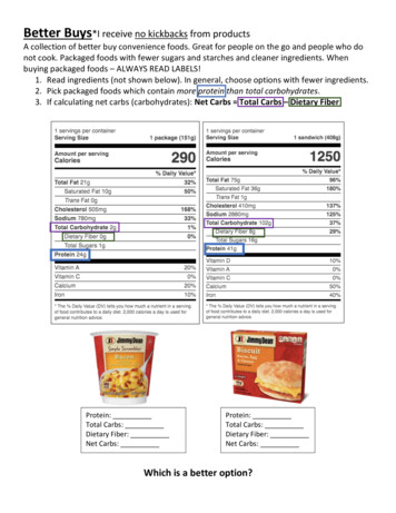
ARTICLE IN PRESSD.R. Berger et al. / Cytotherapy 00 (2020) 1 85Figure 3. Cryopreserved BMAC performs similar to fresh BMAC in the CFU-F Assay. (A) CFU-F frequencies from individual donors (n 98) counted within fresh and frozen BMACsamples and the resultant fold change. (B) Spearman rank order correlation between fresh and frozen BMAC with respect to CFU-F frequency. (C) Mean colony area and densityquantified from high-resolution scans of assay plates obtained from a subset of donors (n 52). (D) Spearman rank order correlation between fresh and frozen BMAC with respectto mean colony area. Violet data points are from BMAC samples plated at 10 103 nucleated cells per cm2; (A and B, n 60 of 98, C and D, n 32 of 52); black data points are fromBMAC samples plated at 30 103 nucleated cells per cm2; (A and B, n 38 of 98, C and D, n 20 of 52); lines represent the median; Wilcoxon signed rank test **P 0.001 versus frozen BMAC. Representative scans and colony analysis of CFU-F within (E) fresh and (F) frozen BMAC samples from a single donor; average CFU-F counts from three independentobservers are displayed above and below the wells on the left; mean colony area and density are displayed above and below the wells on the right; six-well plate averages (mean §SD) are displayed in the center.
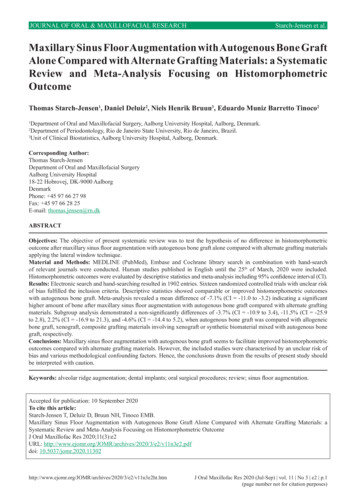
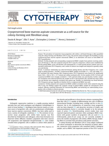
ARTICLE IN PRESS6D.R. Berger et al. / Cytotherapy 00 (2020) 1 8Figure 4. Colony formation is correlated with the total nucleated cell count. (A) Spearman rank order correlation between CFU-F concentration and nucleated cell concentration infresh BMAC. (B) Volume of BMAC following laboratory processing; the line represents the median. (C) Spearman rank-order correlation between total CFU-F and total nucleatedcells; (n 98).Figure 5. Colony formation, but not CFU-F frequency, is negatively correlated with patient age. Spearman rank-order correlations between age and (A) CFU-F frequency, (B) CFU-Fwhen normalized to the volume of bone marrow aspirated, (C) mean colony area and (D) mean colony density in fresh BMAC (A and B, n 98; C and D, n 52).Significant positive correlations were observed between the number of nucleated cells and CFU-F within both fresh and cryopreservedBMAC as well. Several groups have reported improved healingresponses in patients treated with BMAC preparations containinghigher concentrations of CFU-F [11,12]. Together, these data supportour earlier findings of higher BMAC cell concentrations resulting inbetter pain outcomes for the treatment of knee osteoarthritis [25]. Alinear relationship between CFU-F frequency and patient age was notobserved, regardless of whether fresh or cryopreserved BMAC wasused, as concluded in several studies [12,26,27]. Further, unlike otherreports [11,28], no additional correlations with age were found whenpatient samples were separated by gender (data not shown). However, when accounting for the volume of bone marrow aspirated, aweak negative relationship between patient age and the number ofCFU-F became apparent, although the correlation was not statisticallysignificant. The presence of a negative correlation between CFU-F frequency and age has been described in more than one study[17,29,30]. Recently, age-related declines in CFU-F frequency, areaand density within minimally cultured bone marrow aspirates havebeen reported, yet large donor-to-donor variation led the authors toconclude that age was not a reliable predictor of progenitor cell number [31]. Although our data failed to show a correlation betweenCFU-F frequency and age, significant declines were observed in bothmean colony area and density, aligning well with reports of bonemarrow progenitor cell frequencies remaining stable with age whiletheir ability to proliferate declines [27].Variation in CFU-F frequencies can arise intrinsically from patientspecific differences in bone marrow architecture and cellularity [32],whereas extrinsic sources of variation include the bone marrow aspiration site [33], technique [34] and volume [32], as well as the process used to concentrate the progenitor cells. A wide range of bonemarrow volumes was aspirated from the patients included in thepresent study, resulting from the requirements of the different orthopedic procedures involved. To minimize hemodilution and maximize
ARTICLE IN PRESSD.R. Berger et al. / Cytotherapy 00 (2020) 1 8the yield of progenitor cells, additional aspiration sites were usedwhen obtaining larger BMA volumes [35]. Given the inherent variation in CFU-F frequency, a considerable number of BMAC samplesmay need to undergo analysis to establish evidence for a relationshipbetween the prevalence of progenitor cells and patient outcomes.One advantage of using cryopreserved BMAC samples is the capability to analyze all samples of interest at once.Multiple laboratory variables play an important role in colony formation. In addition to cell separation technique [21], plating density[20] and culture duration [22], other contributing factors including basalmedium [7], serum [13], the addition of growth factors or other supplements [36], oxygen concentration [37], culture handling, and countingtechnique can affect overall CFU-F results. The ability to maintain consistency among these factors highlights another important advantage ofperforming retrospective batch analyses on cryopreserved BMAC samples. Efforts were taken to use controlled culture conditions and minimize sources of laboratory variation throughout our studies, andaverage colony counts from three independent observers were used tominimize bias. Nevertheless, manual enumeration of CF
Nucleated cell count and viability measurements were obtained from each BMAC sample post-thaw using 0.4% Trypan Blue stain and an automated cell counter (TC20, BioRad, Hercules, CA, USA) before fur-ther dilution in complete culture medium for cell culture. CFU-F assay Initially, freshly prepared BMAC (n 30) was serially diluted in com-