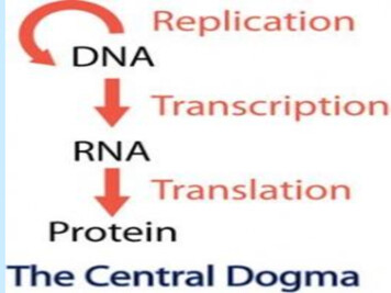
Transcription
Tan et al. Breast Cancer Research (2016) 18:31DOI 10.1186/s13058-016-0692-6RESEARCH ARTICLEOpen AccessA five-gene reverse transcription-PCR assayfor pre-operative classification of breastfibroepithelial lesionsWai Jin Tan1, Igor Cima1, Yukti Choudhury1, Xiaona Wei1, Jeffrey Chun Tatt Lim2, Aye Aye Thike2,Min-Han Tan1* and Puay Hoon Tan2,3*AbstractBackground: Breast fibroepithelial lesions are biphasic tumors and include fibroadenomas and phyllodes tumors.Preoperative distinction between fibroadenomas and phyllodes tumors is pivotal to clinical management.Fibroadenomas are clinically benign while phyllodes tumors are more unpredictable in biological behavior, withpotential for recurrence. Differentiating the tumors may be challenging when they have overlapping clinical andhistological features especially on core biopsies. Current molecular and immunohistochemical techniques have alimited role in the diagnosis of breast fibroepithelial lesions. We aimed to develop a practical molecular test to aidin distinguishing fibroadenomas from phyllodes tumors in the pre-operative setting.Methods: We profiled the transcriptome of a training set of 48 formalin-fixed, paraffin-embedded fibroadenomasand phyllodes tumors and further designed 43 quantitative polymerase chain reaction (qPCR) assays to verifydifferentially expressed genes. Using machine learning to build predictive regression models, we selected a five-genetranscript set (ABCA8, APOD, CCL19, FN1, and PRAME) to discriminate between fibroadenomas and phyllodes tumors.We validated our assay in an independent cohort of 230 core biopsies obtained pre-operatively.Results: Overall, the assay accurately classified 92.6 % of the samples (AUC 0.948, 95 % CI 0.913–0.983, p 2.51E-19),with a sensitivity of 82.9 % and specificity of 94.7 %.Conclusions: We provide a robust assay for classifying breast fibroepithelial lesions into fibroadenomas and phyllodestumors, which could be a valuable tool in assisting pathologists in differential diagnosis of breast fibroepithelial lesions.Keywords: Phyllodes tumors, Fibroadenomas, Fibroepithelial lesions, Prediction, Multigene assay, Core biopsy, FFPEBackgroundFibroadenomas and phyllodes tumors are fibroepitheliallesions of the breast, characterized by proliferation of bothepithelial and stromal components. Fibroadenomas aremore commonly encountered on core biopsies than therarer phyllodes tumors (approximately 20 % and 1 %of breast core needle biopsies respectively) [1, 2]. Thepreoperative distinction between the two lesions hassignificant impact on subsequent treatment. The current* Correspondence: mhtan@ibn.a-star.edu.sg; tan.puay.hoon@singhealth.com.sg1Division of Biodevices and Diagnostics, Institute of Bioengineering andNanotechnology, 31 Biopolis Way, The Nanos, #04-01, Singapore 138669,Republic of Singapore2Department of Pathology, Singapore General Hospital, 20 College Road,Academia, Level 7, Diagnostics Tower, Singapore 169856, Republic of SingaporeFull list of author information is available at the end of the articlerecommended management for phyllodes tumor diagnosedon core biopsy is wide excision without axillary stagingregardless of grade [3]. Conversely, fibroadenomas are observed conservatively or, if tumors are larger than 2 cm,may be simply excised without achieving negative surgicalmargins [3]. This approach is due to the indolent behaviorof fibroadenomas, despite sporadic reports of recurrences[4, 5], while phyllodes tumors have unpredictable outcomeswith malignant tumors potentially progressing to metastasisand mortality [6–10]. It has been challenging separatingcellular fibroadenoma from benign phyllodes tumor due tooverlapping histological features, and this is particularlyproblematic on limited material of core biopsies, whichmay lead to over- or under-treatment for some patients,resulting in unnecessary anxiety and cost. 2016 Tan et al. Open Access This article is distributed under the terms of the Creative Commons Attribution 4.0International License (http://creativecommons.org/licenses/by/4.0/), which permits unrestricted use, distribution, andreproduction in any medium, provided you give appropriate credit to the original author(s) and the source, provide a link tothe Creative Commons license, and indicate if changes were made. The Creative Commons Public Domain Dedication o/1.0/) applies to the data made available in this article, unless otherwise stated.
Tan et al. Breast Cancer Research (2016) 18:31Several studies have proposed differentiating histologicalfeatures such as stromal cellularity, stromal overgrowth,fragmentation, subepithelial condensation and presence ofadipose tissue within stroma on core biopsies being indicative of phyllodes tumor [11–13].However, interpretation ofthese parameters is subjective, with interobserver variationand only moderate reproducibility between pathologists[11, 14]. Varied reports of immunohistochemical markersused in distinguishing phyllodes tumors from fibroadenomas suggest a lack of consensus and objectivity in assessing the expression of these biomarkers. Some authorsreported Ki-67 expression to be helpful in diagnosing phyllodes tumors [15–17] but there are reports to the contrary[18, 19]. Lin et al. suggested a combination immunoscoreof p16-INK4a and retinoblastoma-associated protein (pRB)[20] while Maity et al. reported expression of collagen I, IIIand CD105-positive microvessel density as parameters todifferentiate the two lesions [21]. The vast majority of thesestudies were not conducted using pre-operative biopsies,which is where key management decision is required.We set out to identify a useful molecular signature tohelp differentiate fibroadenomas from phyllodes tumorsusing pre-operative core biopsies to improve predictionof the final diagnosis.MethodsTraining set for assay developmentThe study received approval from the Centralized Institutional Review Board (CIRB 2005/002/F). As this was aretrospective study with anonymized cases, no specificpatient consent was individually required. Forty-eight samples (24 fibroadenomas and 24 phyllodes tumors) were firstemployed as the training set for assay development. Theseincluded 10 paired core biopsies and surgical samples (20samples), and 28 independent core and excisional samplesfrom 38 patients (Table 1 and Additional file 1: Table S1).These formalin-fixed, paraffin-embedded (FFPE) sampleswere randomly selected from cases diagnosed at theDepartment of Pathology, Singapore General Hospitalfrom 2008 to 2012. Hematoxylin and eosin (H&E)-stainedslides were retrieved and reviewed. Phyllodes tumor wasdefined when there were well-developed fronds accompanied by increased stromal cellularity as opposed to fibroadenomas in which epithelial and stromal componentswere arranged in either intracanalicular or pericanalicularpatterns without fronds or stromal hypercellularity.Differences in clinical features between fibroadenomasand phyllodes tumors were assessed with Mann–WhitneyU test and Fisher’s exact test.Expression profiling by Whole-Genome DASL HighThroughput (HT) AssayRepresentative tumor areas were identified of whichthree to seven sections of 10-μm-thick sections from thePage 2 of 9Table 1 Clinical features of the training cohort from 38 patientsFeaturesFibroadenomas(n 19)Phyllodestumors (n 19)p value35 (17–80)44 (18–64)0.0925 (15–50)65 (25–220)AgeMedian (range)SizeMedian (range)Ethnicity, n (%) 0.0010.2Chinese13 (68.4)11 (57.9)Malay0 (0.0)4 (21.0)Indian2 (10.5)1 (5.3)Others4 (21.1)3 (15.8)HistologySimple fibroadenoma15aComplex fibroadenoma4cBenign phyllodes tumor13bBorderline phyllodes tumor3cMalignant phyllodes tumor3caFour paired core biopsies and surgical excisionsThree paired core biopsies and surgical excisionscOne paired core biopsy and surgical excisionbsame FFPE tumor block were obtained, deparaffinizedand macrodissected. RNA was extracted using theRNeasy FFPE kit (Qiagen, Hilden, Germany) and quantified by Nanodrop Spectrophotometer (Thermo FisherScientific, Waltham, MA, USA). A total of 100 ng wasused for quality assessment by real-time amplification ofthe RPL13A gene (forward primer, 5’-CACTTGGGGACAGCATGAG-3’, and reverse primer, 5’-GTAACCCCTTGGTTGTGCAT-3’) using the Power SYBR GreenRNA-to-CT 1-Step Kit (Life Technologies, Carlsbad,CA, USA) on a CFX96 Real-Time PCR instrument(Bio-Rad Laboratories, Hercules, CA, USA). Sampleswith threshold cycle (Ct) below 29 were further subjected to quality assessment on a bioanalyzer. Eligiblesamples were submitted for expression profiling on theWhole-Genome DASL HT Assay (Illumina, Inc., SanDiego, CA, USA) at the Biopolis Shared Facilities A*Star,Singapore. The assay interrogates 29,377 features usingthe HumanHT-12 v4 BeadChip (Illumina, Inc.). Quantilenormalized gene expression data pre-analyzed usingGenomeStudio (Illumina, Inc.) was delivered. Data areavailable through GEO [GEO: GSE78071].Selection of normalization genes and differentiating genesNormalization genes were selected based on the smallestvalue of coefficient of variation among all samples. Differentiating genes were selected using the SignificanceAnalysis of Microarrays package [22] and filtered basedon the following criteria: (1) q-value less than 0.05; (2)mean difference of expression above 500; (3) R-fold
Tan et al. Breast Cancer Research (2016) 18:31Page 3 of 9above 1.5 (for genes highly expressed in phyllodestumors) or less than 0.67 (for genes highly expressedin fibroadenomas).package [27] with inclusion of the interaction terms. Thebest model was selected based on the lowest Akaike information criteria (AIC) value [28].Design of quantitative polymerase chain reaction (qPCR)assayValidation cohort for model validationcDNA was synthesized from 1 μg of RNA using theHigh-Capacity cDNA Reverse Transcription Kit (AppliedBiosystems , Life Technologies, Carlsbad, CA, USA).Each qPCR assay consisted of 1X Power SYBR GreenPCR Master Mix (Life Technologies), 0.5 μM of forwardand reverse primer each and 1 μl of 10-fold dilutedcDNA as a template in a final total volume of 10 μl.Primers were designed using Primer-BLAST (NCBI, Bethesda, MD, USA) [23] with accession number listed inAdditional file 1: Table S2. Non-template control actedas a negative control. Specificity of the amplicons wasverified by melt curve analysis.Data quantification and model buildingDelta Ct (ΔCt) for each gene and sample was quantified as:ΔCt Ct(gene, sample) geomean(Ct(five normalizationgene)). For comparison with expression of the WholeGenome DASL HT Assay, ΔCt data was transformedto 2-ΔCt as a positive linear scale and significance ofcorrelation was analyzed with Pearson’s correlationtest. Similarly to Cima et al. and Kälin et al. [24, 25],we used Random Forest (RF) ensemble classifier [26]to rank the importance of gene transcripts differentiatingfibroadenomas and phyllodes tumors on qPCR assays.The top seven performing genes were used to build predictive logistic regression models using exhaustive searchfor the best model. To this end we used the glmultiThe model of the multigene assay was tested on a separate set of 230 core biopsies with at least 2 years offollow-up. Hematoxylin and eosin (H&E)-stained slideswere retrieved and reviewed. The outcome of the multigene assay was compared against the final diagnosis onthe corresponding surgical excisions. Cases without subsequent surgical excisions were free from progressionfor at least 2 years and diagnosis made based on the initial core biopsy was used as the reference instead.ResultsClinical features of training setThe clinical features and histology of the training set areshown in Table 1. Examples of the histological appearances of fibroadenoma and phyllodes tumor are shownin Fig. 1. Phyllodes tumors were significantly larger thanfibroadenomas (p 0.001). Median age of patients diagnosed with fibroadenomas and phyllodes tumors was35 years and 44 years respectively (p 0.09). No significant differences were observed for ethnicity distributionbetween the two groups of tumors.Expression profiling and correlation with qPCR assaysForty-seven samples (97.9 %) from 37 patients passed thequality control and were profiled successfully. Genesdiscriminating fibroadenomas and phyllodes tumors arelisted in Additional file 1: Table S3. We designed and validated qPCR assays on 43 selected genes. ConcordanceFig. 1 Histology of fibroadenoma and phyllodes tumor on surgical excisions. a, b An example of a fibroadenoma at low-power (a) and high-power(b) magnifications. c, d An example of a benign phyllodes tumor at low-power (c) and high-power (d) magnifications
Tan et al. Breast Cancer Research (2016) 18:31between expression profiling and the qPCR assayswas assessed based on a pilot run on six representativesamples (Additional file 1: Table S4). Twenty-three assayswith Pearson’s r of above 0.6 were further tested on theremaining 40 samples. One case was excluded due to insufficient material after expression profiling.Development of a multigene qPCR panelThe results of ΔCt for all 23 qPCR assays were rankedusing variable importance feature of the Random Forestclassifier (Fig. 2). The seven most important genes inseparating fibroadenomas and phyllodes tumors wereTRIM29, FN1, CCL19, ABCA8, NPTX2, APOD andPRAME. A total of 268,435,456 candidate models wereidentified by glmulti based on these seven genes. Weemployed the genetic algorithm approach in the packageto perform automated screening for the best model basedon AIC value. A final five-gene model encompassingAPOD, ABCA8, PRAME, FN1, and CCL19 with AIC of14.2 was returned with coefficients as listed in Table 2.Performance of the modelPatient demographics of the validation cohort of 230core biopsies are shown in Table 3. Overall the assaywas able to predict 213 (92.6 %) cases accurately. Theprediction accuracy rates for fibroadenomas and phyllodes tumors were 94.7 % (179/189) and 82.9 % (34/41)respectively (Table 4), with positive (PPV) and negative(NPV) predictive values of 77.3 % and 96.2 %. A receiveroperating characteristics curve with an area under thecurve (AUC) of 0.948 (95 % confidence interval (CI)0.913–0.983, p 2.51E-19) was generated, indicating alarge effect size in expected diagnostic performance forthe five-gene assay (Fig. 3).Of the 17 discordant cases (Table 5), seven were diagnosed as phyllodes tumors on pathological reports butPage 4 of 9Table 2 Coefficients of the best model in predicting diagnosisin the 46-sample setGenesCoefficientsAPOD2.95575APOD:ABCA8 0.11934PRAME:FN1 0.43165PRAME:CCL190.08326were predicted as fibroadenomas on our assay. Upon review of these seven cases, two cases contained high epithelium content (Fig. 4), two were heterogeneous onhistology with focal areas suggestive of fibroadenomas,while three other cases were confirmed as phyllodes tumors on review. The other ten of the 17 discordant caseswere diagnosed as fibroadenomas on pathological reports but were predicted as phyllodes tumors on ourassay. Among these ten cases, six cases had postoperative excisional material available as reference whilethe remaining four cases were benchmarked against thepre-operative pathological diagnosis. Of the six with excisional material, four were unequivocally fibroadenomason histology, one was a cellular fibroadenoma withoutprominent fronds, and one was a fibroadenoma withsclerosing adenosis. Of the four pre-operative biopsies,one was unequivocally fibroadenoma, two cases contained features in keeping with fibroadenoma with hyalinized leafy fronds noted albeit without stromalcellularity, and one was an indeterminate case with focalareas of hemorrhage and high cellularity, which couldnot be definitively concluded on review.Table 3 Patient demographics of the validation cohort of 230core biopsiesCharacteristicsNPercentage (%)AgeMedian46 yearsRange15–75 Others3213.9Fibroadenoma18982.2with subsequent surgical excisions5825.2without subsequent surgical excisions13157DiagnosisFig. 2 Box plot of mean decrease in accuracy for 100 RandomForest trees generated for each gene. Mean decrease in accuracymeasures the importance of each gene to the classificationPhyllodes 3
Tan et al. Breast Cancer Research (2016) 18:31Page 5 of 9Table 4 Performance of the five-gene model in predictingdiagnosis in the independent validation cohort of 230 core biopsiesDiagnosis from pathological reportPredicted outcomefrom the five-geneassayFibroadenomasPhyllodes tumorsFibroadenomas1797NPV 0.962Phyllodes tumors1034PPV 0.773Spec 0.947Sen 0.829The five-gene assay has an overall accuracy of 92.6 %, with a sensitivity (sen)of 82.9 % and specificity (spec) of 94.7 %. The positive predictive value (PPV)and negative predictive value (NPV) are 77.3 % and 96.2 % respectivelyAmong the 230 core biopsies, the pre-operative pathological diagnoses were inconclusive for 22 cases wherethe term ‘fibroepithelial lesion’ was assigned, and therewere three cases where the preoperative diagnoses wereincongruous with the post-operative outcome (Table 6).Of these 25 cases, the five-gene assay was 80 % (20/25)accurate in classification with a PPV of 94.7 %.DiscussionClassification of breast fibroepithelial tumors based ondifferentiating morphological and immunohistochemicalfeatures on pre-operative material has been challengingwith variable findings across different groups (see Table 7for summary). Jacobs et al. and Lee et al. first describedindividual pathological parameters which might help todifferentiate fibroadenomas and phyllodes tumors inthese limited samples [11, 16]. Jara-Lazaro proposed acombination of histological and immunohistochemicalmarkers to indicate phyllodes tumors on core biopsies[15] but did not weigh the relative importance of eachparameter in predicting phyllodes tumors. Morganaddressed this question by proposing a predictivetool including coefficient factors for each parameterto distinguish between fibroadenomas and phyllodes tumors but this has yet to be validated in an independentFig. 3 Receiver operating characteristics (ROC) curve of the five-genemodel with an area under the curve (AUC) of 0.948 (95 % confidenceinterval (CI) 0.913–0.983, p 2.51E-19) in predicting diagnosis in theindependent set of 230 core biopsiesTable 5 Seventeen cases with discordant outcomes betweenthe five-gene assay and pathological diagnosis in the validationcohort. Post-operative diagnoses were used as benchmarkreference unless otherwise statedSample IDFive-gene assayPathological diagnosisCB22FABenign PTaCB116FABenign PTCB95FABenign PTCB28FABenign PTaCB68FABenign PTaCB120FABorderline PTaCB130FABorderline PTCB26PTCellular FAb,cCB184PTFAb,cCB251PTFAbCB258PTFAb,dFA fibroadenoma, PT phyllodes tumoraPre-operative pathological diagnoses were inconclusive or discordant withpost-operative pathological diagnoses (see Table 6 asterisked cases)bCases of core biopsies without subsequent surgical excisions. Outcome of thefive-gene assay was benchmarked against the pre-operativepathological diagnosiscFeatures in keeping with fibroadenoma with hyalinized leafy fronds notedalbeit without stromal cellularitydFocal areas of hemorrhage and high cellularity, diagnosis could not bedefinitively concluded on reviewseries of core biopsies [12]. Our study is the first to investigate differentiating features of fibroepithelial lesions onpre-operative material at the molecular level. We have developed a five-gene assay using a systematic approachbased on genome-wide expression profiling data andvalidated the assay in an independent cohort of 230pre-operative core biopsies of breast fibroepithelial lesions, the largest cohort reported so far. The pre-operativecore biopsies were FFPE tissue containing low-qualityRNA. Accordingly, our assay has been developed usingRNA extracted from limited FFPE materials from core biopsies and thus is expected to perform on such materialin the clinical setting.Comparatively in surgical excisional materials, Huanget al. proposed a two-gene test derived from methylationprofiling of an 11-gene panel in 86 samples [29], whichdescribed an elevated RASSF1A and/or TWIST1 methylation observed in phyllodes tumors as compared tofibroadenomas. They further evaluated the test in a separate validation cohort of 19 samples and reported asensitivity and specificity of 0.33 and 0.75 respectively,
Tan et al. Breast Cancer Research (2016) 18:31Page 6 of 9Fig. 4 Example of a discordant case containing high epithelium content. The five-gene assay predicted the core biopsies (a) as fibroadenoma butthe final surgical excision (b) was diagnosed as phyllodes tumor on pathological reportsTable 6 Cases with inconclusive pre-operative pathologicaldiagnoses (n 22), and discordant pre-and post-operativepathological diagnoses (n 3). Among these cases, the five-geneassay was 80 % (20/25) accurate in predicting the finalpost-operative outcomeSample IDPathological reportsOutcome fromfive-gene assayPre-operativePost-operativeCB22FABenign PTFAaCB36FABenign PTPTCB111FABenign PTPTCB21FELBenign PTPTCB31FELBenign PTPTCB40FELBenign PTPTCB43FELBenign PTPTCB57FELBenign PTPTCB59FELBenign PTPTCB96FELBenign PTPTCB99FELBenign PTPTCB107FELBenign PTPTCB114FELBenign PTPTCB20FELBorderline PTPTCB42FELBorderline PTPTCB55FELBorderline PTPTCB73FELBorderline PTPTCB78FELBorderline PTPTCB101FELBorderline PTPTCB115FELBorderline PTPTCB122FELBorderline PTPTCB28FELBenign PTFAaCB68FELBenign PTFAaCB120FELBorderline PTFAaCB77FELFAPTaFA fibroadenoma, PT phyllodes tumor, FEL fibroepithelial lesion withoutdefinitive assignment into fibroadenoma or phyllodes tumoraInaccurate classification by the five-gene assay benchmarked against thepost-operative pathological diagnosiswith a PPV and NPV of 0.83 and 0.23. However, development of the test from a pre-selected panel of 11 genesmay not be representative and the sample size of thevalidation cohort was too small to be conclusive. Incontrast, our assay has a better sensitivity and specificity at 0.83 and 0.95 despite a lower PPV of 0.77. Ina separate study by Kuijper interrogating the transcriptome differences between five fibroadenomas and eightphyllodes tumors, CTAG1/2, PRAME, HOXC13, ELF5 andFABP7 were among 96 other transcripts found to behighly differentially expressed between fibroadenomas andphyllodes tumors [30]. More recently, Vidal et al. reporteda cluster of 47 epithelial- and luminal-related genes wasfound to be more expressed in fibroadenomas than phyllodes tumors among 105 breast cancer-related genes studied [31]. Findings from these studies however, were notfurther deployed as a test to distinguish fibroadenomasfrom phyllodes tumors on pre-operative materials despitethe significant differential expression observed.The training cohort comprised a mixture of surgicalexcisions and core biopsies with varying classifications offibroepithelial lesions, simulating a realistic clinical scenario. Phyllodes tumors comprise benign, borderline andmalignant grades on a continuous spectrum [32]. It isimportant that the assay works across the spectrumalthough one may argue that the malignant grade ofphyllodes tumors is rarely in the histologic differentialdiagnosis between fibroadenomas and phyllodes tumors,and hence the assay may have little utility in the separation of fibroadenomas from malignant phyllodes tumors.The proportion of malignant phyllodes tumors includedin the training cohort concurs with the incidence of malignant phyllodes tumors reported in the literature [33].Nevertheless, even with the exclusion of malignantphyllodes tumors in the training cohort, differences ofexpression between fibroadenomas and phyllodes tumorsfor genes selected for the assay still fall within our selection criteria (R-fold differences above 1.5 and meandifferences above 500) and hence would not havealtered the assay development outcome. Excluding the
Tan et al. Breast Cancer Research (2016) 18:31Page 7 of 9Table 7 Summary of selected studies investigating differential features of fibroadenomas and phyllodes tumors on pre-operativematerialsAuthorNAssessment approachKey findingsJacobs [16]16 FAs12 PTsHistologyImmunohistochemistryStromal cellularity, mitoses, expression of Ki-67 andtopoisomerase IIα might help determining phyllodes tumorsLee [11]38 FAs44 PTsHistologyFeatures indicating phyllodes tumors:(1) increased stromal cellularity in at least 50 % of the specimen;(2) stromal overgrowth; (3)fragmentation and; 4) presence ofadipose tissue within stromaJara-Lazaro [15]21 FAs36 PTsHistologyImmunohistochemistry(1) Marked stromal hypercellularity and nuclear atypia, stromalovergrowth, and ill-defined lesional margins exclusively predictedphyllodes tumor on excision.(2) Expression of Ki67 5 %, topoisomerase IIα 5 % andreduced CD34 correlates significantly with phyllodes tumors.Morgan [12]91a FAs21 PTsHistology37 FAs27 PTsHistologyProposed two putative predictive tools:(1) Logit P(x) 0.9014 (age) 3.61 (mitosis) 11.156(2) Z 0.8909(age) 0.0347(% stroma) 0.5274 (mitoses/10HPF)Yasir [13]Stromal mitoses and/or three or more histological features werehelpful in predicting phyllodes tumors on excisions.FAs fibroadenomas, PTs phyllodes tumorsaDiagnoses not confirmed on excisionsthree malignant tumor cases in the validation cohortonly slightly reduces the sensitivity and PPV from0.829 and 0.773 to 0.816 and 0.756 respectively. It isnot our aim to investigate the differential expressionbetween the phyllodes tumor grades although there isa trend of differences observed between grades in theexpression of these five genes (results not shown).The sample sizes of borderline and malignant phyllodestumors would be too small for meaningful analysis.Several underlying factors which potentially limit theperformance of the assay resulting in 17 discordant outcomes between the assay and pathological diagnosis include tumor heterogeneity and the issue of sampling oncore biopsies. These factors may also have contributedto the three discordant pathological diagnoses betweenpre-operative core and post-operative excision materials.Core biopsies offer insight into only part of a tumor,which may not truly represent its entirety. Also, it is notuncommon for phyllodes tumors to contain areas indistinguishable from fibroadenomas, as seen in two discordant phyllodes tumor cases incorporating focal areassuggestive of fibroadenomas. Two other discordant phyllodes tumors harbored high epithelium content. The contribution of the epithelial component to the performanceof the assay has yet to be ascertained although previousstudies have shown that mutations were found in the stromal but not epithelial component [34, 35].The limitation of our validation cohort is that thesample size for phyllodes tumor is small but the testwas validated on a larger number of fibroadenomas, whichhave higher incidence compared to phyllodes tumors. Weincorporated fibroadenomas on core biopsies which werenot excised surgically although these may theoretically include uncertainty as the diagnoses are based solely on thecore biopsy and not on the excised tumor. However, precluding fibroadenomas without subsequent excisionswould result in a selection bias due to the exclusion of alarge portion of representative cases. Moreover, the incidence of phyllodes tumor subsequent to a fibroadenomadiagnosis on core biopsy is very low [36], with an averageduration of 12 months to the final correct diagnosis.We do not advocate that the current diagnostic framework be replaced by the assay. Apart from the histological findings, clinical decision whether to proceedwith surgical excision takes into account other factorssuch as radiological size and characteristics, as well aspatient symptoms. For instance, a diagnosis of fibroadenoma on core biopsy may still be followed by excision if there is radiologic-pathologic discordance, or ifthe lesion is large or symptomatic. A diagnosis of phyllodes tumor on core biopsy however, warrants excision.Incorporating the results from our assay allows an additional tool that can be integrated into the decisionmaking process, enhancing precision especially when itaffirms the pathological assessment on core biopsy. Thegene assay is also helpful for pathologists in interpretingthese lesions when the histological characteristics are indeterminate or ambiguous. This is exemplified by the 22fibroepithelial lesions without a conclusive classificationon core biopsy in the validation cohort. The multigeneassay was able to classify 82 % of these cases accuratelywith a PPV of 94.7 %. The practicality and utility of theassay however, will need to be further validated in prospective studies.
Tan et al. Breast Cancer Research (2016) 18:31The five-gene assay includes genes of various biologicalfunctions. FN1 (fibronectin 1) encodes a major component of the extracellular matrix. APOD (apolipoprotein D)and ABCA8 (ATP-binding cassette, sub-family Amember 8) encode transporter proteins while PRAME(preferentially expressed antigen in melanoma) andCCL19 (chemokine ligand 19) genes are involved inimmunoregulatory processes. Some of these geneswere reported to be useful in differential diagnosis ofother forms of tumors such as FN1 as a marker forrenal cell carcinoma aggressiveness [37], PRAME as amarker for differentiating Müllerian carcinoma frommalignant mesothelioma [38] and ABCA8 as part of amultigene gene assay for classifying cancer types [39].While the individual functional role of these genes has notbeen implicated in breast fibroepithelial lesions, we foundthat these markers work best in combination for differential diagnosis between fibroadenomas and phyllodes tumors, as derived from our model algorithm.Nonetheless, it would be of interest to investigate thefunctional roles of these genes i
High-Capacity cDNA Reverse Transcription Kit (Applied Biosystems , Life Technologies, Carlsbad, CA, USA). Each qPCR assay consisted of 1X Power SYBR Green PCR Master Mix (Life Technologies), 0.5 μM of forward and reverse primer each and 1 μl of 10-fold diluted cDNA as a template in a final total volume of 10 μl.