Transcription
Manoukian et al. The Journal of Headache and ) 20:44The Journal of Headacheand PainRESEARCH ARTICLEOpen AccessEffects of monoclonal antagonistantibodies on calcitonin gene-relatedpeptide receptor function and traffickingRaffi Manoukian1, Hong Sun2, Silke Miller2, Di Shi2, Brian Chan3 and Cen Xu4*AbstractBackground: Monoclonal antibodies against calcitonin gene-related peptide (CGRP) or its receptor are efficaciousfor the prevention of migraine headaches. The downstream molecular mechanisms following ligand-receptorblockade by which these antibodies prevent CGRP signaling through CGRP receptors have not been demonstrated.Methods: Here we produced tool monoclonal functional antagonist antibodies against CGRP and its canonicalreceptor and developed a novel cellular model using fluorogen-activated protein technology that allows detectionof CGRP receptor internalization by flow cytometry and, for an extended time course, visualization by confocalmicroscopy.Results: Using this cell model we showed that these antagonist antibodies block both CGRP-induced cAMPsignaling and CGRP receptor internalization. At least 10-fold higher concentrations of either antibody arenecessary to block CGRP receptor internalization compared with cAMP accumulation in our cell model.Conclusion: These data reinforce our understanding of how monoclonal functional antagonist antibodiesinterfere with CGRP signaling.Keywords: Calcitonin gene-related peptide, CGRP receptor, CGRP receptor antagonist antibody, Migraine,Receptor trafficking, Receptor recyclingBackgroundMigraine is a highly prevalent neurological conditionwith immense socioeconomic impact on workplaceproductivity and quality of personal life [1]. Prophylactictherapy is recommended for many patients [2], butestablished drugs for migraine prevention have limitedsuccess due to inadequate efficacy, tolerability and patientadherence [3]. Novel monoclonal antibodies that targetthe neuropeptide calcitonin gene-related peptide (CGRP)or its receptor have been approved for use in migraineprevention and have consistently shown positive results inclinical trials (for recent reviews, see [4, 5]). The mechanism by which these monoclonal antibodies are proposedto prevent migraines is by blocking CGRP transmission in* Correspondence: cenx@amgen.com4Department of Neuroscience, Amgen Research, One Amgen Center Dr., MS29-2-B, Thousand Oaks, CA 91320-1799, USAFull list of author information is available at the end of the articlethe trigeminovascular system, a key pathway involved inheadache [6].Monoclonal antibodies that target the neuropeptideCGRP (galcanezumab, eptinezumab and fremanezumab)are proposed to bind and thus deactivate CGRP releasedby trigeminal sensory nerve fibers [7], whereas antibodiesthat target the CGRP receptor (erenumab) presumably actby preventing access of CGRP to its canonical receptor [8].There are two isoforms of CGRP: CGRPα is the predominant form in the central and peripheral nervous systemimplicated in migraine pathology and targeted by monoclonal antagonist antibodies. CGRPβ is mainly found in theenteric nervous system and is less well studied [9, 10]. Thefunctional CGRP receptor comprises the G-protein coupledreceptor calcitonin-receptor-like receptor (CLR) and receptor activity-modifying protein 1 (RAMP1) [11, 12]. Bindingof CGRP to the extracellular binding pocket formed by theheterodimer causes activation of G proteins containingthe Gαs subunit bound to CLR, which in turn activates The Author(s). 2019 Open Access This article is distributed under the terms of the Creative Commons Attribution 4.0International License (http://creativecommons.org/licenses/by/4.0/), which permits unrestricted use, distribution, andreproduction in any medium, provided you give appropriate credit to the original author(s) and the source, provide a link tothe Creative Commons license, and indicate if changes were made.
Manoukian et al. The Journal of Headache and Pain(2019) 20:44adenylyl cyclase and cyclic adenosine monophosphate (cAMP)-dependent signaling pathways [9] thatmediate vasorelaxation in blood vessels [13]. Followingactivation, the CGRP receptor and its ligand CGRP areinternalized in a β-arrestin-dependent fashion and, depending on the temporal characteristics of the activation, are either recycled back to the cell membrane ordegraded: Transient receptor activation leads to internalization followed by receptor recycling back to the cellsurface and resensitization, while sustained activationleads to internalization and receptor degradation [14].Internalization of the CGRP receptor into endosomestriggers a second wave of signaling, which is importantfor pain transmission [15].To investigate the underlying mechanism of action ofantagonistic monoclonal antibodies against the neuropeptide CGRPα (herein referred as CGRP) and the CGRP receptor, we produced human tool antibodies similar to thetherapeutic antibodies used in clinical trials and testedthese antibodies in a novel cellular model using fluorogenactivated protein (FAP) technology to assess functionalcAMP production and CGRP receptor internalization dynamics. FAP-tagged proteins only fluoresce when boundby cognate activating fluorogens of different wavelengths,allowing for assessment of ligand-induced receptor dynamics and internalization [16, 17]. By combining FAPtechnology with flow cytometry and high resolutionconfocal microscopy, we were able to track subcellularlocalization of the CGRP receptor in the presence ofCGRP and CGRP receptor antagonist antibodies inChinese hamster ovary (CHO) cells in vitro. This novelcell model allowed us to explore and characterize therelationship between antagonistic monoclonal antibodiesand CGRP-induced CGRP receptor internalization, adynamic that until now has been unclear.MethodsStudy designRecombinant Chinese hamster ovary K-1 cell lines(CHO-K1, ATCC, Manassas VA, USA, herein referred toas CHO cells) were generated expressing non-taggedand CLR-FAP- and RAMP1-his-tagged chimeras. AcAMP functional assay was used to confirm the activityof both the tagged and CLR-FAP tagged recombinantCGRP receptor complex in response to CGRP, a truncated CGRP receptor peptide antagonist CGRP8–37,which has been widely used in the literature [18, 19],and anti-CGRP (8E11) and anti-CGRP receptor (AA58)antibodies. The tool antagonist antibodies 8E11 andAA58 were generated at Amgen Inc. to recognize CGRPand CGRP receptor epitopes, respectively, similar totherapeutic antibodies used in clinical trials. FAP flowcytometry and high resolution confocal microscopy werethen used to assess CGRP receptor subcellularPage 2 of 12localization after treatment with CGRP alone or in combination with the antagonist antibodies.Cell lines and cell cultureAll cell culture reagents were purchased from Gibco(Thermo Fisher, Waltham, MA), unless otherwise specified.To assess localization of the CGRP receptor complex, weconstructed CGRP receptor chimeras comprised of CLRfused to a Fluorogen Activating Protein (FAP)-tag andRAMP1 fused to a His-tag. These constructs were stablytransfected into CHO cells to create tagged recombinantCGRP receptor expressing cells. CHO cells were transfected using a two-plasmid system with pSLX240.3puroHA-FAP-myc-CALCRL and pSLX240.2hygro H6GS3Ramp1(Selexis, Sunnyvale, CA). Cells stably expressing the taggedCGRP receptor complex were grown in Ham’s F12 mediumwith 10% fetal bovine serum albumin (FBS) and 1X penicillin,streptomycin and glutamine (PSG), 400 μg/mL Hygromycin B (Sigma-Aldrich, St. Louis, MO) and 5 μg/mLPuromycin. CHO cells stably expressing untagged receptors were used as control for cAMP assay and FACSexperiments. These cells were grown in Ham’s F12medium with 10% FBS and 1X PSG, 0.4 mg/mL G418(Geneticin) and 10 μg/mL Blasticidin (Invitrogen).Non-transfected CHO parental cells were maintained inHam’s F12 medium with 10% FBS 1X PSG and used asspecificity control for the antibodies used forimmunocytochemistry.Antibody generationAA58: AA58 is a fully human antibody that recognizesthe CGRP binding site composed of the extracellulardomains of CLR and RAMP1 [20]. AA58 was generatedby immunization of XenoMouse animals (Amgen Inc.,Burnaby, CA, USA) with purified soluble CGRP receptorprotein as the antigen as described by Shi, et al. [20] Inbrief, 293-6E cells were transiently co-transfected withthe N-terminal extracellular domains of human CLR(amino acids 1–138 of GenBank accession no. AAA62158)and human RAMP1 (amino acids 1–117 of GenBank accession no. CAA04472) to generate soluble CGRP receptorpolypeptides. A pool of mice with the highest sera titer wasused to generate hybridomas using a standard protocol [21]and AA58 was identified through competitive binding,functional antagonism and selectivity assays at the humanCGRP receptor. The specificity of AA58 as a tool for detecting the CGRP receptor complex using immunofluorescence techniques has previously been demonstrated [20].8E11:The fully human monoclonal antibody 8E11 againstCGRPα was generated by immunizing transgenic mice(XenoMouse animals) using a conventional immunizationmethod (Amgen Inc., Burnaby, CA, USA). The mice
Manoukian et al. The Journal of Headache and Pain(2019) 20:44received four rounds of immunizations every two weekswith soluble human CGRPα conjugated to peptidesrepresenting T-cell epitopes (TCE peptides). Mice wereimmunized subcutaneously at the base of tail and intraperitoneally with up to 10 μg of antigen emulsified withTiterMax Gold (Norcross, GA, USA). Mice with thehighest detected enzyme-linked immunosorbent assay(ELISA) titer to soluble human CGRPα were selectedfor harvest and subsequent fusion. Four days prior tolymph nodes and spleen harvest, mice were given afinal base of tail and intraperitoneal boost with 5 μg ofsoluble human CGRPα conjugated to TCE peptides inphosphate buffered saline (Cat# SH30256.02, GE Healthcare HyClone, Chicago, IL, USA). Hybridomas were thengenerated using standard techniques [22], plated onto 96well culture plates and cultured for 2 weeks to generateexhausted supernatant for screening. 8E11 was identifiedthrough screening assays including binding, competition,binning, inhibition, kinetics and selectivity against humanCGRPβ.Animal careMice were housed under specific pathogen-free conditions at the Amgen Laboratory Research Facility andcertified by the Canadian Council on Animal Care instrict regulations with associated standards and policies.The protocol was approved by the Animal Care Committeeof Amgen British Columbia. Animals were group-housedon corn cob bedding and have ad libitum access to foodand water via an automatic watering system. Animals weremaintained on a 12:12 h light:dark cycle in rooms at constant temperature and humidity.cAMP functional assayFunctional effects of CGRP, the monoclonal antibodiesAA58 and 8E11, and the truncated peptide antagonistCGRP8–37 were assessed in both recombinant CHO cellsstably expressing untagged or CLR-FAP-tagged CGRPreceptors using a cAMP assay (LANCE Ultra cAMP,Perkin Elmer). Recombinant CHO cells expressingtagged or untagged CGRP receptors were added to96-well half-area white plates (2000 cells per well) andwere treated with CGRP at concentrations from 0.5 pMto 100 nM and with AA58, 8E11 and CGRP8–37 at 5pMto 10 μM to assess agonist activity. To assess antagonistactivity, AA58, 8E11 and CGRP8–37 (positive control)were added to cells at 0.5 pM to 1 μM for 30 min atroom temperature. 20 pM of CGRP corresponding tothe EC70, was then added and cells were further incubated for 15 min at room temperature. The reaction wasstopped by adding detection mix (LANCE Ultra cAMP,Perkin Elmer) to all wells followed by a 45-min incubation at room temperature. The assay plates were read onan EnVision Multilabel Plate Reader (PerkinElmer;Page 3 of 12Waltham, MA, USA) at an emission wavelength of 665nm. The maximal cAMP accumulation response (considered 100% of control) was measured from wells containing agonist only (the highest concentration of CGRPfor agonist curves). Measurements from wells also containing antagonist were normalized to the maximal controlvalue and expressed as percentage of control (POC). Wellscontaining assay buffer without test compounds were considered 0%. All results were analyzed by nonlinear regression curve fit using GraphPad Prism version 6 (GraphPadInc., La Jolla, CA) and data are presented as mean SD.Data from the agonist dose-response curves were used tocalculate half maximal effective concentration (EC50) andhalf maximal inhibitory concentration (IC50) values foragonist and antagonist studies, respectively.Flow cytometryCHO cells expressing FAP-tagged CGRP receptor weredissociated and resuspended in the media describedabove at 2 million/mL. 10 nM of βRed fluorogen wasadded to the cell suspension and incubated for 2 min.100 µL of cell suspension was added directly to wells ofa 96 well plate containing 100 µL of 2X CGRP, AA58 or8E11. Cells were incubated for 35 min in 37 C/5% CO2and then cooled on ice for 5 min to stop reaction. 50µL of βGreen fluorogen was added to the cells for a finalconcentration of 150 nM. Plates were incubated at roomtemperature for 2 min. Plates were directly assayed byflow cytometry using a BD Bioscience HTS sampler unitconnected to a BD Bioscience LSR II flow cytometer(BD Bioscience, San Jose, CA, USA). 488 nm laser linewas used to excite the βGreen fluorogen (membrane-boundCLR) and a 561 nm laser to excite the βRed (internalizedCLR). Healthy cells were determined and gated using thesize (forward scatter) and complexity (side scatter) parameters. Gated cells were measured for their mean fluorescenceintensity, which were collected for each sample and analyzed using DIVA software (BD Bioscience).CGRP induced internalization using immunofluorescencedetectionImmunofluorescence and confocal microscopy were usedto identify and characterize the membrane bound versussubcellular localization of the CGPR receptor.Cells were cultured to sub-confluency in a 96-well,black-walled plate with optical grade glass bottoms (PerkinElmer, Waltham, MA). For sustained exposure, mediacontaining 100 nM CGRP was added to washed wells,over a time course of up to 24 h. Cells were processed(described below) at 0, 5, 10, 60 min and 5 h and 24 h.For transient exposure, the CGRP containing media wasremoved after 5 min and cells remained in culture overthe 24-h time course, sampled at the same time pointsabove. AA58 or 8E11 (each at 70 μg/mL; 479 nM) were
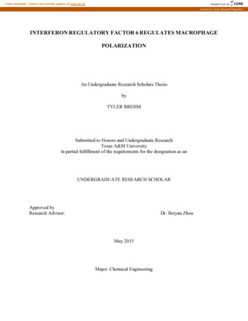
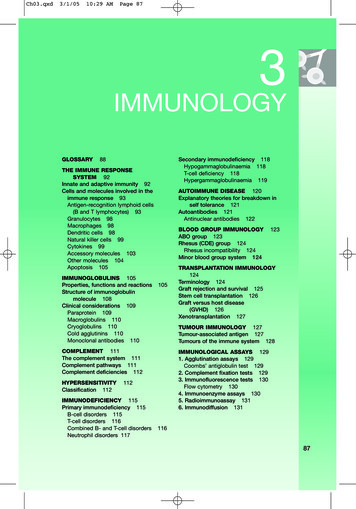
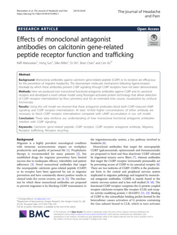
Manoukian et al. The Journal of Headache and Pain(2019) 20:44Page 4 of 12either added alone at the time points above or added atthe same time together with 100 nM CGRP and incubated in sustained or transient mode as described above.For immunofluorescence staining, cell culture mediawas removed from plate wells, cells were washed withwash buffer (phosphate buffered saline (PBS)/0.5% bovine serum albumin (BSA)), and subsequently fixed byoverlaying 10% neutral-buffered formalin (Sigma) for 15min at room temperature. After two washes of 5 mineach with wash buffer (PBS/0.5% BSA), cells werepermeabilized with 0.1% Triton X-100 in PBS for 20min, washed twice again for 5 min and then 100 μL ofdesired antibody cocktail was added: AA58 (5 μg/mL),8E11 (5μg/ml) and RAMP1 rabbit monoclonal(EPR10867, Abcam, Cambridge, MA, USA; 1:1000), wereused to detect the CGRP receptor [20], ie, the CGRPbinding site at the interface of CLR and RAMP1, respectively. Staining of untransfected parental cells, whichlack the CGRP receptor, with AA58 or RAMP1 antibodywas used as negative control. A mouse monoclonal antibody against early endosome antigen 1 (EEA1, 610,457,BD Biosciences, Franklin Lakes, NJ, USA, 5 μg/mL) wasused to label early endosomes. LAMP2 is one of thelysosome-associated membrane glycoproteins [23, 24];therefore, LAMP2 mouse monoclonal antibody(ab25361, Abcam, Cambridge, MA, USA, 5 μg/mL) wasused to identify lysosomes. Secondary antibodies RabbitIgG1 polyclonal AlexaFluor546 (ThermoFisher, Waltham,MA, USA; 1:1000) and Goat anti-human IgG Fc-FITC(Novex, Life Technologies, Waltham, MA, USA; 1:1000)were used with appropriate primary antibodies; primaryantibodies were incubated overnight at 4 C and secondaryantibodies for 2 h at room temperature; FAP fluorogenβRed membrane impermeant was used at 1:1000 prior tofixation (Spectragenetics, Pittsburgh, PA). The finalwash buffer contained 2 μg/mL Hoechst (ThermoFisher,Waltham, MA) solution in PBS as a nuclear counterstain. Concurrent AA58 and 8E11 immunostainingcould not be used due to potential cross reactivity oftwo human primary antibodies with an anti-humansecondary antibody.exported to Photoshop Elements 2.0 (Adobe Inc. San Jose,CA) where they were formatted for publication.Confocal microscopyInhibition of CGRP-induced CGRP receptor internalizationHigh-content imaging was performed on a PerkinElmerUltraview Vox spinning disc confocal microscope(PerkinElmer, Waltham, MA, USA) using laser excitation wavelengths of 405 nm, 488 nm, 561 nm and 633 nm.A Nikon Plan Fluor 40x/0.75 and Apo TIRF 60x/1.49 oilwere used for image capture. Single plane optical slice images were taken and represent a thickness of approximately0.15 µM. Images were acquired using the PerkinElmerVolocity software 6.3 (Perkin Elmer), where some contrastenhancement was used to enhance viewing. Images wereCGRP-induced receptor internalization dynamics werestudied using the quantitative flow cytometry-baseddual-signal FAP assay (Fig. 2). CGRP induced a dosedependent internalization of the CGRP receptor as measured by FAP-tagged CLR localization, with a clearinverse relationship between internalized receptor (redsignal, EC50 7.0 nM) and non-internalized receptor(green signal, EC50 8.8 nM) (Fig. 2a). CGRP-induced receptor internalization at 100 nM CGRP (corresponding tothe EC90) was inhibited by AA58 with an IC50 of 9.8 nMResultsExpression of functional CGRP receptors in recombinantCHO cellsTo examine the functionality of recombinant CGPR receptors, we compared intracellular cAMP concentrationsin response to CGRP, CGRP8–37, anti-CGRP antibody8E11 and anti-CGRP receptor antibody AA58 in bothuntagged (Fig. 1a) and CLR-FAP-tagged (Fig. 1b) CGRPreceptor expressing cells. CGRP was tested at concentrations from 0.5 pM to 100 nM and AA58, 8E11 andCGRP8–37 at 5 pM to 10 μM. The EC50 of CGRP inFAP-CLR-tagged CGRP receptor cells was comparableto that in untagged CGRP receptor cells (8.5 pM and 8.2pM, respectively) indicating that the FAP- and His-tagsdid not affect the binding and functionality of CGRP receptor. It is worth noting that CGRP is more potent inthe current CHO cell model system when compared tothe literature EC50 of 100 pM in the Swiss3T3 orHEK293 cells [12], and in human neuroblastoma cells(SKN-MC) endogenously expressing human CGRP receptors (EC50 670 pM) [20]. The inconsistency is mostlikely attributed to different host cells, and a very highexpression level of the receptor, designed purposefully tovisualize the receptor trafficking in this study. AA58,8E11, and the peptide antagonist CGRP8–37 demonstrated no agonist activity in either cell line (Fig. 1 a andb). Next, we measured the intracellular concentrationsof cAMP in the presence of CGRP (20 pM, corresponding to the EC70) with increasing concentrations (0.5 pMto 1 μM) of the antagonists AA58, 8E11, CGRP8–37 inboth, untagged (Fig. 1c) and CLR-FAP-tagged cells ed and untagged CGRP receptor expressing cells was inhibited by all agents in a dose-dependentfashion and the IC50s were comparable between the twocell lines: The IC50 of AA58 was 0.3 nM and 1.4 nM, ofE811 1.4 nM and 1.9 nM, of CGRP8–37 0.3 nM and 3.2nM in untagged versus tagged CGRP receptor expressing cells, respectively (Fig. 1c and d).
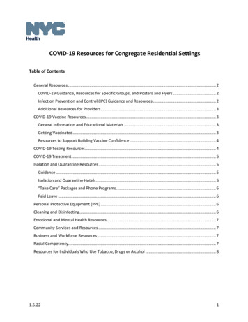
Manoukian et al. The Journal of Headache and Pain(2019) 20:44Page 5 of 12Fig. 1 CGRP-stimulated cAMP production. CHO cells stably expressing un-tagged (a, c) and CLR-FAP-tagged (b, d) CGRP receptors were treatedwith increasing concentrations of CGRP, AA58, 8E11 or CGRP8–37 (a, b). cAMP luminescence was measured and displayed as a percent of control(POC). CGRP produced a dose-dependent increase in both un-tagged (EC50 8.5 pM, a) and CLR-FAP tagged (EC50 8.2 pM, b) CHO cells,whereas AA58, 8E11 and CGRP8–37 demonstrated no agonist activity in either cell line (a, b). Preincubation with AA58, 8E11 or CGRP8–37 inhibitedCGRP-induced (20 pM) cAMP production to a similar degree in both un-tagged (IC50 0.3, 1.4 and 0.3 nM, respectively, c) and CLR-FAP taggedCHO cells (IC50 1.4 nM, 1.9 nM and 3.2 nM, respectively, d)and 8.7 nM for the internalized, red signal and uninternalized, green signal, respectively (Fig. 2b). Similarly, 8E11inhibited CGRP-induced receptor internalization with anIC50 of 40 nM (internalized, red signal) and 18 nM (uninternalized, green signal) (Fig. 2c). There was no receptorinternalization with AA58 or 8E11 in the absence ofCGRP ligand indicating that antibody binding of the receptor or the neuropeptide itself does not induce CGRPreceptor internalization and trafficking (Fig. 2b and c).Subcellular localization of the CGRP receptorsWe next used immunostaining and confocal microscopyto visualize and confirm co-localization of RAMP1 andCLR-FAP fluorescence in the CHO cell model (Fig. 3).AA58 immunostaining was used to visualize the CLR/RAMP1 heterodimer [20] and resulted in prominentmembrane staining of FAP-CLR-tagged cells. Similarly,immunostaining with the RAMP1 antibody resultedmainly in labeling of the plasma membrane. Also,CLR-FAP fluorescence imaging resulted in a faint signalat the membrane. Immunoreactivity of both, AA58 andRAMP1 colocalized with CLR-FAP fluorescence suggesting that CGRP receptors are predominantly localized atthe cell membrane of CHO cells. However, some RAMP1immunoreactivity was seen in the cytoplasm and did notco-localize with AA58 immunoreactivity. This cytoplasmic, non-CGRP receptor-associated RAMP1 immunoreactivity may be an artifact resulting from overexpression,ie, there may be more RAMP1 expressed by the cells thancan co-localize with the amount of CLR expressed. Therewas no FAP fluorescence nor immunoreactivity of eitherAA58 or RAMP1 detected in the parental CHO cells,which lack CGRP receptors. Together, these results suggest that AA58 and RAMP1 immunofluorescence stainingand FAP imaging comprise a specific method for detectingthe subcellular localization of the CGRP receptors in therecombinant CHO cell model.Time course of internalization of the CGRP receptorsWe next used the AA58 and RAMP1 staining, as well asCLR-FAP imaging described above to investigate trafficking of both CGRP receptor components individually
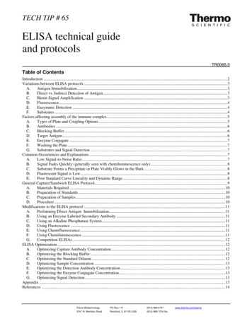
Manoukian et al. The Journal of Headache and Pain(2019) 20:44Page 6 of 12Fig. 2 Inhibition of cAMP-induced CGRP receptor internalization.CLR-FAP tagged CGRP receptor internalization was measured usingflow cytometry and graphed as percent of control meanfluorescence intensity (POC). Increasing CGRP concentrationsresulted in CLR-FAP-tagged CGRP receptor internalization (opentriangles) with a correlative decrease of membrane-bound CLR-FAPtagged CGRP receptors (closed triangles) (a). In the presence of 100nM CGRP, AA58 (b) or 8E11 (c) inhibited internalization (opensquares) and concomitantly increased membrane-bound CLR-FAPtagged CGRP receptors (closed squares), whereas no intrinsic effectwas observed in the absence of CGRP (open and closed circles)and together in the presence of CGRP at 0–5 min, 10min, 60 min, 5 h and 24 h fixed time point intervals(Fig. 4a). Hoechst staining (blue) of the nuclei confirmedcell health during the 24 h experiment. Even at the lasttime point observed, the cells did not present any obvious signs of apoptosis, based on normal nuclearmorphology and lack of condensed DNA. At baseline(0–5 min), AA58, RAMP1 and CLR-FAP staining ismostly co-localized at the plasma membrane with fewtriple-positive, putative endosomal vesicles present inthe cytoplasm of the cells (Fig. 4a and b). 100 nMCGRP, corresponding to the EC90 in FAP-taggedCHO cells based on the FACS experiment describedbefore, induced a time-dependent increase of theseendosomal vesicle-like structures that resulted indense, triple-positive, perinuclear staining over time(Fig. 4a and c): As early as 10 min after CGRP application,there was pronounced formation of these triple-positiveendosome-like vesicles (Fig. 4a). By 60 min, the plasmamembrane was largely devoid of staining and most of thepositive signal was observed in endosome-like vesicles(Fig. 4a and c). Between 60 min and 5 h, the triple-positivevesicular staining increased to span larger perinuclearareas (Fig. 4a). This staining pattern persisted in the cytoplasm up to 24 h, the last time point assessed. The strongtriple-positive plasma membrane immunofluorescenceseen at baseline did not reappear and was not observed upto 24 h, indicating absence of CGRP receptor recyclingwithin the timeframe observed (Fig. 4a).Co-localization of internalized CGRP receptors with EEA1and LAMP2To investigate whether the CGRP receptors that internalized after continuous CGRP exposure were associatedwith endosomes, we used an EEA1 antibody as an endosomal marker together with AA58 staining for the CGRP receptor and compared staining at 0 and 60 min afterexposure to 100 nM CGRP (Fig. 5a and b). At 0 min,CGRP receptors labeled by AA58 were localized to theplasma membrane, whereas EEA1-positive endosomeswere dispersed throughout the cytoplasm (Fig. 5a). At 60min after CGRP exposure, CGRP receptors were internalized and AA58 immunoreactivity was largely co-localized
Manoukian et al. The Journal of Headache and Pain(2019) 20:44Page 7 of 12Fig. 3 Colocalization of CGRP receptor subunits. AA58 (green) and RAMP1 (red) immunoreactivity co-localized (merged) with CLR-FAPfluorescence (magenta) as shown in a confocal optical slice of 0.15 μm with magnification 60x. Note the prominent membrane labeling by allthree CGRP receptor detection agents. Additional, non-co-localized RAMP1 immunoreactivity (red) can be seen in the cytoplasm (merged), likelyrepresenting an artifact from overexpression. No immunoreactivity was observed in parental cells not expressing CGRP receptors (parentalmerged). Scale bar 12 μmFig. 4 CGRP-induced receptor internalization. 24 h time course of CGRP (100 nM) induced internalization of CGRP receptors triple labeled byAA58, RAMP1 and CLR-FAP: Merged images show co-localization in yellow, nuclei are blue. Arrow heads point to endosomal-like vesicles thatappear after 10 min of CGRP stimulation and continue to increase over the 24 h time points (a). Individual channels (AA58 green, RAMP1 red,CLR-FAP magenta) are shown in a 0.15 μm optical slice at 5 min (b) and 60 min (c) after CGRP stimulation (CLR-FAP fluorescence is not visible at0 min). Arrow heads point to endosomal-like vesicles. Scale bars 12 μm
Manoukian et al. The Journal of Headache and Pain(2019) 20:44Page 8 of 12followed by reappearance of the staining at the plasmamembrane (Fig. 6a). Transient, 5 min exposure to 100nM CGRP followed by wash-out with fresh media resulted in initial formation of AA58-, RAMP1-, andCLR-FAP-positive endosome-like vesicles as early as 10min after wash-out with a peak at 60 min (Fig. 6a). Reappearance of CGRP receptor immunoreactivity at theplasma membrane was observed as early as 2–3 h withparallel disappearance of endosome-like vesicular staining from the cytoplasm (not shown). At 5 h after the initial ligand pulse, immunostaining was mainly localizedto the plasma membrane and was maintained there forat least up to 24 h, the last time point assessed (Fig. 6a).Cells remained healthy in this context as suggested bythe healthy morphological appearance of the nuclei asassessed by Hoechst staining.Internalization of CGRPFig. 5 Colocalization of CGRP receptors with endosomes andlysosomes. CGRP receptors were stained with AA58 (green) andendosomes (a, b) with EEA1 (red) or lysosomes (c, d) with LAMP2(red). Nuclei were labeled by Hoechst (blue). At 0 min, green AA58immunoreactivity is localized to the plasma membrane, whereas redEEA1 (a) or LAMP2 (c) immunoreactivity is seen in vesicles throughoutthe cytoplasm (a). At 60 min after stimulation with 100 nM CGRP, theCGRP receptors are internalized and AA58 immunoreactivity is localizedin vesicles in the cytoplasm (b, d). AA58 immunoreactivity is largelycolocalized with EEA1 immunoreactivity (b, yellow, arrowheads). Onlyfew of the vesicles at this time point are double positive for AA58 andLAMP2 (d, yellow, arrowheads), indicating that CGRP receptors are beingdegraded in lysosomes. Scale bars 12 μmwith EEA1 immunoreactivity (Fig. 5b). To investigatewhether some of the CGRP receptors were undergoingdegradation through the lysosomal pathway as describedpreviously [14], we used a LAMP2 antibody as a lysosomalprotein marker together with AA58 staining for the CGRPreceptor and compared staining at 0 and 60 min after exposure to 100 nM CGRP (Fig. 5c and d). At 0 min AA58staining was confined to the plasma membrane, whereasLAMP2 labeled lysosomes in the cytoplasm (Fig. 5c). At60 min after CGRP exposure, AA58 and LAMP2co-staining started to appear, suggesting that some CGRPreceptors were beginning to be degraded by lysosomes(Fig. 5d).Recycling of CGRP receptors after transient exposure to CGRPIn contrast to the persistent perinuclear staining in response to continuous exposure to CGRP, brief exposureto CGRP resulted in transient formation of AA58-,RAMP1-, and CLR-FAP-positive, endosome-like vesiclesAfter demonstrating that CLR and RAMP1 localize together during CGRP receptor internalization in theCHO cell model, we investigated whether CGRP alsointernalized and co-localized with the CGRP receptor(Fig. 6b). Using staining with the anti-CGRP monoclonalantibody 8E11 to detect CGRP, and RAMP1 staining todetect
Receptor trafficking, Receptor recycling Background . One Amgen Center Dr., MS 29-2-B, Thousand Oaks, CA 91320-1799, USA Full list of author information is available at the end of the article The Journal of Headache . (Selexis, Sunnyvale, CA). Cells stably expressing the tagged