Transcription
TECH TIP # 65ELISA technical guideand protocolsTR0065.0Table of ContentsIntroduction .2Variations between ELISA protocols .3A.Antigen Immobilization.3B.Direct vs. Indirect Detection of Antigen.3C.Biotin Signal Amplification .3D.Fluorescence.4E.Enzymatic Detection .4F.Substrates .4Factors affecting assembly of the immune complex.5A.Types of Plate and Coupling Options.5B.Antibodies .6C.Blocking Buffer.6D.Target Antigen.6E.Enzyme Conjugate .7F.Washing the Plate .7G.Substrates and Signal Detection .7Common Occurrences and Explanations .7A.Low Signal-to-Noise Ratio.7B.Signal Fades Quickly (generally seen with chemiluminescence only).8C.Substrate Forms a Precipitate or Plate Visibly Glows in the Dark .8D.Fluorescent Signal is Low .8E.Poor Standard Curve Linearity and Dynamic Range.8General Capture/Sandwich ELISA Protocol.9A.Materials Required .10B.Preparation of Standards.10C.Preparation of Samples.10D.Procedure.10Modifications to the ELISA protocol .11A.Performing Direct Antigen Immobilization.11B.Using an Enzyme Labeled Secondary Antibody .11C.Using an Alkaline Phosphatase System.11D.Using Fluorescence .11E.Using Chemifluorescence.11F.Using Chemiluminescence .11G.Competition ELISAs .12ELISA Optimization.12A.Optimizing Capture Antibody Concentration.12B.Optimizing the Blocking Buffer .12C.Optimizing the Standard Diluent.12D.Optimizing Sample Concentration .13E.Optimizing the Detection Antibody Concentration .13F.Optimizing the Enzyme Conjugate Concentration .13G.Optimizing Signal Detection .13Appendix .13References .14Pierce BiotechnologyPO Box 117(815) 968-07473747 N. Meridian RoadRockford, lL 61105 USA(815) 968-7316 faxwww.thermo.com/pierce
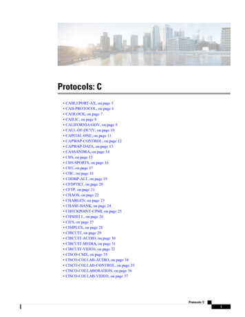
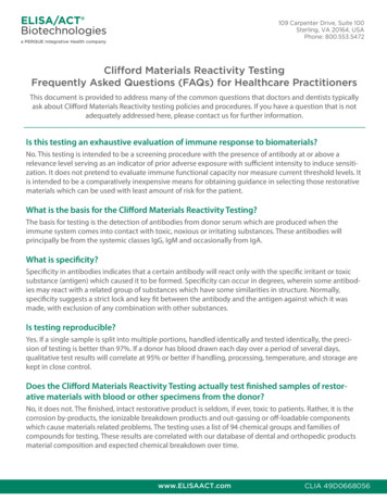
IntroductionThe enzyme linked immunosorbent assay (ELISA) is a powerful method for detecting and quantifying a specific protein in acomplex mixture. Originally described by Engvall and Perlmann (1971), the method enables analysis of protein samplesimmobilized in microplate wells using specific antibodies. The technique has revolutionized immunology and is commonlyused in medical research laboratories. ELISA also has commercial applications, including the detection of disease markersand allergens in the diagnostic and food industries.The ELISA method was made possible because of scientific advances in a number of related fields. Technology enabling theproduction of antigen-specific monoclonal antibodies by Kohler and Milstein (1975) led to their use as probes for detectingindividual molecules in complex protein mixtures or tissue samples. Initially, detection was achieved by radioimmunoassayusing antibodies labeled with radioisotopes, but because of health risks alternatives were sought. Avramais (1966, 1969) andPierce (1967) developed methods to chemically link antibodies to biological enzymes whose activities produce a measurablesignal with solutions containing appropriate substrates. With the development of fluorescence technology, signal generationusing fluorophore-labeled antibodies has also become prevalent, especially in multiplex arrays.Although many variants of ELISA have been developed and used in different situations, they all depend on the same basicelements:1.Coating/Capture: direct or indirect immobilization of antigens to the surface of polystyrene microplate wells.2.Plate Blocking: addition of irrelevant protein or other molecule to cover all unsaturated surface-binding sites of themicroplate wells.3.Probing/Detection: incubation with antigen-specific antibodies that affinity-bind to the antigens.4.Signal Measurement: detection of the signal generated via the direct or secondary tag on the specific antibody.In a typical assay designed to detect an antigen in a complex protein mixture, the antigen is immobilized either by directadsorption or via an antibody adsorbed to the wells of a microplate. The plate is blocked and the antigen is probed with aspecific detection antibody. The detection antibody may be directly labeled with a signal-generating enzyme or fluorophoreor it may be secondarily probed with an enzyme- or fluor-labeled secondary antibody (or avidin-biotin chemistry, see below).For enzymatic detection, the appropriate enzyme substrate is added. The signal observed is proportional to the amount ofantigen in the sample. Washing between steps ensures that only specific (high-affinity) binding events are maintained tocause signal at the final step.Diagram of popular ELISA formats.Pierce BiotechnologyPO Box 117(815) 968-07473747 N. Meridian RoadRockford, lL 61105 USA(815) 968-7316 fax2www.thermo.com/pierce
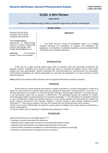
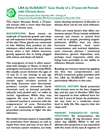
Variations between ELISA protocolsA. Antigen ImmobilizationAntigen immobilization varies between two principle techniques. In a traditional (direct coating) ELISA, antigens are directlyattached to the plate by passive adsorption, usually using a carbonate/bicarbonate buffer at pH 9. Most but not all proteinsbind tightly to the polystyrene surface of microplates in alkaline conditions. However, if antigen is present at low levels ordoes not adhere well to the plastic, then the alternative sandwich or capture ELISA may be used. In capture (indirect coating)ELISA, antigen-specific antibody is adsorbed onto the plastic, which in turn binds and immobilizes the antigen uponincubation with the antigen sample. Attachment of the antibody is typically achieved using the same carbonate/bicarbonatebuffer at pH 9, or in rare instances pre-activated plates are used for a more directed attachment approach.Sandwich ELISAs have become very popular when using complex protein samples because only the specific antigenbecomes immobilized rather than the entire sample of proteins. The more antigen that is immobilized, the higher is thepotential sensitivity of the assay. Sandwich ELISAs require two different antibodies that bind specifically to the antigen (eachreacting with a different epitope). The first antibody (bound to the plate) is called the capture antibody or coating antibody,whereas the second antibody detects the immobilized antigen and is called the detection antibody. Such antibodies are knownas “matched pairs”; they must be validated to work in combination, as they must not compete for binding to the antigen foraccurate results to be possible. Combinations of monoclonal and polyclonal antibodies can be used; it is more common to usethe monoclonal as the coating antibody and the polyclonal as the detection antibody. Sandwich ELISAs sometimes requiremore optimization than traditional ELISAs but usually the signal-to-noise (S:N) ratios are higher.B. Direct vs. Indirect Detection of AntigenDirect detection involves labeling of the detection antibody with an enzyme or an alternative signaling molecule such as afluorophore. Indirect detection involves an additional probing step using another antibody or streptavidin that is labeled witha detectable tag. This additional probe is called the secondary antibody, as its sole purpose is to deliver the measurable signaltag by binding to the detection (primary) antibody. Direct detection is generally faster than indirect detection and potentialbackground signal from secondary antibody cross-reactivity with the coating antibody is also eliminated. Even in the best ofcircumstances however, direct detection cannot provide the signal amplification gained from the use of a secondary antibodyor avidin/biotin systems. Consequently direct detection is generally less sensitive than indirect detection and is best used onlywhen the target is relatively abundant.C. Biotin Signal AmplificationBiotin is a small (MW 244) vitamin molecule that is easily modified so that itcan be chemically attached to proteins, antibodies and other biomolecularprobes of interest. Avidin and streptavidin are proteins that originate fromdifferent sources but have nearly identical functions in binding very stronglyand specifically to the biotin molecule. Thermo Scientific NeutrAvidin Protein is a specially modified form of avidin. Hereafter in this document,“avidin” and “streptavidin” will be used interchangeably to mean any one ofthese biotin-binding proteins. The avidin-biotin affinity system is frequentlyemployed in the design of tagging and detecting systems for ELISA.A common adaptation of indirect detection is to amplify the signal usingavidin-biotin chemistry. There are two approaches. The first method involvesusing a biotinylated detection antibody, which is probed using avidin orstreptavidin protein conjugated to either horseradish peroxidase (HRP) oralkaline phosphatase (AP) enzymes. The second approach also employs abiotinylated detection antibody, but it is probed with a pre-incubated mixtureof avidin and biotinylated enzyme, a process known as “avidin-biotincomplex” (ABC) signal amplification.Diagram of the signal amplification that ispossible with the ABC system.Signal amplification occurs through two mechanisms with these approaches. First, biotinylation (biotin-labeling) typicallyresults in multiple biotin tags per antibody molecule, thus allowing more than one streptavidin molecule to bind to eachantibody. Binding is aided by the fact that avidin-type proteins are tetrameric and have four biotin-binding sites per molecule.Second, the process of either labeling the streptavidin molecule with enzymes or using a pre-incubated mixture ofstreptavidin plus biotinylated enzyme results in conjugates having more than one enzyme. The combined effect of thisPierce BiotechnologyPO Box 117(815) 968-07473747 N. Meridian RoadRockford, lL 61105 USA(815) 968-7316 fax3www.thermo.com/pierce
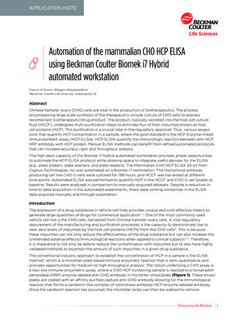
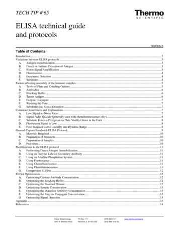
multiple labeling is to increase the number of enzyme molecules in the final immune complex. This increases the catalysis ofappropriate substrate and gives a stronger signal compared to a conventional enzyme-labeled secondary antibody.D. FluorescenceThe relatively recent expansion in the number of stable fluorophores in the visible and infrared ranges has made fluorescentsignal detection an attractive option for ELISA applications. This kind of approach is common when performing multiplexarrays, as more than one antigen can be detected simultaneously with antibodies conjugated to different fluorophores. Influorescence assays, the detection antibody is either labeled directly or the secondary antibody (or occasionally avidin) islabeled for indirect detection. When multiplexing using labeled secondary antibodies it is essential to use detection antibodiesfrom different species in order to distinguish the separate signals. The detection limit is typically around 100pg/well which isless sensitive than colorimetric or chemiluminescent detection.Diagram of a multiplex array ELISA made made possible by using fluorescence. In this case, twelve different capture antibodies arecoated as an array of printed spots on a glass slide. Each antibody captures a different analyte and is detected by its matched detectionantibody, which is biotinylated. Finally, all spots are detected through a fluor-labeled streptavidin conjugate (in this case, the Thermo ScientificDyLight 649 Fluorophore).E. Enzymatic DetectionTwo enzymes are commonly used in ELISA applications. Alkaline Phosphatase (AP) is a large enzyme used in a minority ofassays. Its size (140 KDa) makes it difficult to conjugate more than one or two molecules of the enzyme to each molecule ofan antibody or avidin, and this limits the amount of signal that can be generated. AP is also prone to stability issues unlessstored and handled correctly.Horseradish Peroxidase (HRP) is a more commonly used enzyme. Its small size (40KDa) allows more molecules to becoupled to antibodies or avidin, and this can boost signal generation. HRP is the enzyme of choice for most researchersperforming ELISAs and can be used with a variety of substrates (see below), most of which are more sensitive than APequivalents.F. SubstratesEnzymatic signal generation requires the catalysis of a substrate to produce a colored or fluorescent compound orchemiluminescence (visible light). The signal is measured using a spectrophotometric plate reader, a fluorometer with theappropriate filters or a luminometer set to read total light output. Each type of substrate is discussed below; more informationabout specific products can be found at our website.Colorimetric substrates form a soluble, colored product that accumulates over time relative to the amount of enzyme presentin each well. When the desired color intensity is reached, the product absorbance is either measured directly or in some casesa stop solution is added to provide a fixed end point for the assay. Colorimetric substrates are available for both horseradishperoxidase (TMB, OPD, ABTS) and alkaline phosphatase (PNPP).Pierce BiotechnologyPO Box 117(815) 968-07473747 N. Meridian RoadRockford, lL 61105 USA(815) 968-7316 fax4www.thermo.com/pierce
Chemifluorescent detection is also enzyme-based but the generated product is fluorescent rather than colorimetric. The signalis measured using a fluorometer with the appropriate excitation and emission filters. Chemifluorescence reactions are eithermeasured over time in kinetic assays or halted using a stop solution for direct measurement. Examples of chemifluorescentsubstrates for HRP are Thermo Scientific QuantaRed and QuantaBlu Substrates.Chemiluminescence is a chemical reaction that generates energy released in the form of light. Most chemiluminescentsubstrates are HRP-dependent although some AP equivalents are available. The most common approach is to use luminol inthe presence of HRP and a peroxide buffer. The luminol is oxidized and forms an excited state product that emits light as itdecays to the ground state. Light emission occurs only during the enzyme-substrate reaction, therefore when the substratebecomes exhausted the signal ceases. Chemiluminescent detection is generally considered to be more sensitive thancolorimeteric detection. Chemiluminescent substrates for HRP include Thermo Scientific SuperSignal ELISA Pico andELISA Femto Substrates.Factors affecting assembly of the immune complexAlthough ELISA is a powerful and well-characterized application, attempting to develop and optimize a specific assay can bedifficult. The method involves the assembly of a large immune complex with multiple components, therefore failure tocapture signal can potentially be caused by any of these factors and optimization is essential. A list of such factors andassociated variables is described in Table 1, followed by a discussion of several pertinent issues. The protocol section at theend of this guide includes specific instructions and suggestions about optimization procedures.When setting up an ELISA it is advisable to first generate and optimize a standard curve for the analyte of interest beforeanalyzing multiple samples of unknown composition. If the standard curve displays the correct sensitivity, range andlinearity, the researcher can proceed with confidence to process the samples.Table 1. Factors that affect ELISA signal generation.FactorVariable CharacteristicAssay Platematerial, well shape, pre-activationCoupling Buffercomposition, pHCapture Antibodyspecificity, titer, affinity, incubation time and temperatureBlocking Buffercomposition, concentration, cross-reactivityTarget Antigenconformation, stability, available epitope(s), matrix effectsDetection Antibodyspecificity, titer, affinity, incubation time & temperature, cross-reactivityEnzyme Conjugatetype of enzyme, type of conjugate, activity, concentration, cross-reactivityWashesbuffer composition, volume, duration, frequencySubstratesensitivity, manufacturer lot, ageSignal Detectionfilters, imaging instrument, exposure timeA. Types of Plate and Coupling OptionsPlates used in conventional ELISA applications are typically made of polystyrene. Other materials such as polypropylene,polycarbonate and in some instances nylon are occasionally used; many plates are now gamma-irradiated to impart a positivecharge, which aids coating procedures. The absorbances of colorimetric substrates are measured by shining a laser up throughthe base of each well, so it is essential to use a flat bottomed plate with a clear base. Fluorescent detection requires the use ofan opaque black or white plate, and fluorometric plate readers measure either from above or below the plate.Chemiluminescent detection requires the use of a black or white opaque plate and may also be measured from the top or thebottom. For this reason both fluorescent and chemiluminescent assays must be performed in plates with a clear bottom.Although black plates are preferred for fluorescence (as background is lower) and white plates are preferred forchemiluminescence (as it magnifies the signal), the plates can be used interchangeably.The most common technique for attachment of proteins or peptides to a plate is passive adsorption. This is mediatedprimarily by hydrophobic interactions, but some electrostatic forces may also contribute. A popular coating buffer iscarbonate-bicarbonate buffer (0.2 M sodium carbonate/bicarbonate pH 9.4). The high pH aids solubility of many proteins andpeptides and ensures that most proteins are unprotonated with an overall negative charge, which helps when binding to aPierce BiotechnologyPO Box 117(815) 968-07473747 N. Meridian RoadRockford, lL 61105 USA(815) 968-7316 fax5www.thermo.com/pierce
positively charged plate. Other buffered solutions such as Tris-buffered saline (TBS) or phosphate-buffered saline (PBS) atphysiological pH are sometimes used but coupling is generally not quite as efficient.In some instances researchers require a more directed approach for attachment of the coating antibody or protein sample tothe plate, and several pre-activated plates exist for this purpose. For specific, orientated binding of the coating antibody,plates that are pre-coated with Protein A or Protein G are available (not advised for sandwich ELISAs because of potentialcross-reactivity with detection and/or secondary antibodies). For biotinylated samples or coating antibodies, plates that arepre-coated with streptavidin are ideal. Some assays require direct immobilization of a histidine-tagged protein, in which casenickel- or copper-coated plates are suitable (these plates can also be used to bind and orientate capture antibodies as IgGmolecules have a histidine rich sequence in their Fc domain). For group-specific attachment of molecules via amines or thiolsto form a covalent bond, maleic-anhydride or maleimide-activated plates can be used. (These are especially useful for directattachment of peptide antigens, which do not coat well by passive adsorption because of their small size.) Please visit ourwebsite for a complete listing of our many Thermo Scientific Pierce Coated Microplates.B. AntibodiesNot all antibodies can be used successfully in ELISA applications, and individual antibodies must be evaluated. During theadsorption process, the three-dimensional structure of an antigen might be altered and such that it can no longer bind itstarget epitope. Some antibodies are raised against peptides; if the peptide represents an internal sequence of the antigen, thenthe antibody might not bind if the whole antigen is immobilized on the plate. Furthermore, for an antibody to worksuccessfully in ELISA, it should react specifically with the antigen but not cross-react with a component of the blockingbuffer. For sandwich assays where two different antibodies are required, it is essential that the two antibodies react withdifferent epitopes on the antigen or an epitope that appears several times on the antigen. For example, if the antigen isimmobilized on the plate through the capture antibody, then the detection antibody must be able to interact with its ownepitope without steric hindrance from the first antibody or the plate. In ELISA applications where a secondary antibody isused as part of the detection complex it is also essential that the capture and detection antibodies be raised in different animalspecies so that the secondary antibody does not react with the coating antibody. Antibodies that work in combination witheach other are generally known as “matched pairs”.Another important factor to consider when setting up an ELISA is the concentration of the antibodies. Each will requireoptimization, the optimal range being partially determined by the form and origin and also by the substrate used for signalgeneration. Detection antibody and enzyme conjugate working solutions should ideally be prepared in blocking solution toreduce non-specific interactions. For recommended antibody concentrations see the Appendix at the end of this document.C. Blocking BufferBlocking buffers usually consist of formulations of proteins designed to prevent non-specific binding of proteins to the plate.An optimal blocking buffer maximizes the signal-to-noise ratio and does not react with the antibodies or target protein. Ifcross-reactivity is observed then a different blocker should be tested and if repeated cross-reactivity is observed it may beadvisable to switch to a non-mammalian protein blocker such as salmon serum or a protein free blocking solution. Visit ourwebsite for more information on our wide selection of ready-to-use blocking buffers.Some systems may benefit from the addition of a surfactant such as Tween -20 (a gentle non-ionic detergent) to the blockingsolution. Surfactants can help to minimize hydrophobic interactions between the blocking protein(s) and the antigen or theantibodies. Typically a final concentration of 0.05% (v/v) Tween-20 is used. In addition, blocking buffers should be used insufficient volumes to completely coat the wells; for example 300 l should be used for each well of a typical 96-well plate.D. Target AntigenThe target antigen should be present in a buffer or matrix that allows it to interact with a pre-coated capture antibody or becoupled to the plate directly. Direct coupling may require that the antigen be exchanged into a suitable coupling buffer. Inrare instances the three-dimensional structure of an antigen may be altered during the adsorption process such that it nolonger binds its target epitope. In such cases, the use of a plate pre-coated with a binding protein (such as a capture antibody)may eliminate this problem. If the antigen is present in the form of a biological sample, then the effects of the matrix (i.e.serum, plasma components) should be controlled by performing spike-and-recovery and linearity-of-dilution experiments.For more information on this topic, see the related Tech Tip #58: Linearity and spike-and-recovery experiments for ELISA.If performing a quantitative ELISA it is essential to have an equivalent standard protein (generally a purified recombinant)where the amount of specific protein is known in advance. The standard is used to prepare a series of solutions of knownPierce BiotechnologyPO Box 117(815) 968-07473747 N. Meridian RoadRockford, lL 61105 USA(815) 968-7316 fax6www.thermo.com/pierce
concentration by serial dilution of a protein stock solution followed by construction of a standard curve plotting concentrationversus absorbance. Absorbance values for samples of unknown concentration are extrapolated from this curve to determinethe actual amount of specific protein in the sample.E. Enzyme ConjugateThe concentration of the enzyme conjugate is one of the most crucial parameters in the optimization process. The amount ofenzyme that binds directly influences the amount of signal that is generated. Too little enzyme and the signal may be veryweak with a poor signal-to-noise ratio. Too much and the background may be too high, again resulting in a poor signal-tonoise ratio and little distinction between standards of different concentrations. For recommended enzyme conjugateconcentrations see the Appendix at the end of this document.F. Washing the PlateThe two most commonly used wash buffers in ELI
The ELISA method was made possible because of scientific advances in a number of related fields. Technology enabling the production of antigen-specific monoclonal antibodies by Kohler and Milstein (1975) led to their use as probes for detecting