Transcription
Sukegawa et al. Diagnostic Pathology(2020) SEARCHOpen AccessClinical study on primary screening of oralcancer and precancerous lesions by oralcytologyShintaro Sukegawa1,2* , Sawako Ono3,4, Keisuke Nakano2, Kiyofumi Takabatake2, Hotaka Kawai2,Hitoshi Nagatsuka2 and Yoshihiko Furuki1AbstractBackground: This study was conducted to compare the histological diagnostic accuracy of conventional oral-basedcytology and liquid-based cytology (LBC) methods.Methods: Histological diagnoses of 251 cases were classified as negative (no malignancy lesion, inflammation, ormild/moderate dysplasia) and positive [severe dysplasia/carcinoma in situ (CIS) and squamous cell carcinoma (SCC)].Cytological diagnoses were classified as negative for intraepithelial lesion or malignancy (NILM), oral low-gradesquamous intraepithelial lesion (OLSIL), oral high-grade squamous intraepithelial lesion (OHSIL), or SCC. Cytologicaldiagnostic results were compared with histology results.Results: Of NILM cytology cases, the most frequent case was negative [LBC n 50 (90.9%), conventional n 22(95.7%)]. Among OLSIL cytodiagnoses, the most common was negative (LBC n 34; 75.6%, conventional n 14;70.0%). Among OHSIL cytodiagnoses (LBC n 51, conventional n 23), SCC was the most frequent (LBC n 31;60.8%, conventional n 7; 30.4%). Negative cases were common (LBC n 13; 25.5%, conventional n 14; 60.9%).Among SCC cytodiagnoses SCC was the most common (LBC n 16; 88.9%, conventional n 14; 87.5%). Regardingthe diagnostic results of cytology, assuming OHSIL and SCC as cytologically positive, the LBC method/conventionalmethod showed a sensitivity of 79.4%/76.7%, specificity of 85.1%/69.2%, false-positive rate of 14.9%/30.7%, andfalse-negative rate of 20.6%/23.3%.Conclusions: LBC method was superior to conventional cytodiagnosis methods. It was especially superior for OLSILand OHSIL. Because of the false-positive and false-negative cytodiagnoses, it is necessary to make a comprehensivediagnosis considering the clinical findings.Keywords: Cytology, Pathology, Liquid-based cytology, Screening, InflammationBackgroundHead and neck cancer is one of common malignanciesin the world, and the most common histopathologicaltype is squamous cell carcinoma (SCC). Several patients* Correspondence: gouwan19@gmail.com1Department of Oral and Maxillofacial Surgery, Kagawa Prefectural CentralHospital, 1-2-1, Asahi-machi, Takamatsu, Kagawa 760-8557, Japan2Department of Oral Pathology and Medicine, Okayama University GraduateSchool of Medicine, Dentistry and Pharmaceutical Sciences, Okayama, JapanFull list of author information is available at the end of the articledie every year because of advanced oral SCC [1]. Conversely, studies have reported that early detection andtreatment of oral SCC can reduce mortality and morbidity and increase the likelihood of complete recovery [2,3]. Therefore, it is important to use the simplest and themost accurate method that can to detect early-stage abnormalities in oral mucosal cells. An example of suchmethod is exfoliated mucosal cytology, which involves The Author(s). 2020 Open Access This article is licensed under a Creative Commons Attribution 4.0 International License,which permits use, sharing, adaptation, distribution and reproduction in any medium or format, as long as you giveappropriate credit to the original author(s) and the source, provide a link to the Creative Commons licence, and indicate ifchanges were made. The images or other third party material in this article are included in the article's Creative Commonslicence, unless indicated otherwise in a credit line to the material. If material is not included in the article's Creative Commonslicence and your intended use is not permitted by statutory regulation or exceeds the permitted use, you will need to obtainpermission directly from the copyright holder. To view a copy of this licence, visit http://creativecommons.org/licenses/by/4.0/.The Creative Commons Public Domain Dedication waiver ) applies to thedata made available in this article, unless otherwise stated in a credit line to the data.
Sukegawa et al. Diagnostic Pathology(2020) 15:107making a diagnosis from minimally invasive oral mucosal cells [4].Exfoliative cytology is the microscopic examination ofshed or desquamated cells from the mucus membrane,and it is a simple, safe, and reliable approach. Exfoliativecytology consists of the conventional method and theliquid-based cytology (LBC) method [5]. The conventional method involves chair side scraping of the oralmucosa and then smearing it directly onto a glass slide.This method requires a suitable technique because themorphology of the collected cells would change if handled improperly. Conversely, LBC is a technique inwhich cells are scattered in a fixative liquid to produce athin layer of cells on the slide. Therefore, the LBCmethod has been widely used because of its advantage ofnot requiring complicated operations on the chair side.However, the current gold standard for diagnosing oralepithelial dysplasia and cancer is not exfoliative excisioncytology; rather, it is resection biopsy or histologicalexamination of surgical specimens [6, 7]. Unfortunately,excision biopsy is an invasive diagnostic method, andscrape cytology is suitable for the screening of pathological conditions considering minimal invasiveness.However, the diagnostic capabilities of two types of oralexfoliative excision cytology, the conventional and LBCmethods, remain unclear. Furthermore, the diagnosticaccuracy of the oral scraping cytology compared withthe histopathological diagnosis has not been examinedin detail. Therefore, it is important to consider whetherthe two types of cytology are sufficient to be used as astandard method for the diagnoses of suspicious orallesions.This study was conducted to compare the histologicaldiagnostic accuracy between the conventional methodand the LBC method and to clarify the effectiveness ofcytology.MethodsPage 2 of 6collected, processed, and diagnosed in a single generalhospital.Cytology specimens were processed by conventionalcytology from April 2010 to March 2015 and by theLBC method from April 2015 to March 2019. Cells wereharvested by scraping with a cotton cytobrush device inall cases. In the conventional method, the collected cellswere smeared onto a glass slide to prepare a sample,immersed in 95% ethanol, fixed, and stained with Papanicolaou stain. The LBC method involves dipping a cotton brush containing the sample directly into thetransport medium, which is an alcohol-based preservative (BD CytoRich blue preservative, BD Japan, Tokyo,Japan). The liquid-based cellular material in the vial wasprocessed according to the manufacturer’s protocol (BDJapan). The processing steps included vortexing of thesample, density reagent centrifugation, decantation andresuspension of cell pellets followed by gravity sedimentation on poly-l-lysine coated slides and subsequentstaining with the Papanicolaou stain.Procedure of cytological diagnosisThe specimens were reviewed by raters who had passedthe board examination for cytology of the Japanese Society of Clinical Cytology. Cytology diagnostic expertsconfirmed that the sample was appropriate for cytologydiagnosis. According to the criteria for specimen adequacy, we identified non-diagnosable specimens as inappropriate due to the presence of hypocellular or airdrying artifacts. The specimens were evaluated independently by at least two raters, and a representative cytology result of each case was determined by a majorityvote. Cytological diagnoses were made based on the Bethesda system according to the Japanese society of clinical cytology (JSCC) diagnostic guideline and wereclassified into negative for intraepithelial lesion or malignancy (NILM), oral low-grade squamous intraepitheliallesion (OLSIL), oral high-grade squamous intraepitheliallesion (OHSIL), SCC, and indefinite for neoplasia [8].Study design and sampleWe designed and implemented a cross-sectional studyusing the oral exfoliative cytology results of patients whohad been referred to the Kagawa Prefectural CentralHospital (Takamatsu, Japan) for diagnosis, treatment,and examination of oral lesions. During the period fromApril 2010 to March 2019, a total of 1234 specimenswere obtained from the cytology specimens collected bythe Department of Oral and Maxillofacial Surgery anddiagnosed by the Department of Pathology. Of thesespecimens, nine were insufficient and excluded. Of theremaining 1225 specimens, 251 specimens that underwent histological diagnosis ranging from benign or malignant oral lesions for biopsy and/or surgical resectionwere included in this study. All specimens wereProcedure of histological diagnosisA histological diagnosis was provided by pathologists,and then, the number of biopsy samples was determinedat the investigator’s discretion. These histological slideswere subjected to hematoxylin and eosin staining, andtheir histological findings were divided into two categories as negative group and positive group. Negative wasdefined as non-malignant lesions, including inflammatory ones and mild, or moderate dysplasia. Positive wasdefined as severe dysplasia, carcinoma in situ (CIS),SCC, and other malignancies. Histological diagnosis wasbased on the WHO criteria [9]. CIS was in accordancewith the general rules for clinical and pathological studies on oral cancer [10].
Sukegawa et al. Diagnostic Pathology(2020) 15:107Page 3 of 6The design and methodology of this study have beenapproved by the Ethics Committee of Kagawa Prefectural Central Hospital (Approval No. 946).Statistical analysisData were entered into a database using Microsoft Excel(Microsoft Inc., Redmond, WA, USA). The database wastransferred to JMP version 14.2 for Macintosh computers (SAS Institute Inc., Cary, NC, USA) for statisticalanalysis. To compare between cytological and histological diagnoses, the histological diagnoses were classified into negative and positive, and the cytologicaldiagnoses were also classified into negative (NILM andOLSIL) and positive (OHSIL, SCC, and other malignancies). The diagnostic performance metrics was examinedby comparing the cytological diagnosis against the histological diagnosis, for which the sensitivity, specificity, accuracy, positive predictive value, and negative predictivevalue were calculated, followed.Sensitivity ¼TPTP þ FNðaÞSpecificity ¼TNTN þ FPðbÞAccuracy ¼TP þ TNTP þ FP þ TN þ FNPositive predictive value ¼TPTP þ FPNegative predictive value ¼TNTN þ FNðcÞðdÞðeÞTP and TN indicate true positive and true negativeclassifications, respectively; FP and FN indicate falsepositive and false-negative classifications, respectively.ResultsRate of inappropriate cytological specimensIn this study, there were three cases (3.5%) of inappropriate cytological specimens in the conventional method.In the LBC method, there were no cases of insufficientsample processing.Histological diagnosisThe histological diagnoses of 251 cases were classified asnegative and positive using both the LBC method andthe conventional method (Table 1).Comparison of LBC and conventional cytologicaldiagnoses with histological diagnosesTable 2 shows the distribution of histological diagnosesfrom the viewpoint of cytological diagnosis. Of theNILM cytology cases (78 in total, including 55 using theLBC method and 23 using the conventional method),Table 1 Histopathological categoriesLesionLBC (n 169)Conventional (n 82)Histological negative casesn 103n 53Benign tumor711Inflammation243Leukoplakia196Lichen planus64Others4227Mild Dysplasia4Moderate dysplasia12Histological positeve casesn 66n 29Severe dysplasia51CIS62SCC5526“Others” includes no malignancy, epulis, and mucoselthe most frequent case was negative (50 cases via LBC;90.9%, 22 cases via the conventional method; 95.7%).Among OLSIL cytodiagnosis (total 65, including 45using the LBC method and 20 using the conventionalmethod), the most common case was negative (LBC n 34; 75.6%, conventional n 14; 70.0%). Conversely,among the OHSIL cytology cases (total 74, including 51using the LBC method and 23 using the conventionalmethod), SCC (LBC n 31; 60.8%, conventional n 7;30.4%) was the most frequent and negative cases (LBCn 13; 25.5%, conventional n 14; 60.9%) were common. Among the SCC cytology cases (total 34, including18 using the LBC method and 16 using the conventionalmethod), SCC (LBC n 16; 88.9%, conventional n 14;87.5%) was the most common (Table 2).Diagnostic performance of cytological diagnosesThe positive predictive value of each cytological diagnosis made using the LBC method and the conventionalmethod is shown (Table 3). NILM and SCC demonstrated a high diagnostic accuracy with both methods.However, the positive predictive value of OHSIL forhistological lesions was 34.8% in the conventionalmethod but 72.5% in the LBC method.Regarding the diagnostic results of cytology, assumingthat OHSIL and SCC were cytologically positive, theLBC method and the conventional method showed asensitivity of 81.8 and 79.3%, a specificity of 85.4 and69.8%, a false-positive rate of 14.6 and 30.2%, and afalse-negative rate of 18.2 and 20.7%, respectively(Table 4).False-negative casesThere were false-negative 12 cases in the LBC method.The cytology diagnoses were nine OLSIL cases and threeNILM cases, and the histological types were eight SCC
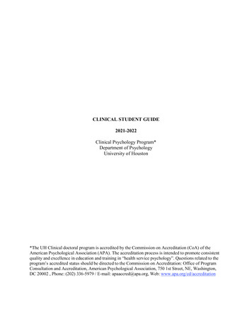
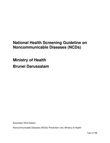
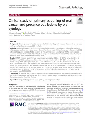
Sukegawa et al. Diagnostic Pathology(2020) 15:107Page 4 of 6Table 2 Results of cytological diagnoses in comparison withhistopathological diagnosesTable 4 Diagnostic performance metrics of cytologicaldiagnosesCytologicaldiagnosisPerfomance metricsLBC mehodConventional curacy84.0%73.2%False positive14.6%30.2%False negative18.2%20.7%PositveNegativeTotalSCC istopathological diagnosis 8Total5511598169Histopathological diagnosis CC1410116Total26325182cases, two CIS cases, and two severe dysplasia cases.There were six false-negative cases in the conventionalmethod. The cytological diagnoses were five OLSIL casesand one NILM case, and the histological types were fiveSCC cases and one CIS case (Fig. 1).DiscussionThe results of our study demonstrate that the LBCmethod is superior to the conventional method for cytodiagnosis. The LBC method was especially superior forTable 3 Diagnostic accuracy of LBC and conventional methodsCytologicaldiagnosisPositive Negative Total Positive predictivevalue (%)Histopathological diagnosis 1711894.4Less thanOHSIL128810088.0OHSIL andabove54156978.3Histopathological diagnosis 334.8SCC1511693.8Less thanOHSIL6374386.0OHSIL andabove23163959.0OLSIL and OHSIL for the positive predictive value. Conversely, because of a certain number of false-positivesand false-negatives in cytodiagnosis, it is important tomake a comprehensive diagnosis considering the clinicalfindings.In this study, there were three cases of inappropriatecytological specimens in the conventional method.Sekine et al. [7] reported that 22.7% of unsuitable specimens were produced by the conventional smearing technique. They reported that the unsuitable samples in theconventional method were those that were considered tobe inadequate for cytodiagnosis due to strong cell or airdrying artifacts. Our inappropriate specimens were similar, but the number of inappropriate specimens was verysmall in our study. We considered that this small number of inappropriate specimens occurred as the slideswere created on the chair side by a specialist cytologytechnician when preparing samples using the conventional method. Conversely, in the LBC method, therewere no cases of insufficient sample processing, andstable sample preparation was possible. This is consistent with previous reports showing that the LBC methodobviously results in fewer inappropriate specimens [11].Thus, it is important to understand and standardize thefeatures and equipment of cytology collection methodsto improve the quality of cytology slides.Regarding the diagnostic efficacy of the methods, theLBC method has been demonstrated to be more effectivethan the conventional method [12]. Although past studyhave reported higher sensitivity (conventional/ LBC;85.7%/ 95.1%) and specificity (conventional/ LBC;95.9%/99.0%)for the LBC method than for the conventionalmethod [6], another study reported that sensitivity (conventional/LBC; 96.3%/97.5%) of the LBC method wasparticularly good, whereas its specificity (conventional/LBC; 90.6%/68.7%) was reduced [13]. Our results indicated an increase in both sensitivity (conventional/LBC;79.3%/81.8%) and specificity (conventional/LBC; 69.8%/85.4%). As the results were judged using the same criteria at the same facility, they were valuable demonstrating the usefulness due to differences in cytodiagnosispreparation. Conversely, there are also some very interesting results in our study. The positive predictive valuewas not different for NILM and SCC between the
Sukegawa et al. Diagnostic Pathology(2020) 15:107Fig. 1 False-negative representative cases. The deep part of theepithelium reveals downward growth and invasion of tumor cells inthe underlying tissue, and surface layer of the epithelium showskeratinocytes without prominent atypiaconventional method and the LBC method. However, alarge difference was observed in the positive predictivevalue between OLSIL and OHSIL. Especially for OHSIL,it was a large difference. We speculate that this is due toa decrease in the number of negative cases misclassifiedas OHSIL and an increase in the number of detectionsof CIS and severe dysplasia. The LBC method contributes to the detection without missing CIS and severedysplasia. These diseases are susceptible to SCC and areeligible for treatment, and appropriate detection of thesediseases is clinically useful.Despite the improvement in efficacy with the LBCmethod, there were a certain number of false-positivesand false-negatives in this study. There were 12 cases offalse-negatives in the LBC method and five cases in theconventional method (false-negative rate; LBC method18.2%, conventional method 20.7%). When we examinedthe histology of cases diagnosed as NILM or OLSIL butwith SCC, all the histological diagnoses of SCC werefound to be well-differentiated SCCs. In addition, allcases were accompanied with well-differentiated keratinocytes lacking strong atypia on the surface. In thisstudy, whether a problem existed with the site of cytology collection cannot be examined. The current diagnostic criteria for the JSCC are limited for the evaluationof atypical superficial keratinocytes. However, even inkeratinized epithelium, which is histologically atypical,cytology has the advantage of examining individual cellsin detail. Therefore, it might be possible to detect highlydifferentiated tumor cells that exhibit a tendency tokeratinize. The diagnostic criteria of the JSCC take thispoint into account, but further examination of the diagnostic criteria is probably necessary in the future [8]. Suzuki et al. reported that false-negative cytology was morelikely to occur in cases where the exposed cell area forPage 5 of 6diagnosis was very small or where very limited growthwas observed [14]. Because it was difficult to collectbasal or parabasal-like atypical cells, cells useful for cytodiagnosis were not sampled, thus leading to falsenegative results. Sekine et al. reported [7] a falsenegative rate of 22.2%, and our result for oral scrapingcytology was acceptable.Unfortunately, we also detected some false-positives.Despite the diagnosis of SCC by the conventionalmethod and the LBC method, the cytodiagnosis was incorrect in each case. Despite the cytological diagnosis ofSCC, one negative case was found with the use of boththe conventional and LBC methods. However, therewere 14 and 15 negative cases of OHSIL with the use ofthe LBC and conventional methods, respectively (falsepositive rate: 14.6% by the LBC method and 30.2% bythe conventional method). Many atypical epitheliumwith large nuclei are often observed, and the presence ofepithelium that is partly suspected to be regeneratingepithelium is suspected to be a malignant tumor; however, this is often confirmed even in cases associatedwith inflammation such as ulcer margin or candida infection [15]. In this study as well, false-positives were observed because there were cases of OLSIL that had to bedifferentiated from SCCs.According to Remmerbach [16], applying the LBCmethod instead of the conventional method slightly reduced the false-negative results but still left a significantnumber of false-negative results. This result was similarto our study results. The false-negative results of cytology may exacerbate an untreated carcinoma, whichmay not be further treated and followed up. The factthat the lesion may subsequently be fatally exacerbatedfrom false-negative results implies that further improvement is required before oral cytology by itself becomes acompletely reliable method. Reducing false-negatives incytology diagnosis is more important. To that end, incases that make the diagnosis difficult, “suspect” resultstend to be considered as “positive” in terms of furtheraction required. In the oral cavity where various conditions are mixed, there is a limit in oral cytology in whichcells of only the surface layer are collected and diagnosed under a microscope. A diagnosis that comprehensively considers clinical information will be necessary.This study being a single-institution cross-sectional research has some limitations, perhaps including a casebias. Conversely, cytodiagnostic technologists and doctors had established the diagnosis on the basis of thesame diagnostic criteria; therefore, it would be better tocompare the accuracy of the conventional method andthe LBC method according to the technology in a futurestudy. The second limitation of this study is the smallnumber of cases. Although the results of histological examinations were correct, the number of histological
Sukegawa et al. Diagnostic Pathology(2020) 15:107examinations was small in cases with NILM diagnosis.Nevertheless, since only a few studies conducted till havecompared histology and the number of cases, our studycould be useful.ConclusionThis study demonstrates that the LBC method is superior to the conventional methods in that histological diagnosis is less discrepant with cytological classification. Inparticular, the LBC method has the advantage of reducing the number of misdiagnosed CIS and severe dysplasia cases in oral cytology. Conversely, because of acertain number of false-positives and false-negatives incytodiagnosis, it is important to make a comprehensivediagnosis considering the clinical findings.AbbreviationsJSCC: Japanese society of clinical cytology; LBC: Liquid-based cytology;NILM: Negative for intraepithelial lesion or malignancy; OHSIL: Oral highgrade squamous intraepithelial lesion; OLSIL: Oral low-grade squamousintraepithelial lesion; SCC: Squamous cell carcinomaAcknowledgementsThis work was supported by JSPS KAKENHI Grant Numbers JP19K19158,JP18K09789, JP18K17224, JP20H03888.Authors’ contributionsSS participated in the study design, acquisition of data, and drafting of themanuscript. SO performed and coordinated the study, collected data, andcontributed to the drafting of the manuscript. KN analyzed the data andcontributed to the drafting of the manuscript. KT and HK performed andcoordinated the study, and contributed to the review and editing of themanuscript. HN managed the study and wrote the manuscript. YF conceivedthe study and its design. All authors have read and approved the finalversion of the manuscript.FundingNone declared.Availability of data and materialsThe datasets generated and /or analyzed during the current study are notpublicly available. The Ethics Committee review board restricts the use of thedatasets to the current study only.Ethics approval and consent to participateThe Ethics Committee of Kagawa Prefectural Central Hospital providedapproval for this study (Approval No. 946).Consent for publicationAll authors have reviewed the manuscript and agreed for its publication.Competing interestsThe authors declare that they have no competing interests.Author details1Department of Oral and Maxillofacial Surgery, Kagawa Prefectural CentralHospital, 1-2-1, Asahi-machi, Takamatsu, Kagawa 760-8557, Japan.2Department of Oral Pathology and Medicine, Okayama University GraduateSchool of Medicine, Dentistry and Pharmaceutical Sciences, Okayama, Japan.3Department of Pathology, Kagawa Prefectural Central Hospital, Takamatsu,Kagawa, Japan. 4Department of Pathology, Okayama University GraduateSchool of Medicine, Dentistry, and Pharmaceutical Sciences, Okayama, Japan.Page 6 of 6Received: 15 June 2020 Accepted: 4 September 2020References1. Bray F, Ferlay J, Soerjomataram I, Siegel RL, Torre LA, Jemal A. Global cancerstatistics 2018: GLOBOCAN estimates of incidence and mortality worldwidefor 36 cancers in 185 countries. CA Cancer J Clin American Cancer Society.2018;68:394–424.2. Stell PM, Wood GD, Scott MH. Early oral cancer: treatment by biopsyexcision. Br J Oral Surg. 1982;20:234–8.3. Kowalski LP, Carvalho AL. Influence of time delay and clinical upstaging inthe prognosis of head and neck cancer. Oral Oncol. 2001;37:94–8.4. Alsarraf A, Kujan O, Farah CS. Liquid-based oral brush cytology in thediagnosis of oral leukoplakia using a modified Bethesda cytology system. JOral Pathol Med. 2018;47:887–94.5. Olms C, Hix N, Neumann H, Yahiaoui-Doktor M, Remmerbach TW. Clinicalcomparison of liquid-based and conventional cytology of oral brushbiopsies: a randomized controlled trial. Head Face Med. 2018;14:9.6. Navone R, Burlo P, Pich A, Pentenero M, Broccoletti R, Marsico A, et al. Theimpact of liquid-based oral cytology on the diagnosis of oral squamousdysplasia and carcinoma. Cytopathology. 2007;18:356–60.7. Sekine J, Nakatani E, Hideshima K, Iwahashi T, Sasaki H. Diagnostic accuracyof oral cancer cytology in a pilot study. Diagn Pathol. 2017;12:27.8. Naito Z. JSCC atlas and guidelines for cytopathological diagnosis 5. Tokyo:Kanehara Shuppan Co. Ltd; 2015.9. Barnes L, Eveson JW, Reichart P. WHO classification of tumours, pathologyand genetics of head and neck tumors. 3rd ed. Lyon: IARC Press; 2005.10. Japanese Society for Oral Tumors. General rules for clinical and pathologicalstudies on oral cancer. 1st ed. Tokyo: Kanehara Shuppan Co. Ltd; 2010.11. Budak MŞ, Sentruk MB, Kaya C, Akgol S, Bademkiran MH, Tahaoğlu AE, et al.A comparative study of conventional and liquid-based cervical cytology.Ginekol Pol Studio K Krzysztof Molenda. 2016;87:190–3.12. Dolens EDS, Nakai FVD, Santos Parizi JL, Alborghetti Nai G. Cytopathology: auseful technique for diagnosing oral lesions?: a systematic literature review.Diagn Cytopathol. 2013;41:505–14.13. Remmerbach TW, Pomjanski N, Bauer U, Neumann H. Liquid-based versusconventional cytology of oral brush biopsies: a split-sample pilot study. ClinOral Investig Springer Verlag. 2017;21:2493–8.14. Suzuki T, Kikuchi T, Yoshida Y, Sato K, Takano N, Tanaka Y, et al. A study ofnew cytodiagnosis report format for liquid-based oral cytology in squamouscell carcinoma. Japanese J Oral Diagnosis / Oral Med. 2018;31:187–92.15. Speight PM. Update on oral epithelial dysplasia and progression to cancer.Head Neck Pathol. 2007;1:61–6.16. Remmerbach TW, Hemprich A, Böcking A. Minimally invasive brush-biopsy:innovative method for early diagnosis of oral squamous cell carcinoma.Schweiz Monatsschr Zahnmed. 2007;117:926–40.Publisher’s NoteSpringer Nature remains neutral with regard to jurisdictional claims inpublished maps and institutional affiliations.
from the viewpoint of cytological diagnosis. Of the NILM cytology cases (78 in total, including 55 using the LBC method and 23 using the conventional method), the most frequent case was negative (50 cases via LBC; 90.9%, 22 cases via the conventional method; 95.7%). Among OLSIL cytodiagnosis (total 65, including 45