Transcription
Total Mandibular Joint Replacement Surgical GuidelinesPeter D. Quinn, DMD, MD
Total Mandibular Joint Replacement Surgical GuidelinesPeter D. Quinn, DMD, MD Vice Dean for Professional Services,School of Medicine Schoenleber Professor of Oral and Maxillofacial SurgerySchool of Dental MedicineDr. Quinn is the Schoenleber Professor of Oral and Maxillofacial Surgeryand Pharmacology. His primary research interests are in the alloplasticreconstruction of the temporomandibular joint and vascularmalformations of the maxillofacial skeleton. He also serves as the ViceDean for Professional Services at the University of Pennsylvania School ofMedicine and as Senior Vice-President in the University of PennsylvaniaHealth System. Dr. Quinn served as the principal and lead developer ofthe Biomet Microfixation Total Mandibular Joint Replacement System.Contributors to this Surgical GuidelineIM. Franklin DolwickDMD, PhDDouglas P. SinnDDSUniversity of FloridaProfessor and ChairmanDepartment of Oral &Maxillofacial Surgery andDiagnostic SciencesCollege of DentistryGainesville, FLClinical ProfessorUniversity of TexasSouthwestern Medical CenterOral & Maxillofacial SurgeryPrivate Practice, Advanced Facialand Oral SurgeryMansfield, Texas
Peter D. Quinn, DMD, MDIntroductionThe anatomy, function and pathology of the temporomandibular joint is clearly themost complex of all the articulations in the human body. The history of alloplastictemporomandibular joint reconstruction has been characterized by multiple failuresbased on inappropriate design, lack of attention to biomechanical principles, andinattention to the exceptional wisdom of the orthopaedic experience.Between 1992 and 1995, the design and testing phase of the Biomet Microfixationtotal mandibular joint replacement system was completed. In the ten year clinicaltrial (from 1995 to 2005), over 400 prosthesis have been implanted with a patientsuccess rate of over 96%.* The current indications for alloplastic jointreconstruction include: Arthritic conditions: osteoarthritis, traumatic arthritis, rheumatoid arthritis Ankylosis, including but not limited to, recurrent ankylosis with excessiveheterotopic bone formation Revision procedures where other treatments have failed (e.g. alloplasticreconstruction, autogenous grafts) Avascular necrosis Multiply operated joints Fracture Functional deformity Benign neoplasms Malignancy (e.g. post-tumor excision) Degenerated or resorbed joints with severe anatomic discrepancies Developmental abnormalityThe Biomet Microfixation total mandibular joint replacement system received FDAapproval in September of 2005.Peter D. Quinn, DMD, MDProfessor and Chair, Oral and Maxillofacial Surgery, University of Pennsylvania*Success rate derived from clinical study conducted for system approvalII
Total Mandibular Joint Replacement Surgical GuidelinesTable of contentsIIIStepDescriptionPage1Exposure of the Ramus12Exposure of the Zygomatic Arch23Exposure of the entire joint34PDQ Retractors to prepare for osteotomy45Dissection medial to condylar neck56Ramal exposure57Dissecting around the mandibular branch of the facial nerve68Masseter reflection69Ramal exposure710Begin osteotomy with 1mm fissure burr711T-bar osteotome to finish osteotomy812Two-step osteotomy - Phase II913Reciprocating rasp flattens fossa eminence1014Sizing and Implantation of Fossa Component1115Inter-maxillary fixation1216Fitting and Placement of the Mandibular Component17Final Screw Placement12/1314
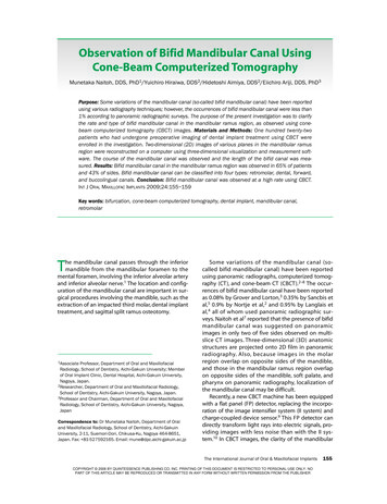
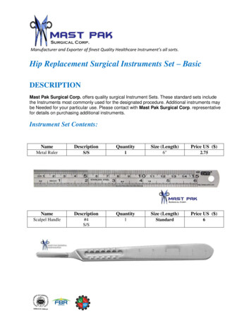
Peter D. Quinn, DMD, MDStep 1: Exposure of the RamusThe Biomet Microfixation total mandibular joint replacementprosthesis is placed through a combination of an endaural incisionand a posterior mandibular incision. It is important to completethe dissections for both the superior and inferior incisions beforethe osteotomies are performed to allow for optimal visualizationand control of hemorrhage from branches of the external carotidshould it occur during the procedure.1c1
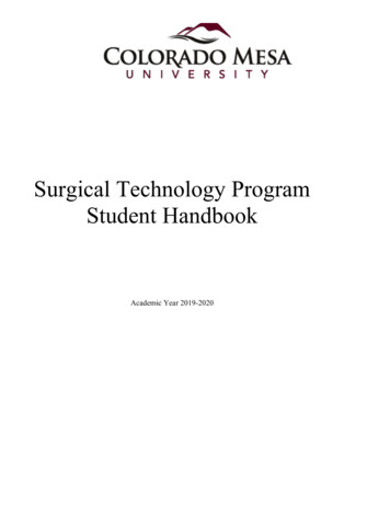
Total Mandibular Joint Replacement Surgical GuidelinesStep 2: Exposure of the Zygomatic ArchDissection is carried down to the posterior root of the zygomaticarch to keep the dissection as far posteriorly as possible to avoid anydamage to the branches of the facial nerve.2
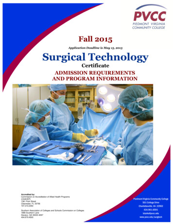
Peter D. Quinn, DMD, MDStep 3: Exposure of the entire jointCondylar retractors are used to isolate the neck of the condyleto avoid damage to the internal maxillary artery and its branches.3
Total Mandibular Joint Replacement Surgical GuidelinesStep 4: PDQ Retractors to prepare for osteotomyWith a combination of condylar retractors and a specificallydesigned condylar neck retractor (PDQ Retractor), the condyleis isolated in preparation for the two-step osteotomy.4
Peter D. Quinn, DMD, MDStep 5: Dissection medial to condylar neckStep 6: Ramal exposureThe superficial temporal artery is ligated as part of the initialdissection down to the root of the zygoma. Adequate dissectionof the soft tissue medial to the neck of the condyle is importantto avoid hemorrhage from the internal maxillary artery and thebranches most commonly involved with hemorrhage duringtemporomandibular joint surgery (middle meningeal artery anddeep temporal artery). This is especially important in the multipleoperated patient where scarring and fibrosis may bring thesevessels in closer proximity to the osteotomy cuts.The posterior mandibular incision is largely a vertical plane ofdissection anterior to the sternocleidomastoid muscle. This givesgreater visualization of the entire ramus for placement of thecondylar prosthesis and allows rapid access to the upper portionof the external carotid in case it is necessary to ligate that vesselto control hemorrhage on the medial surface of the condylarneck and superior ramus. Ligation of the external carotid, abovethe posterior auricular branch and below the transverse facial,has been shown to be more efficacious in decreasing flow to theinternal maxillary artery and its branches compared to ligation atthe bifurcation of the common carotid.5
Total Mandibular Joint Replacement Surgical Guidelines6Step 7: Dissecting around the mandibular branch of the facial nerveStep 8: Masseter reflectionThe posterior mandibular incision maximizes the visibility of the lateral ramus. The marginalmandibular branch of the facial nerve should be retracted superiorly in this dissection.Once the aponeurosis between themasseter and the medial pterygoid hasbeen identified, a scalpel is used to incisethrough the aponeurosis anteriorly fromthe ante-gonial notch and posteriorlyapproximately one-third of the lengthof the posterior ramus.
Peter D. Quinn, DMD, MDStep 9: Ramal exposureStep 10: Begin osteotomy with 1mm fissure burrA periosteal elevator is used to cleanly strip the masseter muscleat its aponeurotic insertion to avoid incising muscle fibers andcausing undue hemorrhage.Once the lateral ramus is stripped from its masseteric insertion andtemporalis insertion, if necessary, attention is directed back to theendaural incision for the first phase of the two-step osteotomy.A 1mm fissure bur is used to perform a standard condylectomywith appropriate protection from the condylar retractor7
Total Mandibular Joint Replacement Surgical GuidelinesStep 11: T-bar osteotome to finish osteotomyApproximately ninety percent of the osteotomy cut is performedwith a 1mm fissure bur and the final portion of the medial corticalbone is separated with a T-bar osteotome to avoid vascular injury.The condyle is then removed with bone holding forceps afterstripping the remnants of the external pterygoid muscle from thefovea of the condyle.8
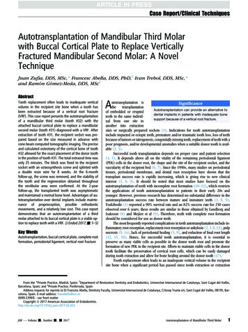
Peter D. Quinn, DMD, MDStep 12: Two-step osteotomy - Phase IISpecially designed bone holding forceps are used to maintain asecure purchase point on the inferior border of the mandible. Theramus is then pushed superiorly up into the space created by stepone of the two-step osteotomy. This now allows better visualizationof the lower portion of the condylar neck and superior ramus forperformance of this second step osteotomy. Approximately5 to 7mm of additional bone is now removed so the osteotomycut is actually below the lowest point of the sigmoid notch. It isimportant to remove adequate bone to allow for the thickness ofthe polyethylene fossa implant. If adequate bone is not removed,then a superior portion of the condyle-ramus may impinge on thefossa prosthesis when the patient is placed in intermaxillary fixation.Again, the retractors are used to protect the medial tissues duringthe osteotomy.9
Total Mandibular Joint Replacement Surgical GuidelinesStep 13: Reciprocating rasp flattens fossa eminenceA coarse diamond reciprocating rasp has been designed to flatten the articular eminence to allow “tri-pod” stability of the fossa implant againstthe base of the skull. The same rasp can also be used to uniformly level the lateral ramus, especially in patients who have had a history of bruxism,where there may be a sharply defined ridge of cortical bone along the inferior border where the hypertrophic masseter muscle had attached.10
Peter D. Quinn, DMD, MDStep 14: Sizing and Implantation of Fossa ComponentWith the use of the fossa templates, the appropriate size of thefossa implant is selected (small, medium, large). It is important tomake sure that the fossa is approximately parallel to the Frankfurthorizontal line. “Tipping” the fossa anteriorly at an excessive anglecould potentially lead to displacement of the condylar prosthesisduring function. It is recommended to place the fossa prosthesiswith two screws initially to make sure the mating between thefossa prosthesis and the condylar prosthesis is optimal beforeplacing the recommended four screws.11
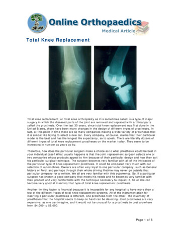
Total Mandibular Joint Replacement Surgical GuidelinesStep 15: Inter-maxillary fixationStep 16: Fitting and Placement of the Mandibular ComponentPrior to the extra-oral incisions, Erich arch bars or Ivy loops wereplaced to allow for intermaxillary fixation in the desired occlusionat this stage of the operation. Once the fossa prosthesis is placed,the wound should be covered with antibiotic-soaked sterilesponges, and the surgeon and assistant now enter the oral cavityto place the patient in rigid maxillary fixation. This is done with aseparate set of instruments specifically for the intra-oral procedure.After the occlusion is secured, the surgical team should changetheir gowns and gloves to return to the extra-oral surgicaldissections.With the patient in intermaxillary fixation, a condylar prosthesisis now selected using the 45, 50 and 55mm templates. Eitherthe narrow or standard design can be used depending onthe adequacy of bone and defects from previous surgeries.As illustrated on the following page (Figure 16d), it is extremelyimportant to position the head of the condyle as farposteriorly as possible so that there can be some degree of“pseudo-translation” of the condylar head in the fossa asthe patient opens to the expected range of 32 to 35mm.Positioning the condyle too far anteriorly in the closedposition, as shown in the right portion of the illustration, couldlead to dislocation of the condyle anterior of the fossa. Alsonote that the fossa is parallel to the Frankfurt horizontal planeand is not tipped anteriorly or superiorly.Fig. 16a12
Peter D. Quinn, DMD, MDFig. 16bFig. 16cFig. 16d13
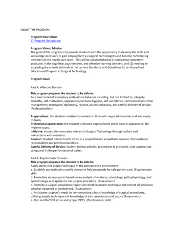
Total Mandibular Joint Replacement Surgical GuidelinesStep 17: Final Screw PlacementThe condylar prosthesis is placed with two 2.7mm screws and again the surgicalincisions are covered, sterile drapes are placed over the wounds and the surgeonand the assistant now return to the oral cavity to remove the intermaxillaryfixation wires to make sure that there is an acceptable range of motion ofthe prosthesis is without any evidence of anterior dislocation or mechanicalobstruction. If the surgical team is satisfied with position of the implant at this time,they again change gowns and gloves and return to the surgical wounds forplacement of the remainder of the ramal condylar implant screws (an averageof 4-6 screws is recommended). The surgeon must note the position of the inferioralveolar nerve in order to place the screws either posterior or anterior to theneurovascular bundle. The standard design with the expanded “foot plate” wasdesigned to allow some flexibility in screw placement. It is preferable to use theheavier bone along the inferior/posterior ramus, if possible, for screw placement.The implant should not be bent and great care should be taken to avoidscratching or damaging the articular surface of the mandibular component.14
Warnings and Precautions for use of the Total Mandibular Joint ReplacementCaution Systemic disease with increased susceptibility to infection.Federal Law (USA) restricts this device to sale, distribution, or use, by or on the orderof a physician. Patients with extensive perforations in the mandibular fossa and/or bonydeficiencies in the articular eminence or zygomatic arch that would severelycompromise support for the artificial fossa component.Description Partial TMJ joint reconstruction.The Total Temporomandibular Joint (TMJ) Replacement System is implanted in thejaw to functionally reconstruct a diseased and/or damaged temporomandibular joint. Known allergic reaction to any materials used in the components. NOTE:Patients with known or suspected nickel sensitivity should not have Co-Cr-Modevices implanted since this material contains nickel.The Total TMJ Replacement System is a two-component system comprised ofmandibular condyle and glenoid fossa components.Both components are available in multiple sizes as right and left side specific designsand are attached to the bone by screws. Included in the system are trials, instrumentsand instrument cases.Materials Mandibular Component—Cobalt-Chromium-Molybdenum (Co-Cr-Mo) alloy withtitanium alloy coating or Titanium (Ti-6Al-4V) alloy with titanium alloy coating Fossa Component—ultra high molecular weight polyethylene (UHMWPE) Screws—titanium alloy Trials: Mandibular—aluminum, Fossa—Radel plastic Instruments: TMJ flat diamond rasp, TMJ diamond burrs, TMJ double-ended drillguide, retractors—stainless steel Instrument Case—stainless steel, silicone, Radel plasticIndicationsThe Total Temporomandibular Joint Replacement System is indicated for reconstructionof the temporomandibular joint. The reconstruction is necessary due to one of thefollowing diagnoses: Arthritic conditions: osteoarthritis, traumatic arthritis, rheumatoid arthritis Ankylosis including but not limited to recurrent ankylosis with excessive heterotopicbone formation Revision procedures where other treatments have failed (e.g. alloplasticreconstruction, autogenous grafts) Avascular necrosis Multiply operated joints Fracture Functional deformity Benign neoplasms Malignancy (e.g. post-tumor excision) Degenerated or resorbed joints with severe anatomic discrepancies Developmental abnormalityContraindications Active or chronic infection. Patient conditions where there is insufficient quantity or quality of bone to supportthe components. Patients with mental or neurological conditions who are unwilling or unable tofollow postoperative care instructions. Skeletally immature patients. Patients with severe hyper-functional habits (e.g. clenching, grinding etc.) Patients with a foreign body reaction due to previous implants.WarningsAdverse EventsAdverse events that may occur following placement of the Total TMJ ReplacementSystem are listed below. See Tables 7 and 8 located in the insert # 01-50-1000for more detailed information on adverse events from the clinical trial. Removal of components(s) including, but not limited to the following:- implant changes caused by loading and/or wear- degenerative changes within the joint surfaces from disease or previousimplants- implant materials producing particles or corroding Loosening or displacement with or without removal of the implant Infection (systemic or superficial) Foreign body or allergic reaction to implant components Fossa wear through Facial swelling and/or pain Mandibular and fossa components are provided STERILE. DO NOT RESTERILIZE. Facial nerve dysfunction Screws, trials, instruments and instrument cases are provided NON-STERILE.CLEAN AND STERILIZE BEFORE USE. Excision of tissue DO NOT USE if there is a loss of sterility of the devices. DO NOT USE damaged implants and only use implants that are packaged inunopened or undamaged containers. DO NOT USE the individual components of this total system (e.g. mandibularcomponents, fossa components, or screws) for partial joint reconstruction.Bone cement or other grouting agents should not be used when implantingthese devices. Safety and efficacy have not been established for the use ofbone cement or other grouting agents with these implants. DO NOT USE IN CHILDREN. The Total TMJ Replacement was designed forskeletally mature patients.PrecautionsThe device is limited to surgeons who are adequately trained in the use of the devicethrough hands-on and educational course work. In all cases sound medical practiceis to be followed and the surgeon must select the type of device appropriate fortreatment. The patient is to be warned that the system does not replace normalhealthy bone in their TMJ and they may continue to have chronic pain and limitedrange of motion. The system can break or loosen as a result of stress, activity, ortrauma. Patients with severe hyper-functional habits may have an undesirableoutcome. The presence of existing mandibular and/or zygomatic arch screws or screwholes may compromise fixation. Note that placement of the implant in one joint onlymay result in harmful effects to the joint on the opposite side. Placement of the implantmay produce an improper relationship between teeth surfaces that should contactduring biting. The patient is to be made aware of surgical risks and possible adverseeffects prior to surgery and warned that failure to follow postoperative care instructionscan cause failure of the implant and the treatment. Specialized instruments/trials aredesigned for use with the Total TMJ Replacement System to aid in the accurateimplantation of the components. DO NOT USE trials/instruments or cases that aredisfigured, cracked, corroded, or otherwise damaged. Instruments/trials are subject towear with normal usage and are susceptible to fracture when exposed to extensiveuse or excessive force. All trials/instruments and cases should be regularlyinspected for wear or disfigurement. These should be disposed of appropriately. Heterotopic bone formation Neuroma formation Ear problems DislocationPatient Counseling Information Discussion of the following points is recommended prior to surgery. The importance of prompt medical attention if they experience unusual swellingin the area of the implant. The risks associated with a Total TMJ System (see Warnings and Adverse Events). Post-operative pain relief and return of function varies from patient to patient. Additional treatment may be required including but not limited to extendedphysical therapy, bite splint, dental braces, and/or orthognathic andreconstructive surgery.SterilityThe Total Temporomandibular Joint Replacement System mandibular and fossacomponents are sterilized by exposure to a minimum of 25 kGy of gammairradiation. DO NOT RESTERILIZE. Screws, trials, and the TMJ Instrument Casecontaining instruments are supplied non-sterile and should be wrapped with an FDAcleared sterilization wrap prior to steam sterilization in order to maintain sterility.15
This brochure is presented to demonstrate the technique and post-operative care regimen of Peter Quinn, MD. As the manufacturer of this device, Biomet Microfixation does not practice medicine and does not recommend this product or surgical technique for use ona specific patient. The surgeon who performs any implant procedure must determine the appropriate device and surgical procedure for each individual patient. Devices shown in this brochure may not be cleared or licensed for use or sale in your individual country.Please contact your local distributor for information regarding availability of this product. Information contained in this brochure is intended for surgeon or distributor information only and is not intended for patient distribution. All surgeries carry risks.For additional information, please see package insert, or visit our web site at www.biometmicrofixation.com or call 1-800-874-7711.r01k0911BMF00-2185
total mandibular joint replacement system was completed. In the ten year clinical trial (from 1995 to 2005), over 400 prosthesis have been implanted with a patient success rate of over 96%.* The current indications for alloplastic joint reconstruction include: Arthritic conditions: osteoarthritis, traumatic arthritis, rheumatoid arthritis