Transcription
HindawiJournal of Immunology ResearchVolume 2020, Article ID 6056373, 10 pageshttps://doi.org/10.1155/2020/6056373Research ArticleRegulation of Innate Lymphoid Cells in Acute Kidney Injury:Crosstalk between Cannabidiol and GILZBabak Baban ,1 Hesam Khodadadi,1 Kumar Vaibhav,2 Cristina Marchetti,3 Carlo Riccardi,3and Mahmood S. Mozaffari 11Department of Oral Biology and Diagnostic Sciences, The Dental College of Georgia, Augusta University, Augusta,Georgia 30912-11282Department of Neurosurgery, Medical College of Georgia, Augusta University, Augusta, Georgia 30912-11283Department of Medicine, University of Perugia, ItalyCorrespondence should be addressed to Babak Baban; bbaban@augusta.edu and Mahmood S. Mozaffari; mmozaffa@augusta.eduReceived 11 October 2019; Revised 23 December 2019; Accepted 31 December 2019; Published 25 February 2020Academic Editor: Patrice PetitCopyright 2020 Babak Baban et al. This is an open access article distributed under the Creative Commons Attribution License,which permits unrestricted use, distribution, and reproduction in any medium, provided the original work is properly cited.Innate lymphoid cells (ILCs) have emerged as largely tissue-resident archetypal cells of the immune system. We tested thehypotheses that renal ischemia-reperfusion injury (IRI) is a contributing factor to polarization of ILCs and that glucocorticoidinduced leucine zipper (GILZ) and cannabidiol regulate them in this condition. Mice subjected to unilateral renal IRI weretreated with the following agents before restoration of renal blood flow: cannabidiol, DMSO, transactivator of transcription(TAT-) GILZ, or the TAT peptide. Thereafter, kidney cells were prepared for flow cytometry analyses. Sham kidneys treatedwith either cannabidiol or TAT-GILZ displayed similar frequencies of each subset of ILCs compared to DMSO or TAT,respectively. Renal IRI increased ILC1s and ILC3s but reduced ILC2s compared to the sham group. Cannabidiol or TAT-GILZtreatment of IRI kidneys reversed this pattern as evidenced by reduced ILC1s and ILC3s but increased ILC2s compared totheir DMSO- or TAT-treated counterparts. While TAT-GILZ treatment did not significantly affect cells positive forcannabinoid receptors subtype 2 (CB2 ), cannabidiol treatment increased frequency of both CB2 and GILZ-positive (GILZ )cells of IRI kidneys. Subsequent studies showed that IRI reduced GILZ subsets of ILCs, an effect less marked for ILC2s.Treatment with cannabidiol increased frequencies of each subset of GILZ ILCs, but the effect was more marked for ILC2s.Indeed, cannabidiol treatment increased CB2 GILZ ILC2s. Collectively, the results indicate that both cannabidiol and GILZregulate ILC frequency and phenotype, in acute kidney injury, and that the effects of cannabidiol likely relate to modulation ofendogenous GILZ.1. IntroductionDysregulation of immune and inflammatory mechanismscontributes importantly to pathogenesis of a variety of disorders including acute kidney injury (AKI) [1–3]. Consequently, better understanding of involved mechanisms andidentification of novel targets of therapy have been the mainstay of research in the field.Glucocorticoids are well-recognized for their widespreadeffects on multiple organ systems and for their regulation ofdiverse physiological functions which contribute to maintenance of basal- and stress-related hemostasis. Therapeutically, glucocorticoids have been used for diverse disorderswith the common denominator of dysregulation of immuneand inflammatory mechanisms [4]. Nonetheless, recognitionof their adverse metabolic effects has prompted unraveling ofmechanisms which contribute to their prominent antiinflammatory effects. These studies have provided strongevidence in favor of glucocorticoid-induced leucine zipper(GILZ) protein as the pivotal regulator of anti-inflammatoryeffects of glucocorticoids [5–7]. Thus, therapeutic GILZ hasraised the prospect of curtailing pathogenic inflammationwhile avoiding adverse metabolic effects of glucocorticoids.Aside from glucocorticoids, the endocannabinoid systemhas emerged as another endogenous pathway regulatingimmune and inflammatory mechanisms, among other
2functions [8–14]. While the use of cannabis dates back toancient history, the discovery of Δ9-tetrahydrocannabinol,as the major psychoactive constituent of cannabis, led to aflurry of research which ultimately established that the endocannabinoid system is comprised of endogenous ligands(e.g., anandamide) and the biochemical machinery responsible for their synthesis, transport, and degradation as well asspecific cannabinoid receptors (i.e., CB1 and CB2); whileCB1 receptors are primarily located in the central nervoussystem, immune cells primarily express CB2 [8]. Nonetheless, both cannabinoid receptor-dependent and receptorindependent effects have been described for cannabinoids[8]. More recently, cannabidiol has received much attentionas a potential therapeutic agent as exemplified by its recentapproval, by the Food and Drug Administration, for treatment of seizures associated with two rare and severe formsof epilepsy [15].We recently showed that both GILZ and cannabidiolexert renoprotective effects, as reflected by reduction in celldeath and improved functional outcomes, in the murinemodel of AKI simulated by ischemia-reperfusion injury(IRI); the renoprotection of GILZ and cannabidiol wasaccompanied with generally similar effects of each agent onpolarization of neutrophils and T lymphocytes [16, 17]. Inlight of these observations, we decided to explore potentialimpact of cannabidiol and GILZ on innate lymphoid cells(ILCs) in AKI.ILCs are largely tissue-resident immune cells whichrespond early in the course of immune response (comparedto T lymphocytes); they are uniquely positioned to respondquickly to pathogens and other environmental stimuli tomaintain homeostasis. ILCs are divided into 3 classes(i.e., ILC1s-ILC3s) depending on their cytokine signalingrequirement and transcription factors for their lineage commitment [18–21]. ILC1s-ILC3s lack adaptive antigen receptors and are functionally considered innate counterparts ofT lymphocytes, mirroring CD4 T helper (Th)1, Th2, andTh17 cells, respectively [18–21]. In mice, and under the influence of cytokine signaling and certain transcription factor(s),innate lymphoid cell precursors differentiate to give rise toeach subset of ILCs; ILC1s utilize T-box transcription factor(T-bet), ILC2s express GATA binding protein 3 (GATA3)and depend on RAR-related orphan receptor α (RORα) fortheir activity and development, while ILC3s require transcription factor RAR-related orphan receptor γt (RORγt)and aryl hydrocarbon receptor (AhR) for their activity anddevelopment [18, 19]. Functionally, ILCs are characterizedby their cytokine profile and their participation in variousimmune responses. For example, ILC1s produce interferonγ (IFNγ) and mediate type 1 immunity while ILC2s produceinterleukin- (IL-) 4, IL-5, IL-9, and IL-13 and mediate type 2immunity, and ILC3s produce IL-17 and IL-22 and mediatetype 3 immunity [18, 19]. Despite their important roles inimmune responses against pathogens/injurious stimuli andin the maintenance of tissue homeostasis, uncontrolled anddifferential activation of ILCs may elicit inflammatoryresponses that further result in pathological conditions.Importantly, similar to T cells, ILCs demonstrate a certaindegree of plasticity thereby allowing them to change theirJournal of Immunology Researchphenotype and functional capacities depending on environmental cues such as polarizing signals in the tissuealong with the expression of cognate cytokine receptors andkey transcription factors in responding ILCs [19–21]. Thisproperty likely contributes to the important roles of ILCsnot only in immune responses against pathogens and sterileinflammation but also in maintaining tissue hemostasis.Nonetheless, differential development and activation of ILCsin a context- and/or disease-specific manner may contributeto inflammatory responses and/or outcome of treatmentmodalities [20–27].In light of the above, we tested the hypothesis that renalIRI is a contributing factor to polarization of ILCs and thatGILZ and cannabidiol regulate them in this condition. Further, in light of evidence indicating an interaction betweenglucocorticoids and endocannabinoid signaling pathways[8], we determined whether an interaction exists betweencannabidiol and GILZ in regulation of ILC subtypes in AKI.2. Materials and Methods2.1. Animals. The studies utilized male, 9-11 weeks of age,Balb/C mice which were obtained from Harlan Laboratoriesand housed in the laboratory animal facilities of the AugustaUniversity with free access to food and water. Rodents ofsimilar age are routinely used for the investigation of variouspathologies and the use of male mice relates to greatersusceptibility of the male gender to AKI [28]. These studiesconformed to guidelines of Institutional Animal Care andUse Committee.2.2. TAT and TAT-GILZ. The protocol for generation of thetransactivator of transcription (TAT) peptide and theTAT-GILZ fusion protein has been described previously[29]. Briefly, TAT and TAT-GILZ, which was constructedby inserting GILZ cDNA in the TAT C vector to producean in-frame fusion protein, were cloned into the pGEX-4T2plasmid (GE Healthcare). The pGEX-4T2 plasmid is aglutathione S-transferase (GST) fusion vector carrying a tacpromoter for chemically inducible high-level expression ofthe protein. GST fusion protein was expressed in lipopolysaccharide- (LPS-) lacking bacteria Clear Coli BL21 (LucigenCorporation, Middleton, USA) which were grown at 37 Cand induced with 1 mM isopropyl b-D-thiogalactopyranosidefor 4 hours [30]; all other materials used in the process weresterile and LPS-free. Following sonication to induce lysis,most of the generated protein was found in the soluble fraction, which was then purified with Glutathione Sepharose4B beads (GE Healthcare) following the manufacturer’sinstructions. Eluted proteins were dialyzed against PBS for48 hrs.2.3. Renal IRI and Treatment Protocols. Animals were anesthetized with ketamine (120 mg/kg, i.p.)/xylazine (16 mg/kg,i.p.) in preparation for the surgical procedure which consisted of two flank incisions and clamping of the left renalpellicle, for 20 min, using a nontraumatic vascular clampwhile the right kidney served as sham control. Thereafter,the vascular clamp was removed and restoration of renal
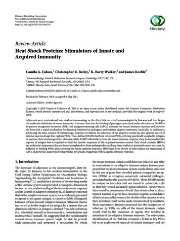
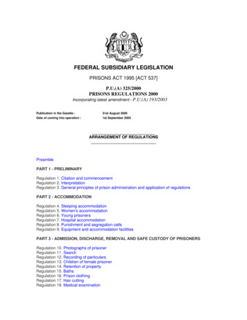
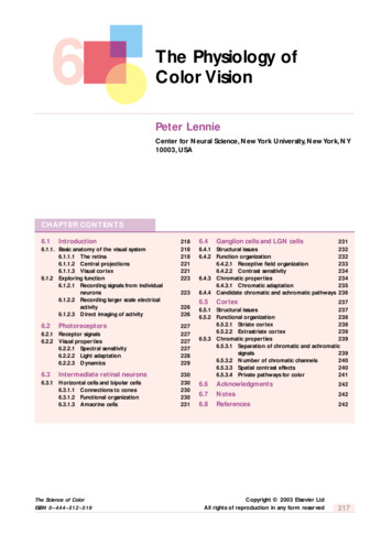
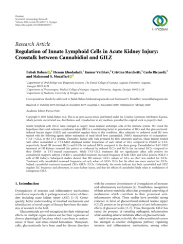
Journal of Immunology Researchblood flow confirmed, visually, prior to closure of muscleand skin layers using 4-0 silk sutures and autoclips, respectively [16, 17]; each independent experiment included 3-6 animals, and the number of animals for each group/condition isindicated under Figure legends. Each of the following agentswas administered, intraperitoneally in a volume of 50 μl,10 min before restoration of renal blood flow: (a) vehicle(DMSO), (b) cannabidiol (10 mg/kg, 16), (c) TAT (0.1 mg/kgin PBS), and (d) TAT-GILZ (0.2 mg/kg in PBS); the latterdosage regimen is based on a two-fold larger molecularweight of GST-TAT-GILZ than GST-TAT as well as studiesindicating resolution of LPS-induced inflammation inresponse to TAT-GILZ treatment and its effectiveness inthe murine model of acute kidney injury [17, 29]. Postoperative analgesia was provided with a single injection of buprenorphine (1 mg/kg, SC). The animals were sacrificed 1-daypostinjury and kidneys procured for cell preparation andflow cytometry-based assays.2.4. Flow Cytometry Analyses. For flow cytometry analysis,renal tissues were sieved through a cell strainer (BD Biosciences, San Diego, CA), followed by centrifugation (1,500 rpm,5 min) to prepare single-cell suspensions. Preparative cell sortswere performed on cells stained with fluorochromeconjugated monoclonal antibodies using a 4-Laser LSR IIflow cytometer as well as a Coulter MoFlo XDP cell sorter[20]. Cells were gated based on forward and side scatter properties and on marker combinations to select cells of interest[20]. Total murine ILCs were gated as Lineage CD45 lymphocytes (lineage cocktail of antibodies included PerCPconjugated anti-CD3, anti-CD4, anti-CD14, anti-CD16,anti-CD19, anti-CD8, anti-CD15, anti-CD20, all from BioLegend) to exclude non-ILCs. Subsequently, ILC1s wereidentified as Lineage CD45 CD127 /-IL-12Rβ2 cells, ILC2sas Lineage CD45 CD127 GATA3 cells, and ILC3s asLineage CD45 CD127 RORγt cells (all antibodies fromBioLegend). The capacity to produce cytokines forILC1s (IFN-γ/TNF-α), ILC2s (IL-5/IL-13), and ILC3s(IL-17/IL-22) was tested, as described previously [20].Further, GILZ expression was assessed in ILCs subset usinganti-GILZ antibody following similar flow cytometry protocol. In addition, whole-kidney cell preparations were alsosubjected to flow cytometry analyses for detection of ILC2sfollowed by further analyses to assess those positive forexpression of CB2 and GILZ using anti-CB2 (Bioss, Catno. BS-2377R-A488) and anti-GILZ (eBioScience, Cat no.12-4033-80) antibodies. Isotype-matched controls were analyzed in each sample to set the appropriate gates; representative panels are reported in relevant Figures. For each marker,samples were analyzed with duplicate measurements.2.5. Sorting of ILC2s, Cytospin Preparation, andImmunofluorescent Staining. Preparative cell sorts to obtainILC2s were performed on kidney cells stained withfluorochrome-conjugated mAbs (sources as detailed, below)using the BD Biosciences FACSCAria II SORP equippedwith 4 lasers and 18 fluorescence detectors. Kidney cellswere gated based on forward and side scatter propertiesand on marker combinations to select cells of interest [20].3Total ILCs were gated as Lineage CD45 CD127 lymphocytes. The lineage cocktail of antibodies included FITCconjugated anti-CD3, anti-CD4, anti-CD14, anti-CD16,anti-CD19, anti-CD8, anti-CD15, anti-CD20, anti-CD33,anti-CD34, anti-CD203 (eBioscience), and anti-FcεRI(BioLegend), with CD127 and Sca-1—both PerCP-conjugated(BioLegend). Subsequently, ILC2s were sorted as Lineage cells positive for GATA-3.Cytospin preparations of 10000 sorted cells per samplechamber were centrifuged (700 r.p.m., 5 min), air-dried, fixedin 10% formalin, and washed twice in PBS. All subsequentprocedures were carried out in the dark and at room temperature. Endogenous peroxidase activity was blocked withhydrogen peroxide (1 : 10 w/PBS, 10 min). Immunofluorescence (GILZ and CB2) for cytospin preparations was stainedas above. To permeabilize, all preparations were incubated in0.2% Triton X-100 for 5 min at room temperature. All slideswere washed three times for 5 min at room temperature andthen incubated in blocking buffer (20% normal donkeyserum, 1% BSA, 0.02% NaN3, 13 PBS) for 45–60 min. Allslides were incubated with anti-CB2 (Bioss, Cat# BS-2377RA488) and anti-GILZ (eBioScience, Cat# 12-4033-80) antibodies for 2 hours in the dark at room temperature. All slideswere washed and then counterstained using DAPI (ThermoFisher Scientific, D3571).2.6. Statistics. Data were analyzed using the analysis of variance followed by Newman–Keuls post hoc test to establishsignificance (p 0:05) among multiple groups. Statisticalcomparison of two groups (i.e., CB2 GILZ ILC2s data) utilized Student’s t-test. Data are reported as means SEM.3. ResultsFigure 1(a) depicts gating strategy based on side scattervs. forward scatter of kidney cells from experimentalgroups followed by identification of each subset of ILCsrelying on phenotypic markers as described in Methods.Figures 1(b)–1(d) show quantitative data for each ILC subtype for each experimental group. Sham-operated kidneysshowed similar frequencies of each subtype of ILCs.However, IRI was associated with significant increases inILC1s and ILC3s but a marked reduction in ILC2s(Figures 1(b)–1(d)). Treatment of IRI kidneys with cannabidiol reversed this pattern as indicated by significant reductions in ILC1s and ILC3s but increased ILC2s. However,while ILC1s and ILC3s of cannabidiol-treated IRI kidneysremained higher than sham-operated kidneys, no differencewas noted in ILC2s between cannabidiol-treated IRI kidneysand sham-operated kidneys (Figures 1(b)–1(d)).The profile of changes in each ILC subsets and theresponse to TAT or TAT-GILZ were very similar to thosedescribed for experiments using cannabidiol or its vehicle.Briefly, IRI was associated with significant increases in ILC1sand ILC3s but significant reduction in ILC2s; treatment withTAT-GILZ restored the frequency of ILC2s to those of shamcontrol groups while ILC1s and ILC3s were partially restoredand remained significantly higher than sham-operated kidneys (Figures 2(a)–2(d)).
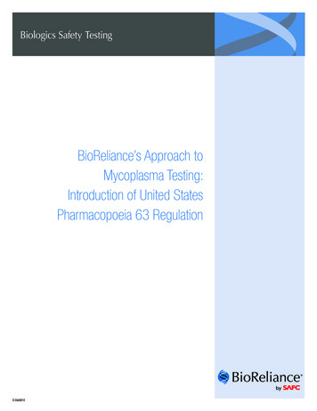
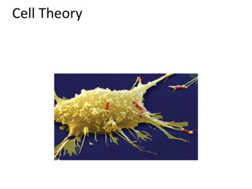
410410410310310310210210210110110Sham; VehicleILC2s200 k150 k100 k11050 k0050 k100 k 150 k 200 k 250 kSham ; CBD010010110210310410200 k310150 k2101100100050 k100 k 150 k 200 k 250 10410010110210310410200 k310310150 k210210210110110110010010010ILC2s50 k100 k 150 k 200 k 250 k010110210310410200 k310210100 k110050 k100 k 150 k 200 k 250 10210310410ILC3s4#, 0210310ILC3s31021001001010⁎6(b)410ILC2sGATA350 k010310150 k10ILC1s8ILC1s410410250 k410ILC1s010410ILC1s010ROR𝛾t031010ILC3s31050 k0210410410100 k110410410250 kIRI; CBDILC1s010ILC2s100 k50 k010410410250 kIRI; Vehicle010ILC3sCells 103 (total kidney cells)250 kCells 103 (total kidney cells)Journal of Immunology ResearchCells 103 (total kidney , †20Sham;DMSOSham;CBDIRI;DMSOIRI;CBD(d)Figure 1: Effects of cannabidiol on ILCs subsets in the murine model of acute kidney injury. Mice were subjected to left renal ischemia(20 min) reperfusion (1 day) injury (IRI) while the right kidney served as sham control; the animals were subdivided to receiveintraperitoneal administration of either vehicle (DMSO) or cannabidiol (CBD), as described under Methods, before restoration of renalblood flow. Thereafter, cell preparations of kidneys from each animal were subjected to flow cytometry analyses for determination of ILCsubsets using specific markers. (a) Gating strategy based on side scatter (SSC) vs. forward scatter (FSC) of kidney cells from experimentalgroups followed by identification of each subset of ILCs relying on phenotypic markers as described in Methods. (b–d) Quantitative datafor each ILC subtype for each experimental group; data are individual values as well as means SEM of n 4‐5 animals/group/condition;each independent experiment included 3-5 animals. p 0:05 compared to other three groups. #p 0:05 compared to their vehicle-treatedcounterparts. †p 0:05 compared to sham-operated groups.We next determined whether treatment with cannabidiolor TAT-GILZ affects expression of CB2 in whole kidney cellpreparations. Induction of IRI did not affect frequencies ofCB2 cells (Figure 3). However, while treatment withTAT-GILZ did not significantly affect CB2 cells, treatmentwith cannabidiol significantly increased CB2 cells in IRIkidneys compared to their TAT-treated and DMSO-treatedcounterparts, respectively (Figure 3).We also determined the effect of cannabidiol on GILZexpression in whole-kidney cell preparations. As shown inFigure 4, vehicle- and cannabidiol-treated sham-operatedkidneys displayed similar frequencies of GILZ cells. However, induction of IRI significantly reduced frequencies ofGILZ cells in vehicle-treated kidneys, an effect abrogatedin cannabidiol-treated IRI kidney (Figure 4(c)).We next determined expression of GILZ in ILCs andwhether treatment with cannabidiol affects each subset ofILCs which also express GILZ. Cannabidiol treatment didnot affect GILZ subsets of ILCs, expressed as percent ofeach ILC subtype, in sham-operated kidneys. However,induction of IRI reduced each subset of GILZ ILCs(Figure 5). Treatment of IRI kidneys with cannabidiolsignificantly increased each subset of GILZ ILCs butthe effect was more marked for ILC2s and ILC3s, achievingvalues similar to those of their sham-operated counterparts(Figure 5).In light of the more profound impact of cannabidiol onGILZ ILC2s (Figure 5) and the demonstration that thetreatment also increases CB2 cells in whole-kidney cellpreparations (Figure 3), we next determined whether cannabidiol treatment increases expression of both CB2 andGILZ in ILC2s; these studies used flow cytometry andimmunofluorescent protocols. As shown in Figures 6(a)and 6(b), compared to vehicle treatment, cannabidiol treatment significantly increased the percent of ILC2s which waspositive for both CB2 and GILZ in the ischemic-reperfusedkidney, an effect also evident from merged immunofluorescent images shown in Figure 6(c).
150 k0050 k 100 k 150 k 200 k 250 k250 kILC2s310210210210110110110100 k50 0210310410410ILC2s200 k150 k050 k 100 k 150 k 200 k 250 k 10110210310410110210310410010250 k410200 k3103103102102102101101104100050 k 100 k 150 k 200 k 250 k010010110210310010410200 k150 k21011050 k050 k 100 k 150 k 200 k 250 kLineageSSC0FSC010310410ILC3s11021031004 110210310ILC3s04 1001010CD127(a)IL-12R𝛽2ILC1s110#, 10⁎6110ILC2s310100 k210410410410250 k110ROR𝛾t100 k010ILC2s150 k50 kIRI; TAT-GILZ010010010GATA3IRI; TAT010ILC1s8(b)ILC1s010ILC3s100 k50 kCells 103 (total kidney cells)Sham; TAT200 kCells 103 (total kidney cells)410250 kSham; TAT–GILZ5Cells 103 (total kidney cells)Journal of Immunology Research410⁎ILC3s43#, †210Sham;Sham;TAT TAT-GILZIRI;TATIRI;TAT-GILZ(d)Figure 2: Effects of GILZ on ILC subsets in the murine model of acute kidney injury. Mice were subjected to renal IRI or sham operation andtreated with either TAT or TAT-GILZ followed by assessment of subsets of ILCs in the kidneys as described under Figure 1 legend(representative flow cytometry data is shown under (a)). (b–d) Individual values and means SEM of n 6 animals/group/condition; eachindependent experiment included 4-6 animals. p 0:05 compared to other three groups. #p 0:05 compared to their vehicle-treatedcounterparts. †p 0:05 compared to sham-operated groups.4. DiscussionThe present study shows that (a) IRI affects the profile of ILCsubsets in the kidney; (b) cannabidiol and GILZ exert similareffects on ILC subsets, restoring their frequencies towardsthose of sham-operated kidneys; (c) cannabidiol increasesboth CB2 and GILZ cells in the kidney subjected to IRI;(d) IRI reduces frequencies of GILZ ILCs, an effect lessprominent for ILC2s; (e) cannabidiol restores GILZ ILCsubsets in the kidney subject to IRI, an effect most markedfor ILC2s but less prominent for ILC1s; and (f) cannabidiolincreases CB2 GILZ ILC2s in the IRI kidney. Collectively,these novel observations indicate that both cannabidiol andGILZ are major regulators of ILC subtypes and that cannabidiol, likely via a mechanism dependent on GILZ modulation, promotes the suppressive/protective ILC2s in acutekidney injury.Utilizing the murine model of AKI, simulated by renalIRI, we now show significant increases in ILC1s and ILC3sbut a significant reduction in ILC2s in the damaged kidneyscompared to their sham-operated counterparts; thesechanges should be conducive to a proinflammatory environment in the damaged kidney. To our knowledge, this is thefirst study which demonstrates the profile of ILC subsets inAKI. Previous studies have focused on therapeutic modula-tion of type 2 immunity, via maneuvers to amplify ILC2s,on the outcome of AKI [22]. Huang et al. [31] showed thatIl-25-treated mice subjected to renal IRI display increasedserum levels of IL-4, IL-5, and IL-13 in association withincreased frequencies of ILC2s and improved renal function.Further, adoptive transfer of ILC2s reduced renal functionaland histopathological features in IRI mice accompanied withinduction of suppressive/regulatory macrophages in the kidney. Further, Cao et al. [32] have shown that treatment withrecombinant IL-33 increases serum and kidney levels of IL-4and IL-13 along with increased frequency of ILC2s (as well asregulatory macrophages and T cells) in mice kidney subjected to IRI; these changes were associated with reducedrenal structural injury and functional abnormalities andimproved survival of mice subjected to AKI. Further, authorsshowed that adoptive transfer of ILC2s also protects thekidney against IRI. Authors concluded that activationIL-33-ILC2 axis represents a therapeutic strategy to conferrenoprotection in AKI. Others have shown that inadriamycin-induced glomerulonephritis, a model of chronicproteinuric renal injury, treatment of animals with IL-33improved the course of the disease via an ILC2-mediatedinduction of protective type 2 response; the treatment alsoincreased IL-5 which promoted accumulation of eosinophilsin the kidney and ameliorated glomerulonephritis [23]. A
Journal of Immunology ResearchCB2 cells (% of total kidney CB2Isotype controlSham; DMSOSham; CBDSham; DMSOIRI; DMSOSham; CBDIRI; CBDIRI DMSOIRI; CBD(b)CB2 cells (% of total kidney m; TATSham; TAT-GILSIRI; TATIRI; TAT-GILS104CB2Isotype controlIRI TATIRI TAT-GILZ(c)(d)5080040600400GILZ302010200000200 400 600 800 1000100101Forward scatter102GILZIsotype controlIRI vehicle(a)103104GILZ cells (% of total kidney cells)1000CountsSide scatterFigure 3: Effects of cannabidiol or GILZ on CB2-positive kidney cells. Kidney cell preparations from experimental groups, as described underMethods, were subjected to flow cytometry analyses for CB2 expression. (a, c) Representative histograms while bar graphs (b, d) showquantitative data for CB2-positive cells for experimental groups. Data are means SEM, 4-5 animals (CBD and vehicle conditions) and 6animals (TAT and TAT-GILZ conditions) for each group; each independent experiment included 3-6 animals. p 0:05 compared toother three groups.151296⁎30Sham; DMSOIRI; DMSOSham; CBDIRI; CBDIRI CBDSham(b)(c)Figure 4: Effects of cannabidiol on GILZ expression in the kidney. Kidney cell preparations from vehicle- and cannabidiol- (CBD-) treatedanimals, as described under Methods, were subjected to flow cytometry analyses for GILZ expression. (a) Side scatter vs. forward scatterfollowed by histogram showing GILZ-positive cells. (c) Means SEM of 3 animals for group/condition; each independent experimentincluded 3 animals. p 0:05 compared to other three groups.
750GILZCounts403020100100101102103GILZIsotype ControlILC2ILC3ILC1(a)104GILZ cells (% of ILC subtype)Journal of Immunology Research15129630⁎#⁎⁎ILC1sILC2sILC3sSham; DMSOIRI; DMSOSham; CBDIRI; CBD(b)Figure 5: Effects of cannabidiol on GILZ-positive ILC subsets. Kidney cell preparations from vehicle- and cannabidiol- (CBD-) treatedanimals, as described under Methods, were subjected to flow cytometry analyses for GILZ expression by each subset of ILCs.(a) Histogram showing GILZ-positive cells; (b) means SEM of 3 animals for each group/condition; each independent experiment included3 animals. p 0:05 compared to other three groups for each ILC subset. #p 0:05 compared to DMSO-treated sham kidneys for ILC1s.more recent study investigated tissue localization of ILC2s inthe normal kidney and whether their deficiency is of consequence for the kidney subjected to IRI [33]. Authors show thatILC2s are located in close proximity to the renal vasculatureand constitutively express high levels of IL-5 under hemostatic conditions. Further, they show that deficiency ordepletion of ILC2s in animals subjected to renal IRI doesnot affect histopathology, collagen deposition, or mRNAexpression of injury-associated (Lcn2), inflammatory (Cxcl1,Cxcl2, and Tnf) or extracellular matrix (Col1a1, Fn1) factors.These observations led authors to conclude the potentialfor ILC2 redundancy and that other components of type2-mediated immunity likely compensate for loss of ILC2sthereby accounting for lack of exacerbated kidney injuryunder these conditions [33].While the focus of recent research has been to unravel therole of ILC2s in pathogenesis of AKI, our demonstration ofmarked increases in ILC1s and ILC3s in this condition is suggestive of their pathogenic roles in determining the outcomeof renal IRI. Thus, there is a need to identify therapeuticoptions that can modulate ILC subsets to those of the normalkidney with the objective of conferring beneficial impact tothe ischemic-reperfused kidney. In light of our recent demonstration that both cannabidiol and GILZ exert significantrenoprotective effects, as reflected by reduction in cell deathand improved functional outcomes, in association with similar changes in polarization of neutrophils and T lymphocytes in favor of their regulatory/suppressive phenotypes[16, 17], we sought to establish the impact of each agent onthe profile of ILC subsets of the kidney subjected to IRI.Interestingly, cannabidiol and GILZ caused similar changesin the profile of ILC subsets in the ischemic-reperfused kidney, restoring their levels towards those of sham-operatedkidneys. However, while cannabidiol and GILZ normalizedILC2s of ischemic-reperfused kidneys, ILC1s and ILC3sremained significantly higher in the ischemic-reperfused kidneys compared to their sham-operated counterparts. Thesenovel observations are suggestive of potential crosstalkbetween cannabidiol and GILZ in determining frequenciesof ILC subtypes in AKI, an aspect further explored asdescribed below.Earlier studies have suggested an interaction betweenendocannabinoid and glucocorticoids signaling pathways asevidenced by high expression of type 1 cannabinoid receptor(CB1) in brain regions that are implicated in actions of glucocorticoids [8]. Further, glucocorticoids are shown to mobilizethe endocannabinoid system and a functional endocannabinoid system is required for many glucocorticoid-mediatedeffects [8]. Given that both glucocor
Research Article Regulation of Innate Lymphoid Cells in Acute Kidney Injury: Crosstalk between Cannabidiol and GILZ BabakBaban ,1 HesamKhodadadi,1 KumarVaibhav,2 CristinaMarchetti,3 CarloRiccardi,3 and Mahmood S. Mozaffari 1 1Department of Oral Biology and Diagnostic Sciences, The Dental College of Georgia, Augusta University, Augusta, Georgia 30912-1128