Transcription
Su et al. Molecular Cancer 2014, /178RESEARCHOpen AccessHMGN2, a new anti-tumor effector molecule ofCD8 T cellsLin Su, Ankang Hu, Yang Luo, Wenjie Zhou, Ping Zhang* and Yun Feng*AbstractBackground: Cytolytic T lymphocytes (CTL) and natural killer (NK) cells have been implicated as important cells inantitumor responses. Our previous research has shown that high mobility group nucleosomal-binding domain 2(HMGN2) could be released by IL-2 and PHA stimulated peripheral blood mononuclear cells (PBMCs) and alsoinduced tumor cells apoptosis at low doses. In this study, we isolated and cultured PBMCs and CD8 T cells toanalyze the expression and antitumor effects of HMGN2.Methods: PBMCs from healthy donors were isolated using Human Lymphocyte Separation tube. CD8 T cells wereseparated from the PBMCs using MoFlo XDP high-speed flow cytometry sorter. Activation of PBMCs and CD8 Tcells were achieved by stimulating with Phytohemagglutinin (PHA) or tumor antigen. In addition, the methods ofELISA, intracellular staining, and fluorescence-labeling assays were used.Results: PHA induced PBMCs to release high levels of HMGN2, and CD8 T cells was the major cell population inPBMCs that release HMGN2 after PHA activation. Tumor antigen-activated CD8 T cells also released high levels ofHMGN2. Supernatants of tumor antigen-activated CD8 T cells were able to kill tumor cells in a dose-dependentmanner. This antitumor effect could be significantly blocked by using an anti-HMGN2 antibody. Fluorescence-labelingassays showed that the supernatant proteins of activated CD8 T cells could be transported into tumor cells, and thetransport visibly decreased after HMGN2 was depleted by anti-HMGN2 antibody.Conclusions: These results suggest that HMGN2 is an anti-tumor effector molecule of CD8 T cells.Keywords: HMGN2, PMBC, CD8 T cells, Antitumor activity, Anti-tumor effector moleculeBackgroundCytolytic T lymphocytes (CTLs) and Natural killer (NK)cells function as antitumor immune cells. CTLs and NKcells are rich in cytoplasmic granules. Upon degranulation,these cells release cytotoxic substances that act on targetcells [1]. Granules in the cytoplasm of CTL contain perforin, granzymes, granulysin, additional effector moleculesinvolved in the antitumor response and several uncharacterized components [2-5].Previously, we isolated and purified an antimicrobialpolypeptide from interleukin (IL)-2 and PHA stimulatedhuman peripheral blood mononuclear cells (PBMCs) andidentified the polypeptide as high mobility group nucleosomal binding domain 2 (HMGN2). When cultured PBMCswere stimulate with IL-2 and PHA, HMGN2 was expressed* Correspondence: pingzhang68@hotmail.com; 953463551@qq.comState Key Laboratory of Oral Diseases, West China College of Stomatology,Sichuan University, Chengdu, Sichuan, Chinain the cytoplasm and then released into the supernatant[6]. HMGN2 is one of the most abundant non-histone nuclear proteins of vertebrates and invertebrates [7], and is ahighly conserved nucleosomal protein thought to be involved in unfolding higher-order chromatin structuresand facilitating transcriptional activation of mammaliangenes [7]. However, until now, the biological role of thisprotein has not been fully defined, and some new research results have revealed that the protein has additional functions.In this study, we isolated and cultured human PBMCsand separated CD8 T cells from PBMCs. PBMCs andCD8 T cells were then activated by PHA or tumor antigen (T-Ag), followed by analysis of HMGN2 protein expression and release. Our results showed that activatedCD8 T cells express high levels of HMGN2 protein,and HMGN2 protein in the culture supernatant of activated CD8 T cells had proved had anti-tumor effect. 2014 Su et al.; licensee BioMed Central Ltd. This is an Open Access article distributed under the terms of the CreativeCommons Attribution License (http://creativecommons.org/licenses/by/4.0), which permits unrestricted use, distribution, andreproduction in any medium, provided the original work is properly credited. The Creative Commons Public DomainDedication waiver ) applies to the data made available in this article,unless otherwise stated.
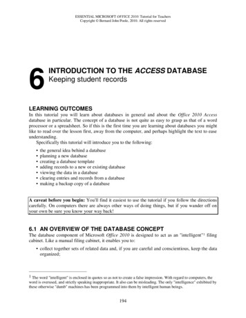
Su et al. Molecular Cancer 2014, /178Page 2 of 10Resultsincreased after PHA stimulation (47.79 11.87%) (Figure 1Band C).PHA-activated T cells released high levels of HMGN2PHA is a lectin found in plants, especially legumes, andis a mitogen that triggers T-lymphocyte cell division andactivation. Supernatants of PBMCs that have been stimulated by PHA contain a series of effector proteins. Inour experiments, PBMCs were isolated from healthy donors and stimulated with 20 μg/ml PHA or 100 IU/mlIL-2 for 72 hours. Supernatants were collected andHMGN2 concentrations were analyzed by ELISA. Theresults showed that PBMCs release high levels of HMGN2(771.33 123.12 ng/ml) following 20 μg/ml PHA stimulation for 72 hours. Compared with the medium control(289.67 98.5 ng/ml) and IL-2 stimulated (397.67 134.15 ng/ml), HMGN2 concentration significantly increased (Figure 1A). In order to make sure that HMGN2was released by activated T cells, we used surface andindirect intracellular staining to analyze HMGN2 expression in CD3 T cells. Consistent with the ELISA results (Figure 1A), compared with the medium control(9.06 3.8%) and IL-2 stimulated (18.85 8.71%), the percentage of HMGN2 CD3 cell population significantlyATo determine which T cell population expressed HMGN2,we stained the PHA pre-activated PBMCs with CD4-PEor CD8-PE surface staining. We then stained intracellularHMGN2 with rabbit anti-human HMGN2/goat antirabbit IgG-FITC. Results showed that HMGN2 expressionsignificantly increased in both CD4 and CD8 T cell populations (Figure 2A, B, C). In addition, HMGN2 expression in CD8 T cells (50.71 10.34%) was significantlyhigher than that in CD4 T cells (16.67 5.61%) afterPHA stimulation (Figure 2D). Finally, we used a MoFloXDP high-speed flow cytometry sorter to purify PHA preactivated CD3 CD8 T cells, CD3 CD8 T cells and CD3 mix cells, followed by culturing in complete medium with2000 IU/ml IL-2 for 5 days and analyzed the expression ofHMGN2 by ELISA and intracellular staining. Resultsshowed that HMGN2 was still expressed at high levels inCD8 T cells (539.00 118 ng/ml; 68.37 15.21%). On theother hand, the expression of HMGN2 in the CD3 CD8 TC*1000HMGN2 was highly expressed in activated CD8 T cellsD*60600400200msIgG-PEHMGN2 positive (%)4020msIgG-FITC02nd-Ab control0MediumcontrolBIL-2CD3-PEPHAMedium ation(ng/ml)800Isotype controlPHAIL-219.5549.162nd-Ab-FITCR5Dot plot graghHMGN2-FITCFigure 1 PHA induced HMGN2 expression in PBMCs. PBMCs were seeded at a density of 1 107 per well in 6 well plates and stimulated with20 μg/ml PHA or 100 IU/ml IL-2 for 72 hours. Normal medium was used as the control. (A) The supernatants were collected and HMGN2 concentrationswere analyzed by ELISA. (B) The cells were removed and stained with anti-CD3-PE and anti-HMGN2-IgG-FITC, followed to run on a Beckmancoulter FC500 Flow cytometry. Data were analyzed by using Submit 5.2 software after gate lymphocytes (R5) group on dot plot graph. Figuresare representative of three independent experiments. (C) Error bars represent HMGN2 intracellular expression positive rate (%) in CD3 T cells.Data are represented as means SD of three independent experiments. *Significantly higher compared to medium control (p 0.05). (D) Representativeplots of the isotype staining and 2nd-Ab-FITC staining control.
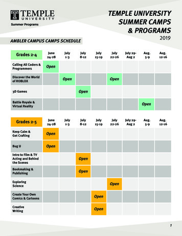
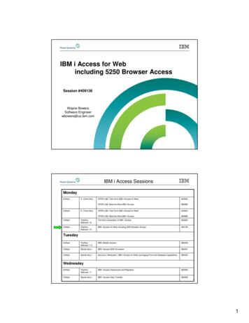
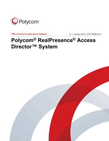
Su et al. Molecular Cancer 2014, /178AMedium controlPage 3 of 10IL -28.48.0CPHA16.2CD4 -PE*0B3.72049.9CD8 -PE2.410*HMGN2 -FITC7005050CD8403020E2 nd -Ab controlIsotype controlCD4 -PECD4100HMGN2 positive(%)#60msIgG -PEHMGN2 positive (%)D30HMGN2 positive(%)100Medium controlIL-2PHAmsIgG -FITC2 nd -Ab -FITCFigure 2 PHA induced HMGN2 expression in different cell populations. PBMCs were seeded at a density of 1 107 per well in 6 well platesand stimulated with 20 μg/ml PHA or 100 IU/ml IL-2 for 72 hours. Normal medium was used as the control. Cells were collected and stained (A)Anti-CD4-PE surface stained and anti-HMGN2/IgG-FITC intracellular stained; (B) Anti-CD8-PE surface stained and anti-HMGN2/IgG-FITC intracellularstained. Figures are representative of three independent experiments. (C, D) Error bars represent HMGN2 intracellular expression positive rate (%)in CD4 or CD8 T cell populations. Data are represented as means SD of three independent experiments. *Significantly higher compared toIL-2 and medium control (p 0.05), #Significantly higher compared to CD4 T cell population (p 0.05). (E) Representative plots of the isotypestaining and 2nd-Ab-FITC staining control.cells (mainly CD4 T cells) (307.67 97.34 ng/ml; 36.23 11.52%) and the CD3 mixed PBMCs population (386.67 105.46 ng/ml; 35.73 13.71%) was significantly lower inboth supernatants and intercellular (Figure 3A, B and C).These results confirmed that activated CD8 T cellsexpressed high levels of HMGN2.Tumor antigen activated CD8 T cells expressed highlevels of HMGN2In order to ensure that HMGN2 was an effector proteinof tumor antigen activated T cells, we used tumor fullprotein as tumor antigen (T-Ag) to stimulate PBMCs for7 days. Supernatants were collected for assaying HMGN2levels by ELISA and cells were collected for HMGN2intracellular staining. Results showed that there was nosignificant change of HMGN2 expression after stimulationwith T-Ag compared with the medium control (Figure 4).In order to ensure that T cells were activated by T-Ag, weused CD44high as the activated marker of T cells. T-Agstimulated PBMCs were stained with CD8-PE/CD44-APCsurface and HMGN2 indirect intracellular staining.CD44high was used as the activated T cell population(Figure 5Aa, gate R2) and CD44low was used as thenaive T cell population (Figure 5Aa, gate R6). T-Ag induced only 18.35 6.20% PBMCs activated (Figure 5Aa,gate R2 and 5B). There was about 43.69 12.51% of activated CD8 T cells expressed HMGN2 (Figure 5Ab andC), where only 18.71 6.34% naive CD8 T cells expressedHMGN2 (Figure 5Ac and C).We used a Beckman Coulter MoFlo XDP high-speedflow cytometry sorter to isolate T-Ag-activated theCD44highCD8 T cell population (Figure 6A, Gate R4).The purified cells were cultured with 2000 IU/ml IL-2 for5 days and then the cells were identified with CD44-APC
Su et al. Molecular Cancer 2014, /178Page 4 of 10C**80MIXCD8-CD3 HMGN2 positive(%)800CD8 CD3 D3-FITCB6.339.970.4CD8 CD3 2nd-Ab-FITC38.6CD8-CD3 MIXHMGN2-FITCFigure 3 PHA-induced HMGN2 was mainly expressed in CD8 T cell population. PBMCs were seeded at a density of 1 107 per well in 6well plates and stimulated with 20 μg/ml PHA for 72 hours. Cells were collected and stained with CD3-FITC and CD8-PE. CD3 CD8 T cells (GateR1), CD3 CD8 T cells (Gate R2) and CD3 mixed cells (Gate R3) were purified with MoFlo XDP sorter. 4 106 CD3 CD8 T cells, CD3 CD8 T cellsand CD3 mixed cells were incubated in 24-well plates respectively for 5 days. (A) The supernatants were collected and HMGN2 concentrationswere analyzed by ELISA. (B) The cells were removed and intracellularly stained for HMGN2. Figures are representative of three independent experiments.(C) Error bars represent HMGN2 intracellular expression positive rate (%) in CD8 T cells populations. Data are represented as means SD ofthree independent experiments. *Significantly higher compared to CD8 CD3 and MIX cells (p 0.05).A3.52nd-Ab-FITC31.941.8HMGN2-FITCMedium controlBT-AgCT-AgT-AgMediumcontrolMedium control02040HMGN2 positive(%)600100200300400HMGN2 concentration(ng/ml)Figure 4 HMGN2 expression in Tumor antigen stimulated PBMCs. PBMCs were seeded at a density of 1 107 per well in 6 well plates andstimulated with 150 μg/ml T-Ag for 7 days. Normal medium was used as the control. (A) The cells were removed and intracellularly stained forHMGN2. Figures are representative of three independent experiments. (B) Error bars represent HMGN2 intracellular expression positive rate (%) inPBMCs after stimulation with T-Ag. (C) The supernatants were collected and HMGN2 concentrations were analyzed by ELISA. Data are representedas means SD of three independent experiments.
Su et al. Molecular Cancer 2014, /178BCD8-PER2CD44highR6CD44lowCD44high positive(%)CD44-APCAPage 5 of 1048.6Aa20100AbMedium controlCFSCCD8-PEHMGN2 MGN2-FITCCD44highCD8 CD44lowCD8 Figure 5 T-Ag activated T cells and induced HMGN2 expression in activated T cell populations. PBMCs were seeded at a density of 1 107per well in 6 well plates and stimulated with 150 μg/ml T-Ag for 7 days. Normal medium was used as the control. Cells were removed and (A) stainedwith CD44-APC/CD8-PE/intracellular HMGN2-2nd-Ab-FITC. (Aa) Gate 1 (CD44high) as the activated T cells, gate R6 (CD44low) as the naïve T cells. (Ab-Ac)Intracellularly expression of HMGN2 in CD44high activated CD8 T cells (Ab) and CD44low naïve CD8 T cells (Ac). (B) Error bars represented thepercentage of activation PBMCs after stimulated with T-Ag. (C) Error bars represented HMGN2 intracellular expression positive rate (%) in T-Ag-activatedCD8 T cell populations. Data are represented as means SD of three independent experiments. *Significantly higher compared to medium control(p 0.05). #Significantly higher compared to CD44lowCD8 naïve T cells (p 0.05).CCD44-APC89.9Cell viability(%)A12010080*604020**0CD8-FITCBDCell ITCFigure 6 T-Ag activated CD8 T cells released HMGN2 to kill tumor cells. PBMCs were seeded at a density of 1 107 per well in 6 well platesand stimulated with 150 μg/ml T-Ag for 7 days. (A) Cells were removed and stained with CD44-APC/CD8-FITC. CD44high/CD8 T activated T cells(Gate R4) were purified with MoFlo XDP sorter. The purified CD44high/CD8 T cells were cultured in complete medium with 2000 IU/ml IL-2 for5 days. (B) The cells were removed and stained with Anti-CD8-PE surface stained and anti-HMGN2/IgG-FITC intracellular stained. (C) The antitumoreffects of the supernatants at different concentration. (D) The antitumor effects of the 20% (v/v) supernatants after blocking HMHN2 using anti-HMGN2antibody. Figures are representative of three independent experiments. Data are represented as means SD of three independent experiments.*Significantly decreased compared to medium control (p 0.05). #Significantly decreased after with anti-HMGN2 antibody (p 0.05).
Su et al. Molecular Cancer 2014, /178and CD8-FITC staining (Figure 6A). Cells were collectedand stained with CD8-PE surface and HMGN2 intracellular staining. A total of 69.35 12.13% of CD8 T cells stillexpressed HMGN2 (Figure 6B).HMGN2 as an anti-tumor effector molecule of CD8 TcellsTca8113 cells were seeded at a density of 1 103 cellsper well in 96-well plates. After overnight growth, themedium was replaced with maintenance medium containing the desired concentrations (v/v) of T-Ag activatedCD8 T cell supernatants. To one group, anti-HMGN2antibody (10 μg/ml) was added, in order to block HMGN2.Human HMGN2 protein was used as the positive control,and the same volume of normal medium as the negativecontrol. Cell viability was assessed after 72 hours using theCCK8 colorimetric assay. The supernatant was able to killtumor cells in a dose-dependent manner; 20% supernatantcould significantly kill tumor cells, as could the HMGN2positive control (Figure 6C). The effect of killing tumorcells by the supernatant could be significantly inhibited bythe anti-HMGN2 antibody (Figure 6D).HMGN2 protein could transmembrane transported intotumor cellsTo confirm that the HMGN2 in the supernatant of T-Agactivated CD8 T cells could be transmembrane transported into tumor cells, we used fluorescence FITC tolabel the proteins in the supernatant, before adding tothe medium. Human FITC labeled HMGN2 protein wasused as the positive control. Results showed that theprotein in the supernatant could effectively be transportedinto the tumor cells, as could the human HMGN2 proteincontrol (Figure 7A, Figure 7B b&e, Figure 7C b&d). AfterHMGN2-depleted by anti-HMGN2, the number of FITCpositive cells visibly decreased comparing with HMGN2undepleted samples analyzed by both fluorescentmicroscope (Figure 7B b vs c, e vs f ) and Flow Cytometry (Figure 7C b vs c, d vs e).DiscussionHigh mobility group (HMG) proteins have been describedto be an abundant family of nonhistone proteins in cellnucleus of vertebrate and invertebrate organisms [7]. TheHMG protein family is subdivided into three subfamilies:HMGB, HMGA and HMGN. Each subfamily appears toexert a single characteristic nuclear function [7]. However,peptides in the HMG protein family also exhibit adjunctroles. For example, HMGbox1 (HMGB1) is an abundant,highly conserved cellular protein, widely known as a nuclear DNA-binding protein [8,9]. A decade-long search hasculminated in HMGB1 as a late toxic cytokine of endotoxemia. HMGB1, released by macrophages upon exposure toendotoxin, activates a number of other proinflammatoryPage 6 of 10mediators and is lethal to otherwise healthy animals [8,9].And, HMGB proteins 1, 2 and 3 had been found functionas universal sentinels for nucleic-acid-mediated innate immune responses [10].The HMGN family includes five chromatin architectural proteins that are present in higher vertebrates [11].Of these proteins, HMGN1, 2, and 4 are expressed ubiquitously [12,13], whereas HMGN3 and 5 are expressed inspecific tissues [14,15]. Initially, HMGNs were regarded astranscription co-regulators; their roles in DNA repair andcancer progression have, however, recently been established. Recent studies suggest that the archetype ofHMGN1 has characteristics of a tumor suppressor gene[16]. In addition to HMGN1, the expression of HMGN5(formerly NSBP1) was found to be elevated 4-fold inhighly metastatic breast cancer cells compared with thatin low metastatic cells [17]. In mice, overexpression ofHMGN5 in the uterus was associated with the development of uterine adenocarcinoma [18,19]. These studiesare consistent with the involvement of HMGN5 in cancer progression.The HMGN2 gene is located at chromosome 1p36.1and contains six exons [20], with an extremely high GCcontent and an “HpaII tiny fragment” island. These hallmarks are indicative of a housekeeping gene that maybe crucial to the basal functioning of cells [7]. However,biological role of this protein has been poorly defined.HMGN2 is preferentially associated with chromatinsubunits [7], and abnormal HMGN2 gene or protein expression is associated with development of neoplasmsand autoimmune diseases [21,22]. Porkka et al. [23] examined phage-displayed cDNA libraries in vivo to search forphages capable of homing to the vascular endothelia of tumors. This revealed a remarkably potent homing peptide,F3, which corresponded to a 17–48 amino acid fragmentin HMGN2. The 31-residue peptide was shown to selectively bind to tumor cells both in vitro and in vivo. Andour previous showed that HMGN2 significantly inhibitsthe growth of Tca8113 cells, adenoid cystic carcinomacell-2 line (ACC-2), human lung adenocarcinoma epithelial cell line A549 and bladder cancer cell line T24, whichacted by promoting apoptosis in vitro and in vivo [24,25].CTLs and NK cells are rich in cytoplasmic granules.Following degranulation, the cells release specific biologically active substances, which have a cytotoxic effecton target cells [1]. The granules in the cytoplasm ofCTL contain perforin, granzymes, granulysin and othereffector molecules involved in the anti-tumor effect, aswell as certain unidentified components [2,3]. In ourprevious study [6], we found that HMGN2 is released byPBMCs in the presence of IL-2 and PHA. HMGN2 mayrepresent an effector molecule for CTL or NK cells.In the present study we isolated and cultured PBMCsand separated CD8 T cells from PBMCs. PBMCs and
Su et al. Molecular Cancer 2014, /178APage 7 of 10ab(X200)Bc(X200)(X200)cbaFITC labeledHMGN2protein control(X200)(X200)(X200)edfFITC labeledCD8 T cellsSupernatant(X200)(X200)(X200)HMGN2 (X200)depletedHMGN2 undepletedCaf0.0HMGN2 Depletedb71.7cd67.4e27.044.6FITC labeledCD8 T cellsSupernatant**HMGN2FITC labeledHMGN2protein controlCD8 T cells SupernatantHMGN2 undepleted0HMGN2 undepletedFigure 7 (See legend on next page.)HMGN2 depleted20406080100FITC positive Tca8113 cells (%)
Su et al. Molecular Cancer 2014, /178Page 8 of 10(See figure on previous page.)Figure 7 HMGN2, released by T-Ag activated CD8 T cells, transmembrane transported into tumor cells. HMGN2 protein and thesupernatant of T-Ag activated CD8 T cells were pre-labeled with FITC. Tca8113 cells were seeded at a density of 3 104 per well in24-well plates. After overnight growth, the cells were cultured in medium with FITC pre-labeled samples. (A) HMGN2 transport intotumor cells analyzed with fluorescence microscope. The three figures are the same area. (a) Light micrographs of Tca8113 cells. (b)Fluorescent micrographs of Tca8113 cells of Hoechst 33258 nuclear staining. (c) Fluorescent micrographs of FITC labeled HMGN2protein distribution in Tca8113 cells. (B) The Tca8113 cells were analyzed with fluorescent microscope. (a, b, c) FITC pre-labeled HMGN2 as the positivecontrol. (d, e, f) FITC pre-labeled CD8 T cells supernatant. (a, d) Cells under a light microscope. (b, e) Cells under a fluorescent microscope. (c, f) Cellsunder a fluorescent microscope after cultured in medium with HMGN2 depleted samples. (C) The Tca8113 cells were analyzed with Flow Cytometry.(a) Untreated Tca8113 control. (b, d) Tca8113 cultured in medium with FITC labeled samples. (c, e) Tca8113 cells cultured in medium withHMGN2 depleted samples. Figures are representative of three independent experiments. (f) Error bars represent FITC positive rate (%) ofTca8113 cells after cultured in medium with FITC labeled or HMGN2 depleted sample for 1 hour. Data are represented as means SD of threeindependent experiments. *Significantly decreased compared to HMGN2 undepleted (p 0.05).CD8 T cells were then activated by PHA or tumor antigen, followed by analysis of HMGN2 protein expressionand release. Our results demonstrated an enhanced expression of HMGN2 protein in activated T cells, especiallyin activated CD8 T cells. In addition, the supernatants ofactivated CD8 T cells were able to kill tumor cells in adose-dependent manner, and the tumor-killing effect ofthe supernatant could be significantly inhibited by antiHMGN2 antibody. Fluorescence-labeling assays showedthat HMGN2 in the supernatant of activated CD8 T cellscould be significantly transported into tumor cells. Our results suggest that HMGN2 is an anti-tumor effector molecule of CD8 T cells.Biotech Company Limited, China). PBMCs were platedat a density of 1 107/well in six-well plates and culturedin RPMI 1640 medium containing 10% fetal bovine serum,100 IU/ml penicillin, and 100 μg/ml streptomycin). Thecells were stimulated with 150 μg/ml T-Ag for 7 days, or20 μg/ml Phytohemagglutinin (PHA, Sigma, USA), 100 IU/ml IL-2 for 72 hours to activate T lymphocytes. Supernatants were collected and HMGN2 concentration was analyzed, along with its effect on tumor cell survival. Cellswere removed to analyze HMGN2 expression by intracellular staining.Purifying T cell populations by flow cytometric sortingConclusionsHMGN2 protein was enhanced express in activated Tcells, especially in activated CD8 T cells. In addition,HMGN2 protein in the culture supernatant of activatedCD8 T cells had antitumor effects. These results suggestthat HMGN2 is an anti-tumor effector molecule of CD8 T cells.MethodsPreparation of T-Ag, activated T cells, and the cell culturesupernatantsThe human tongue squamous cell carcinoma cell lineTca8113 cells were obtained from the State Key Laboratory of Oral Disease (Sichuan University, Chengdu,China). Cells were cultured in RPMI 1640 medium supplemented with 10% FBS, 100 IU/ml penicillin, 100 μg/mlstreptomycin, 3% L-glutamine, and 7.5% sodium bicarbonate (GIBCO Life Technologies). Cells were maintained asa monolayer in 25 cm plastic tissue culture flasks at 37 Cin a humidified atmosphere containing 5% CO2. Tumorfull protein (Tumor antigen, T-Ag) was prepared by lysingTca8113 cells in PBS, the lysate was collected and storedat 80 C as T-Ag.PBMCs from healthy human donors were isolated usingHuman Lymphocyte Separation tube (Beijing DakewePHA stimulated PBMCs were stained with CD8-PE/CD3-FITC at 4 C for 30 minutes. Cells were washedthree times with sterilized PBS. The CD8 CD3 T cells,CD8 CD3 T cells, and CD3 Mix cells populations weregated and isolated using MoFlo XDP high-speed flow cytometry sorter (Beckman). The purified T cell populations were cultured in complete medium with 2000 IU/ml IL-2 for 5 days. Cells were collected for analyzingHMGN2 expression.T-Ag-stimulated PBMCs were stained with CD8FITC/CD44-APC at 4 C for 30 minutes. CD44highCD8 cells were gated as the activated CD8 T cell population.CD44highCD8 activated CD8 T cells were isolated usinga MoFlo XDP high-speed flow cytometry sorter (Beckman).The purified T cells were cultured in complete mediumwith 2000 IU/ml IL-2 for 5 days. Supernatants and cellswere collected for analyzing anti-tumor effect andHMGN2 expression.ELISA analysis of HMGN2 concentration in thesupernatantELISA plates were coated with 100 μl of supernatants,standards made from different concentrations of humanHMGN2 protein, or PBS (negative control). The plateswere left at 4 C overnight. After washing three times
Su et al. Molecular Cancer 2014, /178with wash buffer, 100 μl of rabbit anti-human HMGN2antibody (Proteintech Group, USA) (1:500) were addedand plates were incubated at 37 C for 1 hour. Then,100 μl of HRP-conjugated anti-rabbit IgG secondaryantibody (1:1000) were added after thorough washing ofthe first antibody, and plates were incubated at 37 C for1 hour. Add 100 μl TMB substrate solution to each well.After 20 min incubation, reactions were stopped with2 N H2SO4 and measured at 490 nm in an ELISA platereader (VARIOSKAN FLASH, Thermo Fisher Scientific,Vantaa, Finland).Intracellular staining analysis of HMGN2 expression byflow cytometryPHA or T-Ag stimulated PBMCs or purified T cell populations were collected and stained with fluorescence-labeledCD4, CD8, or CD44 surface marker or msIgG-PE/FITCisotypes (Biolegend, USA) control. After washing threetimes with wash buffer, cells were analyzed for HMGN2expression by using an intracellular staining kit (Invitrogen, USA). Briefly, a volume of 100 μl fixation buffer wasadded to fix cells, which were then left at 4 C overnight,followed by washing with permeabilization buffer threetimes in order to permeabilize cells. All of the sampleswere divided into two tubes, rabbit-anti-human HMGN2antibody (1 μg/ml) was added into one tube and the samevolume PBS was added into the other as the 2nd-AbFITC control. The samples were incubated at 4 C for 1 hour.The cells were washed three times with permeabilizationbuffer and then incubated with goat anti-rabbit-IgG-FITCsecondary antibody (2nd-Ab-FITC) at 4 C for 1 hour. Finally,the cells were washed with Flow Cytometry buffer and runedon a Beckman coulter FC500 Flow cytometry. Dates wereanalyzed by using Submit 5.2 software (Beckman Coulter,USA) after gate lymphocytes group on dot plot graph.Anti-tumor effects of HMGN2 in the supernatant ofT-Ag-activated CD8 T cellsTca8113 cells were seeded at a density of 1 103 cells perwell in 96-well plates. After overnight growth, the mediumwas replaced with maintenance medium containing the desired concentrations (v/v) of supernatant of T-Ag-activatedCD8 T cells. Human HMGN2 protein was used as thepositive control and medium as the negative control.Blocking of HMGN2 was achieved by adding 10 μg/mlanti-HMGN2 antibody to 20% (v/v) supernatants. Cellviability was assessed after 72 hours using the CCK8colorimetric assay. Briefly, the cells were washed with300 μl of PBS, followed by incubation with 100 μl of5 mg/ml CCK8 in RPMI 1640 at 37 C for 1 hour, andquantified by measuring the optical absorbance (OA)at 450 nm in a plate reader (VARIOSKAN FLASH,Thermo Fisher Scientific, Vantaa, Finland).Page 9 of 10Fluorescence-labeled HMGN2 transmembranetransported assayHuman HMGN2 protein (2 μg/ml) and the supernatantsof T-Ag activated CD8 T cells were labeled with FITC.Briefly, FITC was added to 2 mg/ml protein solution andincubated for 5 hours at room temperature. The unconjugated FITC was removed using a 3 kDa filter. Half ofthese FITC-labeled samples were depleted of HMGN2by using anti-human HMGN2 antibody adsorption in96-well plates. Briefly, 10ug anti-human HMGN2 antibody was coated in 96-well plates at 4 C for overnight.The free antibody was removed by washing the wellswith PBS three times. The FITC labeled samples wereadded into the wells and incubated at 37 C for 1 hour.The samples were collected and store at 80 C as theHMGN2 depleted samples.Tca8113 cells were seeded at a density of 3 104 per wellin 24-well plates. After overnight growth, 2 μg FITClabeled HMGN2, 20% (v/v) FITC-labeled CD8 T cellssupernatants and the same volume HMGN2-depletedsamples were added to the mediums, respectively. Plateswere incubated at 37 C in a humidified atmosphere containing 5% CO2 for 1 hour. Nucl
PHA stimulation (Figure 2D). Finally, we used a MoFlo XDP high-speed flow cytometry sorter to purify PHA pre-activated CD3 CD8 Tcells, CD3 CD8 Tcells and CD3 mix cells, followed by culturing in complete medium with 2000 IU/ml IL-2 for 5 days and analyzed the expression of HMGN2 by ELISA and intracellular staining. Results