Transcription
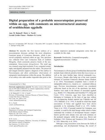
Naturwissenschaften (2008) 95:493–500DOI 10.1007/s00114-008-0347-2ORIGINAL PAPERDigital preparation of a probable neoceratopsian preservedwithin an egg, with comments on microstructural anatomyof ornithischian eggshellsAmy M. Balanoff & Mark A. Norell &Gerald Grellet-Tinner & Matthew R. LewinReceived: 26 September 2007 / Revised: 17 December 2007 / Accepted: 12 January 2008 / Published online: 27 February 2008# Springer-Verlag 2008Abstract We describe the first known embryo of aneoceratopsian dinosaur, perhaps the most ubiquitousLaurasian group of Cretaceous dinosaurs, which is preserved completely enclosed within an egg. This specimenwas collected from Late Cretaceous beds of southernMongolia, which commonly preserve fossils of the neoceratopsian, Yamaceratops dorngobiensis. The small eggwas scanned using high-resolution X-ray computed tomography and digitally prepared from the matrix. The preservedand imaged elements support a diagnosis of the embryo toNeoceratopsia and allow preliminary observations ofontogenetic transformations within this group. The additionof an embryo also adds another important data point to theCommunicated by G. MayrElectronic supplementary material The online version of this article(doi:10.1007/s00114-008-0347-2) contains supplementary material,which is available to authorized users.A. M. Balanoff (*) : M. A. NorellDivision of Paleontology, American Museum of Natural History,Central Park West at 79th Street,New York, NY 10024, USAe-mail: abalanoff@amnh.orgM. A. Norelle-mail: norell@amnh.orgG. Grellet-TinnerDepartment of Geology and Geological Engineering,South Dakota School of Mines,Rapid City, SD 57701, USAe-mail: Gerald.grellet-tinner@sdsmt.eduM. R. LewinDepartment of Emergency Medicine, University of California,San Francisco School of Medicine,San Francisco, CA 94143, USAe-mail: aplysia99@yahoo.comalready impressive postnatal ontogenetic series that areavailable for this clade.Keywords Ornithischia . Computed tomography .Eggshell microstructure . EmbryoIntroductionNeoceratopsia is a diverse group of ornithischian dinosaurs thatincludes basal, relatively primitive forms like Liaoceratops, aswell as the more familiar large, derived ceratopsids (e.g.,Triceratops). Despite often being one of the most commonelements of Laurasian Cretaceous dinosaur faunas (Dodsonet al. 2004), Neoceratopsia completely lacks a record ofindisputable embryos (i.e., found in ovo—contained within anegg). Previous neoceratopsian material was consideredembryonic based on the size of the specimens, but identifications were made in the absence of associated eggshell(e.g., Bohlin 1953; Maryańska and Osmólska1975; Dong andCurrie 1993). Likewise, eggs have been classified asbelonging to Ceratopsia on the basis of egg type withoutany accompanying skeletal material (see Carpenter and Alf1994; Mikhailov et al. 1994). It should be noted that simpleassociations of bones and eggshell have been shownpotentially to be misleading (Norell et al. 1995, GrelletTinner et al. 2006). If prenatal material for Ceratopsia wereavailable, it would be the ideal lineage within which to betterunderstand the evolution of development because comprehensive growth series already exist, and work continues onthe postnatal ontogeny within this group (e.g., Brown andSchlaikjer 1940; Erickson and Tumanova 2000; Goodwin etal. 2006; Makovicky et al. 2006).Although a long association exists between the fields ofpaleontology and developmental biology, the record of
494described embryonic non-avian dinosaurs remains sparse(e.g., Horner and Currie 1994; Norell et al. 2001;Varricchio et al. 2002; Reisz et al. 2005; Salgado et al.2005). Moreover, the connection between these disciplinestypically is restricted to observations of developmentalpatterns in living taxa used to explain patterns seen in thefossil record. Relatively few studies concentrate on understanding the patterns of developmental trajectories acrossclades. Nevertheless, the recent surge in the number ofdescriptive works on dinosaur embryos bodes well for ourunderstanding of the evolution of development within thislong and interesting lineage. An increased concentration indevelopmental morphology on the part of paleontologists,the discovery of new fossil embryos, as well as newtechnologies with which to visualize these unique specimens will aid in the continued endeavor to understandfossil dinosaur embryos.In this study, we use high-resolution X-ray computedtomography (HRCT) to digitally prepare and subsequentlydescribe a fossilized egg, which contains an embryonicskeleton (Fig. 1). This specimen represents the firstindisputable embryonic specimen that can be diagnosedwith any confidence to Neoceratopsia. Furthermore, ananalysis of the microstructural anatomy of the eggshell wasperformed to associate this specimen with a specificeggshell type. This embryo in ovo, preserved within theegg, provides a new and important data point on theontogenetic trajectory of Neoceratopsia with which we canbegin to make observations of this lineage’s growth anddevelopment that eventually could be used to shed light onthe larger dinosaurian clade.Fig. 1 a Photograph of IGM100/2010; b three-dimensionaldigital rendering of IGM 100/2010 in same orientation presented in a, with eggshell andmatrix rendered semitransparent. Skeletal elementsare opaqueNaturwissenschaften (2008) 95:493–500Materials and methodsIGM 100/2010 (Institute of Geology, Mongolia) wasdiscovered as an isolated egg in the Khugenetslavkant locality(eastern Gobi Desert, Dorngobi Aimag, Mongolia) that is LateCretaceous in age (Eberth et al., unpublished). The sedimentsof this locality consist largely of a coarse sandstone that isinterpreted as being fluvial in nature (see Makovicky andNorell 2006). The fauna at this locality is dominated by theneoceratopsian, Yamaceratops dorngobiensis, and alsoincludes remains of primitive ornithopods, troodontids,dromaeosaursids, and several kinds of mammals and lizards.The egg is small (total volume of 28.5 cm3), symmetricaland elongate ( 4.75 cm greatest length, 2.23 cm greatestwidth; Fig. 1), and is essentially complete except for onebroken pole. The break reveals the presence of fragile bonescontained within a coarse sandstone matrix that fills the egg.Several other isolated eggs of identical morphology, but notknown to contain embryos, also have been recovered fromthe locality.The small size and fragility of the skeletal remainspreclude physical preparation of IGM100/2010. Instead, thespecimen was scanned using HRCT, and the embryonicskeleton was digitally removed from the matrix (Fig. 2).Digital preparation revealed a large number of bonesrecognizable in the HRCT slices by their hollow naturethat could not otherwise be observed.The entire egg was HRCT-scanned along its long axis atThe University of Texas at Austin. The scan resulted in atotal of 1,296 sequential images. Scanning parametersinclude a slice thickness of 0.036 mm and interslice spacing
Naturwissenschaften (2008) 95:493–500495Fig. 2 Two views of the threedimensional digital rendering ofskeletal elements of IGM 100/2010 with eggshell and matrixrendered transparent. bc braincase, cv cervical vertebra, ffemur, fib fibula, h humerus, pdpredentary, qu quadrate, sqsquamosal, tar tarsals, tib tibia.Scale bar equals 5 mm(i.e., z-spacing) of 0.036 mm. The field of reconstruction is32 mm, and resultant image size is 1,024 1,024 pixels.These parameters yield an interpixel spacing (i.e., x-spacingand y-spacing) of 0.031 mm.Image processing was done in the program VGStudioMax 1.2.1. The two-dimensional slice images werereduced from 1,024 1,024 pixels by one third to 768 768 pixels so that they could more easily be loaded andmanipulated. Image processing of these data resulted inseparate three-dimensional volume renderings of eachisolated embryonic bone without any physical preparationof the egg (see Balanoff and Rowe 2007 for a morecomprehensive description of the methods). All originaldata and image processing are available for viewing on thewebsite Digital Morphology (http://www.DigiMorph.org/specimens/neoceratopsian egg).For the microstructural analysis of the egg, eggshellfragments were removed from the broken pole of IGM 100/2010. Two fragments were gently cleaned with air and thenbroken into two smaller fragments that were immediatelygold-coated to preserve their integrity, avoid contaminationbefore examination, and facilitate scanning electron microscopy (SEM) observations. SEM examinations wereperformed at 12–15 KVs on a Hitachi H-7000 FAtransmission electron microscope and scanning transmission electron microscope.ResultsMuch of the embryonic skeleton is present, and approximately 40 elements were digitally prepared from all regionsof the skeleton including the skull, vertebral column, andfore- and hindlimbs (Fig. 2). Because of the extremelysmall size of the specimen (many bones being on the scaleof 1 mm) and early stage of development, some of theelements are not recognizable. The majority of the skeletonhas settled to one side of the egg yet remains semiarticulated, a taphonomic feature indicating that IGM 100/2010 was filled with sediment before extensive decomposition or resorption could occur (Dong and Currie 1993;Sato et al. 2005). Although many skeletal elements are inan advanced state of ossification, the relatively small size ofthe embryo compared to the egg volume suggests thathatching was not imminent (Ricklefs and Starck 1998;Starck and Ricklefs 1998); however, a more rigorousanalysis of the ontogenetic maturity of this specimen isforthcoming. The hindlimbs are preserved in articulation(Electronic supplementary material; Fig. 2). Other regionsof the skeleton, although not in articulation, are positionedroughly where they would be expected in life.Cranial elements such as part of the braincase, quadrate,and predentary are easily discernable. Skeletal immaturityis particularly evident in the recovered braincase, whichconsists of the exoccipitals and supraoccipital. Theseelements are closely associated, but not yet fused (Fig. 3eand f), and have been identified as being a portion of thebraincase based on their overall morphology, lack of aneural spine, and the extremely large opening (foramenmagnum). Cervical vertebrae are also present in thisspecimen and have elongate neural spines and relativelysmaller openings for the neural canal (Fig. 2). The generalmorphology of the braincase is similar to immatureProtoceratops andrewsi in that the paroccipital processes
496Naturwissenschaften (2008) 95:493–500Fig. 3 a Digital rendering oflateral view of quadrate of IGM100/2010. Scale bar equals1 mm. b Photograph of lateralview of quadrate of adultProtoceratops (IGM 100/1246).Scale bar equals 3 cm. c Digitalrendering of right lateral view ofpredentary of IGM 100/2010.Scale bar equals 500 μm.d Photograph of right lateralview of predentary of adultProtoceratops (IGM 100/1113).Scale bar equals 1 cm. e Digitalrendering of posterior view ofbraincase of IGM 100/2010.Scale bar equals 5 mm.f Photograph of posterior viewof braincase of adultProtoceratops (IGM 100/2012).Sutures between the exoccipitaland supraoccipital are outlinedin red. Scale bar equals 1 cmdo not reach as far laterally relative to the adult and theforamen magnum remains relatively large. The braincase issimilar to other described juveniles (see Goodwin et al.2006) as well as that of fully mature P. andrewsi andYamaceratops dorngobiensis in that the supraoccipitalforms the dorsal margin of the foramen magnum (Brownand Schlaikjer 1940; Makovicky and Norell 2006; Fig. 3eand f). It is not apparent if the exoccipital contribution tothe occipital condyle is present at this early ontogeneticstage. The predentary of IGM 100/2010 closely resemblesthat of Y. dorngobiensis in that it is a relatively solid bonewith a slightly concave buccal surface. The ventral processis missing probably due to its young ontogenetic age. Thequadrate possesses a dorsal shaft that is elongate andcompressed rostrocaudally, as seen in other basal neoceratopsians. The pterygoid flange extends medially fromthe ventral portion of the quadrate, but does not extend thelength of the shaft as it does in the adult of Y. dorngobiensis(Makovicky and Norell 2006).The postcranium is better ossified than the cranium, andcross-sectional data derived from HRCT slices suggest thatthe epiphyses of the long bones are formed in calcifiedcartilage (Horner and Currie 1994). Recovered vertebraeare derived from the anterior part of the cervical series asindicated by the size and morphology of the neural arches,particularly that of the expanded neural spines (Fig. 2).Included among the anterior cervical vertebrae is the axis(Fig. 2) that possesses an expanded, hatchet-shaped neuralspine that extends over the succeeding vertebra. Theproximal ends of the femora and distal ends of the tibiaeand fibulae are broken, and therefore, measurements werenot obtainable from the hindlimb. Although these measurements are not available, the femur clearly exceeds thelength of the humerus, which is 15.6 mm. Postcranialelements are morphologically similar to neoceratopsians,and the limbs possess a gracile aspect that is strikinglysimilar to that exhibited by younger specimens of P.andrewsi (Brown and Schlaikjer 1940). There is a smallprocess on the midshaft of the femur that we interpret as thefourth trochanter based on shape and spatial arrangement.This feature is difficult to discern in the three-dimensionalreconstructions but much more easily seen in the originalCT slices (slice 259 of the horizontal movie; http://www.DigiMorph.org/specimens/Neoceratopsian egg).
Naturwissenschaften (2008) 95:493–500Phylogenetic diagnosisComprehensive growth series are only known in extinctdinosaurs in a few exceptional cases (e.g., Protoceratops,Brown and Schlaikjer 1940; Maiasaura, Horner et al.2000). The lack of comprehensive ontogenetic data inclosely related clades combined with the generally poor stateof preservation (due to the presence of poorly ossified bonesand a lack of developed morphology) makes it extremelydifficult to diagnose particular fossil embryos to specific taxa(see Norell et al. 2001). The digitally isolated elements ofIGM 100/2010, however, allow the specimen to be diagnosed to the successively nested clades of Ornithischia andNeoceratopsia based on derived cranial features. Thepresence of a predentary bone (Fig. 3c and d) unequivocallydiagnoses the specimen as an ornithischian (Sereno 1986).IGM 100/2010 is diagnosed further to Neoceratopsia basedon a quadrate shaft that is straight in lateral view (Fig. 3a andb; Makovicky 2002; Xu et al. 2002). This feature isdescribed by Makovicky (2002) as being diagnostic ofNeoceratopsia and consisting of a quadrate shaft that isstraight in lateral view from articulation with the squamosalto the condylar end. This character has not been surveyed inall ornithischians, but rather only ceratopsians and a fewoutgroups (i.e., Ornithopoda and Pachycephalasauria).Therefore, the diagnosis to Neoceratopsia is tenuous,pending a more comprehensive study of this character withinall of Ornithischia.It should be noted that the above identification is madeusing a strictly apomorphy-based approach. Other featuresprovide additional support for IGM 100/2010 as a neoceratopsian. Most notably, the dorsally concave shape ofthe predentary is shared with all other neoceratopsians(Fig. 3c and d), in contrast to the much straighterpredentary of ornithopods and basal ceratopsians (Normanet al. 2004; You and Dodson 2004). The predentary alsolacks the severe scoop-like shape characteristic of formssuch as Leptoceratops gracilis, P. andrewsi, and ceratopsids(Makovicky and Norell 2006), although this feature couldbe correlated with the young ontogenetic maturity of thespecimen. The lack of this character also does not prohibitthe specimen from being a more derived ceratopsian.On the postcranial skeleton, the presence of a fourthtrochanter on the midshaft of the femur further supports adiagnosis to Ornithischia (Sereno 1986). Furthermore, thereduction of the fourth trochanter distinguishes IGM 100/2010 from ornithopods, which possess well-developedfourth trochanters even at the earliest known stages of theirdevelopment (Horner and Currie 1994). The fourth trochanter, however, also is reduced in immature specimens ofPsittacosaurus; therefore, this feature is useful only todistinguish the specimen as a ceratopsian.497These similarities suggest that this specimen is aneoceratopsian. However, the similarities are not includedin the explicit diagnosis of this specimen because theyhave not been established as derived for these taxa in amore global phylogenetic analysis. Any questionsaddressed using this specimen must be restricted toNeoceratopsia or a more inclusive clade to avoid introducing circularity.Embryonic growth within NeoceratopsiaAlthough a more refined diagnosis cannot currently besupported, even with this less refined diagnosis to the levelof Neoceratopsia, observations concerning the ontogeneticdevelopment of neoceratopsians in general still can bemade. The paroccipital processes have a positive correlationwith ontogenetic maturity within Ceratopsia, as there is amarked distal expansion of the processes through timebeginning with the rudimentary processes present in theembryo (see Maryańska and Osmólska 1975 and Makovicky2002). There is a negative allometry between the size of theforamen magnum relative to the rest of the braincase throughontogeny (Fig. 3e and f). This pattern supports a broaderpattern that is derived for vertebrates in general in which thematuration of the nervous system is delayed relative to theremainder of the skull (see Emerson and Bramble 1993).The morphology of the predentary undergoes perhaps one ofthe most dramatic changes (Fig. 3c and d). The ventralprocess is not present in the embryo, yet is present in theearliest postnatal examples of ceratopsians, which suggeststhat this process, if not broken, may ossify sometime in lateprenatal development or the earliest stages of postnataldevelopment.Several transformations in the postcranial developmentof Neoceratopsia also are made evident by IGM 100/2010(Fig. 2). The anterior cervical vertebrae of the embryo arefree and not yet fused into a syncervical, as in basalneoceratopsians, indicating that this fusion occurs in lateprenatal or early postnatal development. The fore- andhindlimbs are very gracile elements in the embryo withlittle curvature along their long axis, whereas the sameelements of adult basal neoceratopsians are more curved,robust, and relatively short. The limbs thus demonstrate anegative allometry with the body as a whole (see alsoChinnery 2004). Although this study presents only a fewexamples of changes in characters throughout ontogeny,work continues to be done on growth series withinCeratopsia (e.g., Erickson and Tumanova 2000; Makovickyand Norell 2006; Makovicky et al. 2006; Goodwin et al.2006; Horner and Goodwin 2006), and this specimenprovides an important new addition to the comprehensiveseries that are already available.
498Naturwissenschaften (2008) 95:493–500The eggshell of IGM 100/2010 is very friable and coveredby siliciclastic sediments that likely are the cause ofmoderate acidification of the carbonate shell (Faccio1994). Nevertheless, sparse and randomly distributed nodes(Fig. 4a and b) are preserved on the outer eggshell surface,but no pore canals or apertures were observed on thestudied samples. A secondary epitaxial growth blankets theinner shell surface and probably is due to bacterialmediation originating from the decomposing embryo andmobilization of calcium ions (Grellet-Tinner 2005; Fig. 4c).We estimate, based on four digital measurements, the totalthickness of the eggshell to be 183–184 μm.The eggshell is divided into three structural layers(Fig. 4c and d). The innermost layer averages 61–62 μmand displays semicircular spherulites at its base thatsurround the loci of eggshell units (Fig. 4c). The spherulitesform layer 1 and consist of blocky, blade-shaped crystals(Fig. 4c) which are vertically elongate. Layer 2 (81 μm inthickness) is not clearly demarcated from layer 1 but isdifferentiated by larger and more numerous vesicles(although the latter may be exaggerated by dissolutionduring diagenesis) and a sub-horizontal crystal orientation.Layer 2 is capped by a third structural layer that is 40 μmwith a vertical crystallographic orientation similar to that oflayer 1.Eggs with clear taxonomic association that exhibit atrilaminated eggshell structure have been referred to aviantheropods (Schweitzer et al. 2002; Buffetaut et al. 2005;Grellet-Tinner 2006; Grellet-Tinner and Makovicky et al.2006). However, these theropod eggs are always asymmetric (Grellet-Tinner et al. 2006). The symmetry found inIGM 100/2010 is a feature normally associated with basallydiverging reptiles (Reptilia sensu Gauthier et al. [1988])such as crocodilians, snakes, and turtles. Because the dataFig. 4 a SEM of surficial ornamentation. Note the node on the leftand the overall scaled appearance of the egg surface due to post-burialacidification. The dimensions of the nodules approximate 2.9 μm (atthe base) and 1.9 μm in height. b SEM of layer 1 (L1), the innermoststructural layer. Although very thin, fragile, and partially weathered,the crystals in L1 still display a vertical orientation and a bladedshape. c SEM of the entire eggshell thickness. Note that L1 isdistinguishable from layer 2 (L2) by the orientation and shape of itscalcite crystals and also by lesser amount of vesicles. Despitemoderate diagenetic alteration, some of the spherulites (SP) at thebase of the eggshell units are still visible. L2 differs from L1 by itsnearly horizontally orientated smaller crystals and by more numerousand larger vesicles. Layer 3 (L3) is proportionally wide and differsfrom L2 by its vertically orientated crystals. Note the epitaxial calciticgrowth (EP) that underlies L1, a feature likely due to mobilization ofCaCO3 during diagenesis. d Transmitted light micrograph of a thinsection through the entire eggshell thickness illustrating the threestructural layers of the eggshellEggshell microstructure
Naturwissenschaften (2008) 95:493–500from eggshell microstructure and egg shape conflict, it isevident that more than one character system is required toaccurately diagnose fossil eggs that are not taxonomicallyidentified by clearly associated skeletal remains (i.e.,embryos, brooding adults, or eggs present in the bodycavity; Grellet-Tinner 2006; Grellet-Tinner and Makovickyet al. 2006).ConclusionsFossil embryos are difficult to study in a phylogeneticcontext because their early state of ossification and smallsize complicates the identification of apomorphic characters. Yet, even if taxonomic identifications cannot be madeto refined levels, embryonic specimens can still be used tosupport statements about broader patterns of development.In particular, they provide an unique opportunity to addresslarge-scale evolutionary questions such as the evolution ofdevelopmental constraints, the evolution of growth rates,the acquisition and allometric growth of osteologicalfeatures, and the paleobiological properties of organisms(e.g., altricial vs. precocial), but these specimens must beplaced within a larger context of where they sit within theirown developmental progression as well as where they sit onthe tree of life (phylogenetic diagnosis). These issues wouldseem to be intuitive to any analysis that includes embryonicfossils; however, such matters have not been dealt withexplicitly in the literature.Using a strictly apomorphy-based approach, IGM 100/2010 is diagnosed unequivocally to Ornithischia based onthe presence of a predentary and to Neoceratopsia based on astraight quadrate shaft. Thus, this unique specimen representsthe first associated embryo and egg for this importantdinosaur clade and provides a basis to begin to understandthe earliest parts of development within Neoceratopsia.The taxonomic identification allows the association ofthis clade of dinosaurs with an eggshell type, whichpreviously was identified based on eggs that lacked skeletalremains (Mikhailov 1995, 1997). Eggshell charactersusually are fairly stable within higher taxa of dinosaurs(Grellet-Tinner et al. 2006). The presence of three layerssuggests that the three groups of asymmetric and largereggs with two microstructural layers that previously wereassigned to protoceratopsids (Mikhailov et al. 1994;Mikhailov 1995) cannot presently be assigned with certainty to Neoceratopsia.This study illustrates that despite the long history ofdescriptive embryology (extending back well over150 years; see Russell 1916), future attention should bedirected toward this endeavor. There still exists a paucity ofavailable descriptions useful to providing both an ontogenetic and phylogenetic diagnosis for fossil embryos. The499wider the taxonomic distribution of these data, the morerefined the conclusions that can be drawn from the fossilrecord. If the fossil record can actually shed light on theevolution of development, then such refinement will bebeneficial to paleontologists, neontologists, and biology ingeneral. Fossil embryos provide a direct window into theevolutionary history of development. Ornithischians, andspecifically the growing number of ontogenetically informative specimens of ceratopsians, provide an excitingopportunity to better understand the evolution of development in dinosaurs.Acknowledgments GGT would like to acknowledge E. Duke at theAnalytical Facilities of Engineering and Mining Experiment Station ofSDSMT for the use of the SEM. MRL would like to thank E.Kernberg, UCSF, for preliminary radiological analyses of thespecimen. Mick Ellison provided much help in the preparation offigures. Jessie Maisano assisted in the preparation of the web-basedimagery accompanying this paper. Thanks to Gabe Bever, PeterMakovicky, and three anonymous reviewers for their useful comments. CT scanning was performed by the HRCT Facility at TheUniversity of Texas at Austin. Support for this project came from theAmerican Museum of Natural History, Division of Paleontology andDEES, Columbia University.ReferencesBalanoff AM, Rowe T (2007) Osteological description of anembryonic skeleton of the extinct elephant bird, Aepyornis(Palaeognathae: Ratitae). Soc Vert Paleontol Memoir 9, J VertPaleontol 27(suppl. to 4):1–53Bohlin B (1953) Fossil reptiles from Mongolia and Kansu. The SinoSwedish Expedition, Publication no. 37, pp 1–105Brown B, Schlaikjer EM (1940) The structure and relationships ofProtoceratops. Ann NY Acad Sci 40:133–266Buffetaut E, Grellet-Tinner G, Suteethorn V, Cuny G, Tong H, KoširA, Cavin L, Chitsing S, Griffiths PJ, Tabouelle J, Le Loeuff J(2005) Minute theropod eggs and embryo from the LowerCretaceous of Thailand and the dinosaur-bird transition. Naturwissenschaften 92:477–482Carpenter K, Alf K (1994) Global distribution of dinosaur eggs, nests,and babies. In: Carpenter K, Hirsch KF, Horner JR (eds)Dinosaur eggs and babies. Cambridge University Press, Cambridge,pp 15–30Chinnery B (2004) Morphometric analysis of evolutionary trends inthe ceratopsian postcranial skeleton. J Vert Paleontol 24:591–609Dodson P, Forster CA, Sampson SD (2004) Ceratopsidae. In:Weishampel DB, Dodson P, Osmólska H (eds) The Dinosauria.University of California Press, Berkeley, pp 494–513Dong Z-M, Currie PJ (1993) Protoceratopsian embryos from InnerMongolia, People’s Republic of China. Can J Earth Sci 30:2248–2254Emerson SB, Bramble DM (1993) Scaling, allometry, and skulldesign. In: Hanken J, Hall BK (eds) The skull, volume 3:functional and evolutionary mechanisms. University of ChicagoPress, Chicago, pp 384–416Erickson GM, Tumanova TA (2000) Growth curve of Psittacosaurusmongoliensis Osborn (Ceratopsia: Psittacosauridae) inferred fromlong bone histology. Zool J Linn Soc–Lond 130:551–566
500Faccio G (1994) Dinosaurian eggs from the upper Cretaceous of Uruguay.In: Carpenter K, Hirsch KF, Horner JR (eds) Dinosaur eggs andbabies. Cambridge University Press, Cambridge, pp 47–55Gauthier J, Kluge AG, Rowe T (1988) Amniote phylogeny and theimportance of fossils. Cladistics 4:105–209Goodwin MB, Clemens WA, Horner JR, Padian K (2006) Thesmallest known Triceratops skull: new observations on ceratopsid cranial anatomy and ontogeny. J Vert Paleontol 26:103–112Grellet-Tinner G (2005) The membrana testacea of titanosauriddinosaur eggs from Auca Mahuevo (Argentina): implicationsfor the exceptional preservation of soft tissue in Lagerstätten. JVert Paleontol 25:99–106Grellet-Tinner G (2006) Phylogenetic interpretation of eggs andeggshells: implications for oology and Paleognathae phylogeny.Alcheringa 30:130–180Grellet-Tinner G, Makovicky PJ (2006) A possible egg of thetheropod Deinonychus antirropus: phylogenetic and biologicalimplications. Can J Earth Sci 43:705–719Grellet-Tinner G, Chiappe L, Bottjer D, Norell M (2006) Paleobiologyof dinosaur eggs and nesting behaviors. Palaeogeogr PalaeoclPalaeoecol 232:294–321Horner JR, Currie PJ (1994) Embryonic and neonatal morphology andontogeny of a new species of Hypacrosaurus (Ornithischia,Lambeosauridae) from Montana and Alberta. In: Carpenter K,Hirsch KF, Horner JR (eds) Dinosaur eggs and babies. Cambridge University Press, Cambridge, pp 312–336Horner JR, Goodwin MB (2006) Major cranial changes duringTriceratops ontogeny. Proc R Soc B 273:2757–2761Horner JR, de Ricqlés A, Padian K (2000) Long bone histology of thehadrosaurid dinosaur Maiasaura peeblesorum: growth dynamicsand physiology based on an ontogenetic series of skeletalelements. J Vert Paleontol 20:115–129Makovicky PJ (2002) Taxonomic revision and phylogenetic relationships of basal neoceratopsia (Dinosauria: Ornithischia). Ph.D.dissertation, Columbia University, p 279Makovicky PJ, Gao KQ, Zhou CF, Erickson G (2006) Ontogeneticchanges in
The University of Texas at Austin. The scan resulted in a total of 1,296 sequential images. Scanning parameters include a slice thickness of 0.036 mm and interslice spacing Fig. 1 a Photograph of IGM 100/2010; b three-dimensional digital rendering of IGM 100/ 2010 in same orientation pre-sented in a, with eggshell and matrix rendered semi .