Transcription
Supplement to May/June 2019USINGPHOTODYNAMICTHERAPYIN 2019:Current Concepts forReal-World UseRishi P. Singh, MD, ModeratorVivek Chaturvedi, MDDavid S. Dyer, MD, FACSAmani Fawzi, MDDistributed withProvided byA CME activity provided by Evolve Medical Education LLCSupported by a grant from Bausch Lomb
Using Photodynamic Therapy in 2019:Current Concepts for Real-World UseRelease Date: May 15, 2019Expiration Date: May 15, 2020FACULTYRISHI P. SINGH, MDMODERATORStaff Physician, Cole Eye Institute,Cleveland ClinicAssociate Professor ofOphthalmologyCleveland Clinic LernerCollege of MedicineCleveland, OhioVIVEK CHATURVEDI, MDIllinois Retina AssociatesAssistant Professor of OphthalmologyRush Medical CenterChicago, IllinoisCONTENT SOURCEThis continuing medical education (CME) activity capturescontent from a virtual round table discussion that occurred onMarch 28, 2019.ACTIVITY DESCRIPTIONVerteporfin for photodynamic therapy (PDT) has beenapproved for decades as a treatment for retinal disorders. Eyecare professionals need to understand all the real-world clinicalscenarios in which the use of PDT alone or in combination withanti-vascular endothelial growth factor (VEGF) therapy can be amore efficacious treatment option than anti-VEGF monotherapy.TARGET AUDIENCEThis certified CME activity is designed for retina specialists andophthalmologists involved in the management of retinal diseases.LEARNING OBJECTIVESUpon completion of this activity, the participant should beable to: Summarize the clinical benefits of PDT in patients with retinaldisorders Design a treatment regimen based on a personalizedmedicine approach for patients who do not respond adequatelyto anti-VEGF injections2 SUPPLEMENT TO RETINA TODAY MAY/JUNE 2019DAVID S. DYER, MD, FACSRetina AssociatesShawnee Mission, KansasAMANI FAWZI, MDCyrus Tang and Lee Jampol Professorof OphthalmologyNorthwestern UniversityEvanston, Illinois Identify methods for effective PDT delivery in clinic settings,including dosing, infusion periods, and determination oftreatment size Differentiate the benefits of half fluence PDT and full fluencePDT on a real-world populationGRANTOR STATEMENTSupported by a grant from Bausch Lomb.ACCREDITATION STATEMENTEvolve Medical Education LLC (Evolve) is accredited bythe Accreditation Council for Continuing Medical Education(ACCME) to provide continuing medical education for physicians.CREDIT DESIGNATION STATEMENTEvolve designates this enduring material for a maximum of1 AMA PRA Category 1 Credit . Physicians should claim only thecredit commensurate with the extent of their participation inthe activity.TO OBTAIN CREDITTo obtain credit for this activity, you must read the activity in its entirety and complete the Pretest/Posttest/ActivityEvaluation/Satisfaction Measures Form, which consists of a seriesof multiple choice questions. To answer these questions online
and receive real-time results, please visit evolvemeded.com andclick ent.Upon completing the activity and self-assessment test, you mayprint out a CME credit letter awarding 1 AMA PRA Category1 Credit Alternatively, please complete the Posttest/ActivityEvaluation/Satisfaction Form and mail or fax to Evolve MedicalEducation LLC, 353 West Lancaster Avenue, Second Floor,Wayne, PA 19087; Fax: (215) 933-3950.DISCLAIMERThe views and opinions expressed in this educational activity are those of the faculty and do not necessarily represent theviews of Evolve, Retina Today, or Bausch Lomb.DIGITAL EDITIONTo view the online version of the material, please visit go ement.DISCLOSURE POLICYIt is the policy of Evolve that faculty and other individualswho are in the position to control the content of this activity disclose any real or apparent conflict of interests relating tothe topics of this educational activity. Evolve has full policies inplace that will identify and resolve all conflicts of interest priorto this educational activity.The following faculty/staff members have the following financial relationships with commercial interests:Rishi P. Singh, MD, and/or spouse; has had a financialagreement or affiliation during the past year with thefollowing commercial interests in the form of Consultant: CarlZeiss Meditec, Genentech, Novartis/Alcon and RegeneronPharmaceuticals/Bayer. Grant/ Research Support: Apellis.Vivek Chaturvedi, MD, and/or spouse; has had a financialagreement or affiliation during the past year with the followingcommercial interests in the form of Advisory Board: Genentech.Consultant: Allergan plc. Stock/Shareholder: Covalent and USRetina.David S. Dyer, MD, FACS, and/or spouse; has no financialrelationships with commercial interests.Amani Fawzi, MD, and/or spouse; has no financialrelationships with commercial interests.EDITORIAL SUPPORT DISCLOSURESErin K. Fletcher, MIT, director of compliance and education;Susan Gallagher-Pecha, director of client services and projectmanagement; Cassandra Richards, director of education development, Evolve; and Michelle Dalton, writer, have no financialrelationships with commercial interests. Jaya Kumar, MD, peerreviewer, has had a financial agreement or affiliation during thepast year with the following commercial interests in the form ofConsultant: Allergan.OFF-LABEL STATEMENTThis educational activity may contain discussion of publishedand/or investigational uses of agents that are not indicated bythe FDA. The opinions expressed in the educational activity arethose of the faculty. Please refer to the official prescribing information for each product for discussion of approved indications,contraindications, and warnings.MAY/JUNE 2019 SUPPLEMENT TO RETINA TODAY 3
PRETEST QUESTIONSPlease complete prior to accessing the material and submit with Posttest/ActivityEvaluation/Satisfaction Measures Instructions for CME Credit.1. PLEASE RATE YOUR CONFIDENCE ON YOUR ABILITY TO APPLY UPDATES INUSING PHOTODYNAMIC THERAPY (PDT) IN THE CLINIC. (BASED ON A SCALEOF 1 TO 5, WITH 1 BEING NOT AT ALL CONFIDENT AND 5 BEING EXTREMELYCONFIDENT.)a. 1b. 2c. 3d. 4e. 52. PLEASE RATE HOW OFTEN YOU INTEND TO APPLY ADVANCES IN USING PDT INTHE CLINIC. (BASED ON A SCALE OF 1 TO 5, WITH 1 BEING NEVER AND 5 BEINGALWAYS.)a. 1b. 2c. 3d. 4e. 53 PDT SHOULD BE CONSIDERED IN PATIENTS WITH THE FOLLOWING CONDITIONSEXCEPT:a. Polypoidal choroidal vasculopathyb. Myopic choroidal neovascularizationc. Dome-shaped maculad. Central serous chorioretinopathy (CSR)4. ACCORDING TO THE EVIDENCE, IN WHICH CLINICAL SITUATION WOULD FULLFLUENCE PDT BE APPROPRIATE?a. Full-fluence PDT is never appropriate due to the potential adverseevents.b. Full-fluence PDT is appropriate in all clinical scenarios because it’smore effective than half fluence.c. For patients with central serous chorioretinopathy.d. For patients with polypoidal choroidal vasculopathy who did notrespond to anti-VEGF treatment.5. THE PANELISTS ADVISE OPTIMIZING THE PDT WORKFLOW MAY BEST BEACCOMPLISHED WHEN .a. There is a PDT laser in at least one office locationb. There is a dedicated PDT expert in your practice, such as a nurse ortechnicianc. Patients needing PDT are scheduled on the different daysd. Angiogram and PDT occur on separate days of the month4 SUPPLEMENT TO RETINA TODAY MAY/JUNE 20196. THE FOOD AND DRUG ADMINISTRATION RECOMMENDS PATIENTS AVOID THESUN FOLLOWING PDT TREATMENT FOR .a. 2 daysb. 3 daysc. 4 daysd. 5 days7. ACUTE VISION LOSS CAN OCCUR AFTER PDT. WHAT PERCENTAGE OF PATIENTSHAVE ACUTE VISION LOSS FOLLOWING PDT ACCORDING TO THE VERTEPORFININ PHOTODYNAMIC THERAPY STUDY GROUP?a. 5%b. 4%c. 2%d. 1%8. WHEN DIAGNOSING A PATIENT FOR CSR, CLINICIANS SHOULD ASSESS FOR ALLTHE FOLLOWING RISK FACTORS EXCEPT:a. Caffeine useb. Steroid use, both by the patient and in the homec. Hypotensiond. Excessive stress/Type A personality9. AN ELECTRICIAN WITH RECURRENT CSR PRESENTS IN YOUR OFFICE. HE HASA HISTORY OF TOPICAL STEROID USE, GYNECOMASTIA, AND HYPERTENSION.HE IS VERY CONCERNED ABOUT HIS ABILITY TO WORK BECAUSE THE CSR ISLOCATED IN HIS DOMINATE EYE WITH BETTER VISION. WHAT IS AN ACCEPTABLEEVIDENCE-BASED COURSE OF ACTION TO FIRST TAKE WHEN TREATING THISPATIENT?a. Recommend half-fluence PDT immediatelyb. Take him off steroids, reemphasize the need to control hishypertensionc. Prescribe spironolactoned. Recommend half-fluence with anti-VEGF10. AN APPROPRIATE TIME FOR REEVALUATION AFTER PDT IS .a. 3 monthsb. 2 yearsc. 2 to 4 weeksd. 4 to 6 weeks
USING PHOTODYNAMIC THERAPY IN 2019:Current Concepts for Real-World UseUsing Photodynamic Therapy in 2019:Current Concepts for Real-World UseVerteporfin photodynamic therapy (PDT) has been approved for decades as a treatment for retinal disorders such as predominantly classic subfovealchoroidal neovascularization due to age-related macular degeneration (AMD), pathologic myopia, or presumed ocular histoplasmosis. Over the past fewyears, its use and role in ophthalmic care has evolved to include central serous chorioretinopathy, polypoidal choroidal vasculopathy, and peripapillarychoroidal neovascularization. Although PDT may not be a treatment clinicians use daily, it is a valuable tool to have in their armamentarium. Thefollowing CME-approved activity gathers thought leaders to discuss its real-world utility, clinical benefits, and pearls for use.—Rishi P. Singh, MD, ModeratorREAL-WORLD USES OF PHOTODYNAMIC THERAPYQRISHI P. SINGH, MD: PDT is a selective vasoocclusivetreatment targeting choroidal vascular abnormalities.Although it has utility in other medical specialties, it wasoriginally developed for use in ophthalmology to treat neovascular age-related macular degeneration (AMD).1,2 Over the years, itsusage has evolved to treat a range of other chorioretinal conditions such as choroidal hemangioma,3 central serous chorioretinopathy (CSR),4,5 polypoidal choroidal vasculopathy (PCV),6,7 andperipapillary choroidal neovascularization (CNV).8 There are manyvariables that impact the effectiveness of PDT, including verteporfin dosing, fluence dosing, infusion periods, and the accuratedetermination of lesion size.9-13 What role does PDT have in yourpractice today?DAVID S. DYER, MD, FACS: PDT doesn’t have a large role in mypractice, but it is important to have at your disposal. I use it inpatients with CSR and PCV, and as a rescue therapy for lesions thataren’t fully responding to anti–VEGF treatment.14 It can also be usedin combination with anti-VEGF.The EVEREST study showed that PDT alone or in combinationwith ranibizumab can achieve a complete regression of polyps inpatients with symptomatic macular PCV compared with ranibizumab alone.6,7,15,16 The PLANET study compared intravitreal afliberceptmonotherapy with or without rescue PDT and found a 10.8 lettergain after 3 months, with polyp closure rates of 38.9%. Further, 81.7%of eyes had no active polyps.17,18Several studies have shown that the use of PDT in juxtafoveal andextrafoveal CNV can achieve long-term stabilization and an improvement of visual acuity.19,20 Han et al evaluated the use of anti-VEGFand PDT for juxtafoveal and extrafoveal CNV and found that combination therapy preserves good visual function and is well suited forsome cases of nonfoveal CNV.21I’ll also use PDT for either choroidal or capillary hemangioma.These aren’t common issues, but they do come up. I typically don’tuse PDT for myopic CNV because I feel the risk for developing atrophy is too significant,22 and I’ve had success with anti-VEGF therapyin these patients. That being said, I would consider PDT in a myopicpatient if they were unresponsive to anti-VEGF therapy and therewas not a subfoveal or juxtafoveal lesion. I’ll also consider PDT inpatients on anti-VEGF therapy who have had a recent stroke, just toget them through that 90-day window before we can resume antiVEGF treatments.23VIVEK CHATURVEDI, MD: On the whole, 75% of my PDT use is forchronic CSR and 25% is for polypoidal AMD or refractory, wet AMD. Ido not use PDT for patients with myopic CNV. For patients with wetAMD, I recommend walking them through how the medication worksand how it’s different from the monotherapy they’ve been receiving.AMANI FAWZI, MD: The bulk of my PDT patients have CSR andpolypoidal lesions. I also use it in hemorrhagic lesions and peripherallesions that are causing substantial exudate. I’ve also had successwith it for proliferative tumors that are causing a lot of exudatethat’s tracking through the macula, as well as in patients with vonHippel–Lindau peripheral lesions.24 If those lesions become toolarge and they are too peripheral to laser completely with thermallaser, then PDT is an option. Finally, I also use PDT in patients with adome-shaped macula. Those eyes sometimes have chronic subretinal fluid that doesn’t respond to other treatment. Data have shownthat myopic eyes associated with a dome-shaped macula and fovealserous retinal detachment may be responsive to PDT.25,26OPTIMIZING THE PDT WORKFLOWQDR. SINGH: The PDT workflow can be somewhat complicated due to the number of steps necessary. The typicalworkflow is as follows: A technician or nurse preps thepatient with an IV, performs the IV fluorescein angiography(IVFA)/indocyanine green (ICG), maintains the IV’s patency, mixesthe drug, and supervises the infusion. The clinician sees thepatient after the infusion, reviews the angiogram, and places thelaser. The technician or nurse then removes the IV and reviewsthe postoperative instructions with the patient. How do you optimize the workflow and scheduling for maximum efficiency?MAY/JUNE 2019 SUPPLEMENT TO RETINA TODAY 5
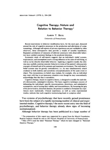
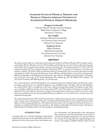
USING PHOTODYNAMIC THERAPY IN 2019:Current Concepts for Real-World UseCASE 1: CHRONIC CENTRAL SEROUS RETINOPATHY Presented by Dr. ChaturvediA 55-year-old man with chronic CSR came to me on a referralsuperior to the macula, and the ICG, which showed leakage all around(Figure 1). His scans showed a small amount of subretinal fluid, and we the fovea. I did a continuous set of four different targeted areas justdecided to observe. He was very concerned about his ability to worksuperior to the fovea in a box pattern over the course of the 83 secbecause his right eye was his dominant and better eye.onds. Four months after PDT, his VA was 20/80, but his fluid was goneHe was taking dextroamphetamine-amphetamine for adult(Figure 5). Figure 5 shows substantial loss of the outer retina as well asattention-deficit/hyperactivity disorder. During his next visit, we foundirregularities to the EZ junction.increased fluid. He was asymptomatic but his vision dropped to 20/30.How would you treat this patient?After discussing it with some of my partners, we decided to take himoff dextroamphetamine-amphetamine, feeling that there could beDr. Fawzi: I would have pushed for the PDT about a year earlier andmineralocorticoid effect in its use. His fluid resolved (Figure 2) a couple explained that we could treat all the areas outside the fovea in stagesof months later.and then reassess to see if he needs more. Unfortunately, when the fluidFour or 5 months later, however, his fluid returned. He was, again,sits for a long time, the photoreceptors suffer. I would have tried to treatasymptomatic but with 20/30 vision. This pattern continued the folthe superior areas and stay outside the fovea to see what happens.lowing year: the fluid resolved and then returned. At this point werecommended that he start rifampin,1 but he elected not to becauseDr. Dyer: I agree. I would have done multiple treatmentsof its contraindication with alcohol.around the fovea since it’s his best eye and since he’s hesitantWe then recommended reduced-fluence PDT,which he also declined out of worry about thepotential side effects and abrupt vision loss. This fluidcontinued to worsen (Figure 3), and we decided tostart him on oral eplerenone to reduce the fluid,which the literature supports.2-4 We know thatspironolactone works better for fluid reduction,but the risk profile is higher, especially in males withdecreased libido and gynecomastia.5 Spironolactoneis associated with dose-dependent sexual sideeffects. Both eplerenone and spironolactoneincrease potassium concentrations, although theeffect with spironolactone appears to be greaterthan with eplerenone.6His angiogram showed multiple areas of RPEstaining and small areas of leakage. The ICG showedFigure 1. Chronic CSR in a 55-year-old male, left and right eyes, respectively.diffuse congestion, diffuse leakage, and diffuse dilation throughout the posterior pole with multiplehot spots (Figure 4). We decided to increase theeplerenone dosage from 50 mg to 100 mg, and hecontinued to have persistent fluid. He’s still asymptomatic, with 20/30 vision. Seven months later, he stillhas persistent fluid, and we’re beginning to see someRPE changes; we decided to stop oral eplerenone. I’mpushing PDT strongly at this point, but he is still concerned about abrupt vision loss.On his next follow-up appointment, we see thathis visual acuity (VA) has dropped to 20/60 and heis symptomatic. We continue to monitor him foranother year and see no changes in the FA or ICG.He finally agrees to low-fluence PDT. My strategywas to develop a treatment based on what I saw onthe angiogram, which showed most of the leakageFigure 2. Fluid resolution after discontinued Adderall use.Figure 3. Increased fluid in after eplerenone 50 mg.6 SUPPLEMENT TO RETINA TODAY MAY/JUNE 2019
USING PHOTODYNAMIC THERAPY IN 2019:Current Concepts for Real-World UseCASE 1 (Continued)Figure 4. Angiogram and indocyanine green angiography.Figure 5. After PDT treatment.about treatingthe fovea directly.His vision loss isprobably becausehe just waited toolong before havingtreatment. I’ve triedmultiple medications,including the threethat you mentionhere, and no one hasresponded.Dr. Fawzi: I thinkthe only medicationthat works is withdrawing the steroidsif they’re on any. Wehaven’t had a goodexperience with anyrifampin or eplerenone either.Dr. Chaturvedi: I completely agree. I have not had much successwith systemic medications.Dr. Singh: Let’s talk a little bit about the diagnostic dilemma herewith this case. What do you ask about when you’re interviewingpatients with chronic CSR?Dr. Chaturvedi: I try to assess what, if anything, they are encountering or interacting with that could potentially exasperate their symptoms. I always ask the patient about steroid use, not just about thesteroids they are using but if anyone in the home I using steroid creamor if they have any children with a steroid inhaler. Although the linkbetween steroids and CSR is poorly understood, a number of caseshave reported CSR development or worsening in conjunction withtopical, oral, or inhaled steroids.7-9DR. DYER: Because we don’t use it on a daily basis, we bring thepatient back once we identify the need for PDT. It’s scheduledwithin a week to 10 days. We can do the PDT in any office, but wedo have a room in our main location that’s set up for PDT specifically. We try to centralize PDT unless it’s a long drive for the patient.I use half fluence for almost every indication, and base the lesion sizeon the angiogram.DR. SINGH: Something I’ve done to streamline the process is notremoving the IV if I’m doing a same-day angiogram. That way, I canuse the same IV for the PDT.I also talk to them about their personality, especially if there is nosteroid use, and how endogenous cortisol levels may impact theirdisease.10,11 Type A personality and stress are known risk factors forCSR.12 Most people do carry some degree of stress in their life, and it’sdifficult to reduce it unless you feel like your stress level is exceedingwhat your typical stress level is. I don’t dive into that too deeply anymore, although I used to.Dr. Singh: I have three or four patients with CSR that isexacerbated by caffeine.13 I have one patient in particular whocan drink green tea but can’t drink coffee. I’ve also had somepatients with the same experience eating chocolate. I now askmy patients how much caffeine they consume, and some people have responded by simply cutting back. Although the linkbetween CSR and caffeine abuse has not been well reported inthe literature, clinicians are starting to make this connection.14Dr. Fawzi: I also talk to them about hypertension because that’s theother strong association with CSR.11 If they have hypertension, I encourage them to control it. Sometimes, that enough to reduce the fluid.1. Shulman S, Goldenberg D, Schwartz R, et al. Oral Rifampin treatment for longstanding chronic central serous chorioretinopathy. Graefes Arch Clin Exp Ophthalmol. 2016;254(1):15-22.2. Chatziralli I, Vlachodimitropoulou A, Daoula C, et al. Eplerenone in the treatment of central serous chorioretinopathy: a reviewof the literature. Int J Retina Vitreous. 2018;4:33.3. Cakir B, Fischer F, Ehlken C, et al. Clinical experience with eplerenone to treat chronic central serous chorioretinopathy. GraefesArch Clin Exp Ophthalmol. 2016;254(11):2151-2157.4. Zucchiatti I, Sacconi R, Parravano MC, et al. Eplerenone Versus Observation in the Treatment of Acute Central Serous Chorioretinopathy: A Retrospective Controlled Study. Ophthalmol Ther. 2018;7(1):109-118.5. un X, Shuai Y, Fang W, et al. Spironolactone versus observation in the treatment of acute central serous chorioretinopathy. Br JOphthalmol. 2018;102(8):1060-1065.6. Struthers A, Krum H, Williams GH. A comparison of the aldosterone-blocking agents eplerenone and spironolactone. ClinCardiol. 2008;31(4):153-158.7. Chan LY, Adam RS, Adam DN. Localized topical steroid use and central serous retinopathy. J Dermatolog Treat.2016;27(5):425-426.8. Nicholson BP, Atchison E, Idris AA, Bakri SJ. Central serous chorioretinopathy and glucocorticoids: an update on evidence forassociation. Surv Ophthalmol. 2018;63(1):1-8.9. Shah SP, Desai CK, Desai MK, Dikshit RK. Steroid-induced central serous retinopathy. Indian J Pharmacol. 2011;43(5):607-608.10. Scarinci F, Ghiciuc CM, Patacchioli FR, et al. Investigating the Hypothesis of Stress System Dysregulation as a Risk Factor forCentral Serous Chorioretinopathy: A Literature Mini-Review. Curr Eye Res. 2019:1-7.11. Chatziralli I, Kabanarou SA, Parikakis E, et al. Risk Factors for Central Serous Chorioretinopathy: Multivariate Approach in aCase-Control Study. Curr Eye Res. 2017;42(7):1069-1073.12. Yannuzzi LA. Type-A behavior and central serous chorioretinopathy. Retina. 1987;7(2):111-131.13. al’Absi M, Lovallo WR, McKey B, et al. Hypothalamic-pituitary-adrenocortical responses to psychological stress and caffeinein men at high and low risk for hypertension. Psychosom Med. 1998;60(4):521-527.14. Brian W. Toussaint; Adeel H. Shaikh; Robert A. Sisk M. Caffeine and CSCR: Is There a Link? Retina Specialist. 2015.DR. FAWZI: We do PDT once a month in my practice, and weschedule all the patients on the same day to maximize efficiency. Wehave a nurse that comes on that day specifically for the PDT, andgets every patient prepped individually.DR. CHATURVEDI: We have one laser for our 11 offices, andarrangements are made to bring the laser to an office if we have apatient who is a candidate for PDT. Most of the offices have onePDT-dedicated nurse because it’s a complex set up. PDT’s requirepreapproval from insurance, so we end up scheduling patients within2 or 3 weeks of identifying that they need the treatment.MAY/JUNE 2019 SUPPLEMENT TO RETINA TODAY 7
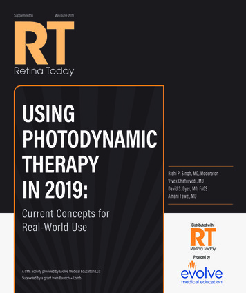
USING PHOTODYNAMIC THERAPY IN 2019:Current Concepts for Real-World UseCASE 2: POLYPOIDAL LESIONS Presented by Dr. FawziA 58-year-old Hispanic woman came in for examination ofa macular lesion. She was 20/80 in her left eye, and she did notuse steroids or have relevant medical history. Her baseline OCThad subretinal fluid with debris in it (Figure 1). The shape of thedebris looked like CSR, but there’s also a shallow lying pigmentepithelium detachment with material in it that was suspicious fora polypoidal-type lesion. We did an ICG, which showed polypson the outside, plaque in the middle, low-lying PED, and featuresof CSR. Figure 2 illustrates the autofluorescence dripping pattern,which is indicative of a pachychoroid spectrum. There’s thick choroid, neo blood vessels, and a long-standing history of CSR thatcomes and goes.This case is from 2016, and at that point I had had some anecdotal success for polypoidal with aflibercept, which was laterborne out later with the PLANET trial.1 I started with aflibercept to see how she would respond. Would anyone have gonestraight to PDT or combination therapy? How would you treatthis patient?Dr. Singh: You’re pointing out the big difference between thePLANET study and the EVEREST trial. Whereas PLANET studyshowed that aflibercept monotherapy was sufficient for treatmentof polypoidal, EVEREST did not.1,2,3,4 I agree with starting withaflibercept in this case; I would have started with aflibercept aswell. But PDT could have a role in reducing the frequency of treatments for the patient in trying to resolve subretinal fluid faster.Figure 1. Baseline OCT in a 58-year-old hispanic female with macular lesion.Figure 2. Baseline indocyanine green angiography in a 58-year-old hispanic female withmacular lesion.Dr. Fawzi: We started with aflibercept, and a year later she was20/40; a lot of the fluid was gone. I extend the treatment interval over the next year and it fails; fluid comes and goes despitetherapy every 4 weeks. The fluid is not responding to aflibercept(Figure 3). I then decide to add PDT to the treatment.Dr. Singh: Where was your lesion treatment in this case?Dr. Fawzi: It was subfoveal. The ICG showed that we shrunkslightly the branching vascular network, but the polyps are exactlythe same size (Figure 3).Dr. Singh: Did you use full fluence or partial fluence in thiscase?Dr. Fawzi: We used full fluence because of how poorly she wasresponding to treatment.Dr. Dyer: I would have started with monotherapy as well,but treated the patient with partial fluence PDT to see how sheresponded. I don’t think it’s wrong to select full fluence, however.Dr. Fawzi: We did the PDT guided by ICG so that we cap-8 SUPPLEMENT TO RETINA TODAY MAY/JUNE 2019Figure 3. Macular lesion after aflibercept treatment.tured the polyps and the branching vascular network (Figure 3).We did pure PDT only, and did not include anti-VEGF therapy.Four months post therapy she was dry, with no extra anti-VEGFneeded (Figure 4). There’s some outer retinal changes and hervision is 20/80, but the PED and polyps shrunk. We are waiting tosee if we can recover some of her vision.Dr. Singh: When you manage these patients, are you constantlyscreening them with OCT and ICG? I often wonder in many ofthese patients if there’s a CNV component.Dr. Fawzi: You should be suspicious of CNV if the patient haslarge choroidal vessels abutting the RPE (so called pachy-vessels),combined with a shallow irregular PED (so called double-layersign), as this case showed. OCT angiography (Figure 5) is my
USING PHOTODYNAMIC THERAPY IN 2019:Current Concepts for Real-World UseCASE 2 (Continued)nibizumab or alone versus ranibizumab monotherapy in patients with symptomatic macular polypoidal choroidal vasculopathy.Figure 4. Four months after full-fluence PDT.go-to screening method because it helps differentiate a CSR pigment epithelial detachment that has no vessels in it versus a neovascular PED that has neo vessels. If the OCT angiography doesn’treveal much, my next step is ICG to look for polypoidal.1. Lee WK, Iida T, Ogura Y, et al. Efficacy and Safety of intravitreal aflibercept for polypoidal choroidal vasculopathy in the PLANETstudy: A randomized clinical trial. JAMA Ophthalmol. 2018;136(7):786-793.2. Koh A, Lee WK, Chen LJ, et al. EVEREST study: efficacy and safety of verteporfin photodynamic therapy in combination with ra-DETERMINING DOSING AND LESION SIZEQDR. SINGH: Dr. Dyer, you mentioned that most of yourpatients get partial fluence. Is that correct? How do youdetermine lesion size?DR. DYER: I do use half fluence; I only use full fluence if the halfwasn’t adequate. The standard PDT regimen includes IV verteporfin at 6 mg/m2 for 10 minutes. Five minutes later, the patientreceives a diode laser at a wavelength of 689 nm and energy of50 mJ/cm2 over 83 seconds to the target lesion.27 Half fluence is25 mJ/cm2 over 83 seconds. Both full and half fluence have beenshown to be effective,12,28-31 but full-fluence PDT is the only treatment modality that will achieve 70% to 80% closure rates for polyps.6,7 However, half fluence has significantly fewer adverse eventsthan full fluence, including atrophic changes, choroidal ischemia,retinal pigment epithelium (RPE) atrophy, or secondary choroidalneovascularization.32-34 Further, some studies have shown that standard-dose PDT may be associated with choriocapillaris hypoperfusion, which could result in decreased vision.35I base the size of the lesion off FA, ICG, or color fundus photography. FA is the most commonly used tool to identify PDTcandidates,9,36 but ICG is recommended in patients with PCV,especially if they are of African or Asian desent.36 In general, thecalculation is 1,000 μm plus the lesion. If I have a CSR patient andthe leakage is not subfoveal, I’ll try to get the spot size in an areaso that I don’t have to treat the fovea. Studies have shown thatamong patients with myopic CNV, those treated with fovealsparing PDT had significantly better visual acuity compared withFigure 5. OCT angiography shows branching vascular network but not the polyps.Retina. 2012;32(8):1453-1464.3. Tan CS, Ngo WK, Chen JP, et al. EVEREST study report 2: imaging and grading protocol, and baseline characteristics of arandomised controlled trial of polypoidal choroidal vasculopathy. Br J Ophthalmol .2015;99(5):624-628.4. Tan CS, Ngo WK, Lim LW, et al. EVEREST study report 3: diagnostic challenges of polypoidal choroidal vasculopathy. Lessonslearnt from screening failures in the EVERES
The EVEREST study showed that PDT alone or in combination with ranibizumab can achieve a complete regression of polyps in patients with symptomatic macular PCV compared with ranibizum-ab alone.6,7,15,16 The PLANET study compared intravitreal aflibercept monotherapy with or without rescue PDT and found a 10.8 letter