Transcription
MANSOURA ENGINEERING JOURNAL, (MEJ), VOL. 47, ISSUE 2, APRIL 2022E: 17Mansoura UniversityFaculty of EngineeringMansoura Engineering JournalPhotoacoustic Imaging in Medicine – A ReviewMohammed T. GadAllah*, Abd El-Naser A. Mohamed, Alaa A. Hefnawy, Hassan E. Zidan, Ghada M.El-Banby and Samir M. sticTomography(PAT),Multispectral OptoacousticTomography(MSOT),Photoacoustic Microscopy(PAM),Raster-ScanOptoacoustic ging-Guided Surgery(IGS), Non-Contact LaserUltrasound (LUS).Abstract — This photoacoustic imaging (PAI) in medicine paper started withan introduction to PAI and the famous photoacoustic techniques includingphotoacoustic tomography (PAT), multispectral optoacoustic tomography(MSOT), photoacoustic microscopy (PAM), raster-scan optoacoustic mesoscopy(RSOM), and photoacoustic elastography (PAE). A modest review about noncontact laser ultrasound (LUS), having the advantage of operator-independentimage quality, has been also demonstrated. A concise review of most of PAI’smedical applications is demonstrated including cancer screening (for breast,thyroid, ovarian, prostate, lung, and skin), tissue oxygenation measurements,brain imaging, imaging-guided surgery (IGS), and the guidance of high intensityfocused ultrasound (HIFU). Some safety considerations contributed withmedical ultrasound and lasers have been then presented. In conclusion, morescientific and clinical development in the field of PAI is expected, and an increasein approved devices that utilize PAI's techniques in medical applications is alsoexpected to serve wide sectors of medicine, whether diagnostic or therapeutic. I. INTRODUCTIONPHOTOACOUSTIC imaging (PAI), or optoacousticimaging, is a biomedical hybrid imaging modality basedon the use of laser-generated ultrasound due to thephotoacoustic effect [1-2]. The photoacoustic effect as a physicalphenomenon was reported in 1880 by A. G. Bell [3-6]. Thereason is the energy exchange process which transforms theabsorbed light energy into kinetic energy, which in role results ina temperature rise thus a pressure wave or sound [6]. Measuringthe sound at different wavelengths produced the origin of thephotoacoustic spectroscopy (PAS); also, called optoacousticReceived: (28 November, 2021) - Revised: (20 February, 2022) - Accepted:(05 March, 2022)*Corresponding Author: Mohammed Tarek GadAllah, (Ph.D. Student),Assistant Researcher with Computers and Systems Department, ElectronicsResearch Institute (ERI), Joseph Tito St, Huckstep, El Nozha, Cairo, Egypt.(Emails: mohammed.tag.1986@eri.sci.eg; mohammed.tag.1986@gmail.com)Abd El-Naser A. Mohamed, Professor Emeritus with Electronics andElectrical Communications Engineering Department, Faculty of Egypt.(e-mail:abd elnaser6@yahoo.com)Alaa A. Hefnawy, Researcher Professor with Computers and SystemsDepartment, Electronics Research Institute (ERI), Joseph Tito St, Huckstep, ElNozha, Cairo, Egypt. (e-mail: alaa@eri.sci.eg)(Ser. NO.BFEMU-2111-1199)spectroscopy (OAS). PAS can be applied to gases, liquids, andsolids [6-8]. PAS directly measures the absorbed power of light,which in role serves as a highly sensitive technique with noscattering losses [9]. Photoacoustics’ uses have been emergedfirst in the field of gas spectroscopy and later in biomedicalapplications [10]. In the last two decades; PAI has rapidly gainedwide popularity in more biomedical applications [1]. PAItechnique can be roughly expressed as “light in and sound out”,merges high-contrast of optical imaging with high spatialresolution and penetration depth of ultrasonography [11-12]. Anillustration of the basic principle of PAI is presented in Fig. 1. InPAI: tissue components having different absorptioncharacteristics can be separated spectrally [13]. PAI is sensitiveHassan E. Zidan, Researcher with Computers and Systems Department,Electronics Research Institute (ERI), Joseph Tito St, Huckstep, El Nozha,Cairo, Egypt. (e-mail: hbanna@eri.sci.eg)Ghada M. El-Banby, Associate Professor with Industrial Electronics andControl Engineering Department, Faculty of Electronic Engineering, MenoufiaUniversity, Menoufia, Egypt. (e-mail: ghadaelbanby75@gmail.com)Samir M. Badawy, Professor Emeritus with Industrial Electronics andControl Engineering Department, Faculty of Electronic Engineering, b@gmail.com;Samir.badawy@el-eng.menofia.edu ).
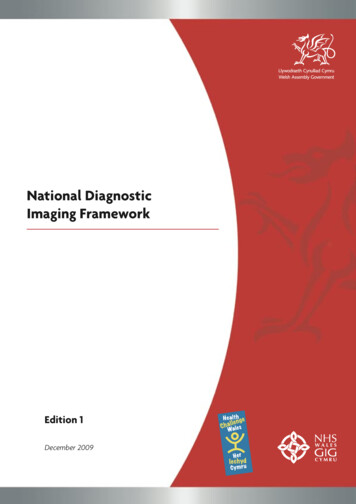
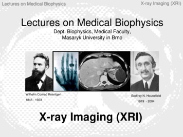
E: 18MOHAMMED T. GADALLAH*, ABD EL-NASER A. MOHAMED, ALAA A. HEFNAWY, HASSAN E. ZIDAN, GHADA M.EL-BANBY AND SAMIR M. BADAWYfor chromophore mapping, whereas these chromophores can bemoreover separated into two categories: the 1st like hemoglobin,melanin, and cytochromes, absorbs ultraviolet-visible light byelectronic transitions, while the 2nd like lipids and water, absorbslight ranges from near-infrared to mid-infrared by vibrationaltransitions [14]. In contrast to conventional ultrasonography, PAIhas the potential to produce speckle-free images [3]. Functionalinformation can be provided also by PAI as blood flow,temperature, and oxygenation [2]. Recently, a photoacoustic(PA) topography through an ergodic relay (PATER) system, hasbeen introduced to achieve a wide field of view (FOV), snapshot,and high frame rate in a low-cost system [15-17]. At very deepregions, where PAI cannot achieve adequate resolution, the PAprobe should be positioned close to the area of interest by the aidof endoscopy [18]. PA endoscopy is aiming to outdo theresolution limitations of endoscopic ultrasound (EUS) which is aclinically available tomographic tool utilized for diagnosingmore diseases [19]. From a market point of view, it is forecastedthat the PAI market will increase in 2022 [20].In this review paper the utilized strategy for achieving it wasperforming a literature search through the Internet, selecting 189references to build our review, and contacting with two authorsof two different references [71, 111] for giving us permission toprocess and publish their figures’ data.talk about safety considerations for both laser and ultrasound.Finally, in section XI, we concluded that PAI is a promisingbiomedical imaging tool in more applications in medicine.II. MOST FAMOUS PHOTOACOUSTIC STRATEGIESPAI has more techniques to be implemented. Here, wedivided PAI’s techniques into five main categories: PAT, PAM,PA endoscopy, PATER, and PAE. A concise chart for the PAI’smain techniques mentioned in this paper is illustrated in Fig. 2.Where; OD: optical detection, OR: optical resolution, AR:acoustic resolution, MS: multispectral, fcPAT: functionalconnectivity PAT, LD: light-emitting diode (LED). In thefollowing sub-sections, we will talk in some detail about A. PAT,B. PAM, and C. PAE. PA endoscopy (the third main category) isa very prospective field in PAI, providing molecular contrast atlarge depths, allowing for simultaneous visualization of structuraland functional information [21]. PA endoscopy presents the samestrength in spatial resolution of routinely used clinical EUS,whereas introducing more functional information atphysiological sites [22]. For more about PA endoscopy, we referthe reader to [18, 19, 21, 23-28]. PATER (the fourth maincategory) could be considered a combination of computed PATtheory and PAM, trying to achieve a wide-field image with onlya single-element ultrasonic detector upon a single laser shot [1517]. For more about the PATER technique, we refer the reader to[15-17].A. PATPAT technique can be briefly described in four steps [29,30]:1. Illumination of the tissue by a pulsed broad laser beam.2. A small but rapid temperature rise will be generated.3. The emitted short-wavelength pulsed ultrasonic waves dueto thermoelastic expansion will be then detected byunfocused ultrasonic transducers.4. Image reconstruction process produces tomographicimages of optical contrast and high resolution.Fig. 1. Illustration of PAI’s basic principle.In the following section II: we will talk about the most famousPA techniques including A. PAT, A.1. MSOT, B. PAM, and C.PAE. In section III, we talk about the recent development of noncontact laser ultrasound (LUS) medical imaging. In section IV, abrief introduction to PAI’s applications in medicine mentioned inthe following five sections (sections V, VI, VII, VIII, and IX) ispresented. In the following section V, most of PAI’s applicationsin cancer screening are presented including A. breast, B. thyroid,C. ovarian, D. prostate, E. lung, and F. skin cancer. Sections VI,VII, VIII, and IX; present in some detail the applications of PAIinto tissue oxygenation measurements, brain imaging, ImagingGuided Surgery (IGS), and PAI’s guidance of High IntensityFocused Ultrasound (HIFU), respectively. In section X we willFig. 2. A concise chart for the PAI’s main techniques mentionedin this review.
MANSOURA ENGINEERING JOURNAL, (MEJ), VOL. 47, ISSUE 2, APRIL 2022PAT can image nearly all molecules, fluorescent or notbecause it doesn’t rely on fluorescence emission [31, 32]. PATis speckle-free unlike ultrasound imaging and optical coherencetomography (OCT) [33]. PAT has the potential to producefluid‐dynamic, functional, molecular, and anatomical imaging,playing an efficient role in biomedical research [34]. PAT hasintroduced a super-depth combined with high-resolution opticalimaging exceeding the optical transport mean free path whichis the depth limitation of OCT, confocal microscopy, and twophoton microscopy ( 1 mm in the skin) [35]. Anatomical andvascular structures imaging can be provided by endogenoushemoglobin contrast. Molecular, functional, and reporter geneimaging can be provided by using exogenous optical contrast[29, 30, 36]. A contrast agent called black mesoporous silicon(BPSi), has been introduced, by Wujun Xu et al [37], for thelight-emitting diode (LED) based three-dimensional (3D) PAT,resulting in strong contrast. A United States patent applicationby James B. Pitner et al [38], has been introduced forHydroporphyrins as contrast agents for PAI. Another advanceto PAT was published by M. Nasiriavanaki et al [39]:developing a functional connectivity photoacoustictomography (fcPAT) system, allowing non-invasive imaging ofthe resting-state functional connectivity (RSFC) in a mousebrain, with a large FOV as well as high spatial resolution. RSFCwas alerted in many brain disorders [39]. A different techniqueof PAT is the optical-detection photoacoustic tomography (ODPAT) in which optical approaches have been used to detect thephotoacoustic signals instead of piezoelectric detection inclassical ultrasound probes [22].A.1. MSOTMSOT, a Multispectral PAT (MS-PAT) technique, has beenable to introduce a generation of biomedical imaging. Aparticular advantage of MSOT is its ability to scale withdifferent tissue sizes [40]. MSOT has been succeeded tovisualize fluorochromes in tissues that are not visible withconventional single wavelengths’ PAI [41]. A fast acquisitionMSOT whole-body scanner has been introduced by Rui Ma etal to visualize small animals’ molecular markers with multiwavelength illumination [42]. Razansky et al [43], haveintroduced their protocol of volumetric real-time MSOT ofbiomarkers; assuring MSOT’s ability in visualizing tissuebiomarkers and optical contrast with speed and resolutionrepresentative of ultrasound. A fast MSOT platform fordynamic imaging of pharmacokinetics and bio-distribution inmultiple organs has been demonstrated by Adrian Taruttis et al[44]; introducing fast and high-resolution in vivo imaging’scapabilities. Semi-quantitative MSOT for volumetricPharmacokinetic (PK) imaging of gastric emptying had beendemonstrated by S. Morscher et al; using MSOT in monitoringgastrointestinal motility in mice given indocyanine green (ICG)by oral gavage aiming fate of ICG be monitored in thegastrointestinal tract [45]. Not only ICG, but MSOT alsovisualize a range of exogenous contrast agents e.g., methyleneblue (MB) [46]. The performance of an MSOT system equippedwith 2D vs. 3D handheld probes has been demonstrated byNeuschmelting et al [47], for potential clinical translation. FirstE: 19in vivo clinical use of MSOT non-invasively has beenintroduced by S. Y. Chuah et al [48], for Structural andfunctional 3D mapping of skin tumors, confirming the benefitof that imaging method for surgical intervention guidance. Overthe last few years, MSOT has succeeded in more than oneapplication in medicine such as imaging of breast cancer [49],particle size-dependent intratumoral distribution of polymericmicelles [50], thyroid disorders [51], drug-induced liver injury(DILI) [52], neural dynamics and organization of the intactmouse brain [53], and orally-administered particles within thegastrointestinal tract of murine models [54]. MSOT has thepotential to achieve real-time tracking in vivo of magneticnanoparticles quantitatively [55]. A co-registration of MSOTand magnetic resonance imaging (MRI) data from murinetumor models, has been introduced by M. Gehrung et al [56],assuring feasibility of hardware and software-based registrationframework for MRI and MSOT images.B. PAMPAM technique can be briefly described as focusing apulsed laser beam into the tissue, ultrasonic waves will be thengenerated, and finally, a focused ultrasonic transducer willdetect the generated waves to form a depth-resolved onedimension (1D) image [29, 30]. Also, three-dimension (3D)high-resolution tomographic images can be generated by rasterscanning [29, 30]. L. V. Wang has illustrated in [57] that: PAMcan image up to 3 mm into scattering tissue with 15micrometers axial resolution while working at 50 MHzultrasonic detection frequency. Whereas working at 5 MHz,PAM can image 10 times as deep but with 10 times axialresolution [57].PAM has the potential to image the microvascular networkin the skin, which is invaluable in dermatology and relatedcancer research [58]. Weak acoustic scattering in tissue is anadvantage of PAM, unlike pure optical microscopic techniques[59]. PAM breaks through the optical propagation limit ( 1mm in soft tissue); providing images with high-resolution atimaging depths up to a few millimeters, with excellentscalability [59].There are two different techniques of PAM: Opticalresolution photoacoustic microscopy (OR-PAM) and Acousticresolution photoacoustic microscopy (AR-PAM).OR-PAM: this technique uses focused light to spatiallylimit the stimulation, producing an optical diffraction-limitedresolution in the lateral direction.AR-PAM: in this technique a relatively large area isilluminated, rather than focus light to an optically diffractionconfined spot, so more laser energy is allowed in AR-PAM thanin OR-PAM, increasing the chance of photons to reach a muchgreater depth [22].In AR-PAM, the illumination can be either dark-field orbright: The dark-field approach has the advantage to reducesurface interference to deeper PA signals, whereas the brightfield method can carry higher fluence to a targeted volume [22].A most famous technique that belongs to the AR-PAMfamily is the RSOM (raster-scan optoacoustic mesoscopy).
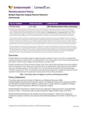
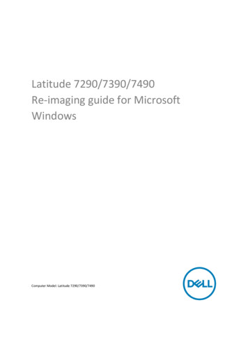
E: 20MOHAMMED T. GADALLAH*, ABD EL-NASER A. MOHAMED, ALAA A. HEFNAWY, HASSAN E. ZIDAN, GHADA M.EL-BANBY AND SAMIR M. BADAWYOptoacoustic mesoscopy is, optoacoustic (or photoacoustic)imaging having acoustic resolution and wide-bandwidthultrasound detection. RSOM’s placement is like a bridge inbetween optoacoustic microscopy and optoacousticmacroscopy [60]. Optoacoustic mesoscopy or macroscopyqualifies deeper imaging than optical or optoacousticmicroscopy methods which use focused light [46]. Aclassification of microscopy, mesoscopy, and macroscopyconcerning their penetration depth, had been stated by V.Ntziachristos in [61], as: ( 1 mm), (0.5 mm to 10 mm), and ( 1 cm); respectively. Another penetration-based classificationhad been stated by A. Taruttis, G. M. van Dam, and V.Ntziachristos in [46], as: ( 1 mm), (1–5mm), and (beyond 5mm); respectively. Over the last decade, more papers have beenpublished concerning optoacoustic mesoscopy [62-69].In recent years, several applications in medicine have beenreported using advanced PAM imaging systems (Such as LaserDiode (LD) based PAM systems) towards a low-cost andaiming for a portable PAM system for point-of-care andwearable applications [70, 71]. LD-based PAM imaging hasbeen investigated to reduce the size of bulky lasers usually usedin PAM and also for economic issues [71]. A sample imagegenerated from an original 4060 by 2700 pixels data (taken withpermission from [71]) for in vivo LD-based PAMmicrovasculature imaging of an 8.12 mm by 5.4 mm area froma mouse ear is illustrated in Fig. 3.phase projection [74]. In the same year 2011; K. J. Parker et al,have stated their 20 years’ perspective on Imaging the elasticproperties of tissue [75]. In 2015; Pengfei Hai et al, haveproposed Vascular elastic photoacoustic tomography (VEPAT) to measure blood vessel compliance in humans [76]. APAT imaging system called quantitative photoacousticelastography (QPAE) imaging system helping in tissue elasticproperties’ recovery nondestructively and noninvasively, hasbeen developed by Pengfei et al [72, 77, 78]. Till now, PAE isin the upbringing stage of its development [72].III. LUS MEDICAL IMAGINGThe non-contact laser ultrasound (N-CLUS) medicalimaging technique is based on the concept of optical detectionof ultrasound (OD-US) [79, 80]. OD-US can play an importantrole in PAI for biomedical study and clinical diagnosis [80]. Infull N-CLUS technique; PA sources are employed on the skinsurface, in combination with laser interferometric detectioninstead of piezoelectric ultrasound detectors used in most PAsystems [81]. The main difference between PAI and N-LUS isthe detection mechanism (piezoelectric or optical), we canname the technique of N LUS by optical detection PAI (ODPAI). Historically, ultrasound generation with lasers nearlygoes back to the first laser invention (the ruby laser) [82], whichwas used for generating ultrasound or shock waves in materials[83]. Recently, more studies have been emerged clarifying themedical applicability of N-CLUS's concept [81-90].IV. PAI’S APPLICATIONS IN MEDICINEPAI has a wide variety of applications in more branches inbiomedicine [184-189]. In the following sections V, VI, VII,VIII, and IX, a concise review is presented for most PAI’sapplications in cancer screening, tissue oxygenationmeasurements, brain imaging, guiding surgeries, and HIFU’sguidance, respectively.V. PAI’S APPLICATIONS IN CANCER SCREENINGFig. 3. A sample image produced with permission from [71], illustrating anin vivo LD-based PAM imaging of a mouse ear. Where the numerical valueson each X-axis and Y-axis are referring to the pixels’ coordinates in theimage (where each pixel has a size of 2 µm by 2 µm).C. PAEElastography imaging can be described as art to measureand posterior graphical representation of the spatial allocationof tissue’s stiffness [72]. The elasticity distribution in biologicaltissue can be noninvasively mapped by Elastography to revealdisease conditions [73]. Published studies on PAE imaging(PAEI) are so limited [72]. The start was in 2011, when G. Gaoet al, had published their development of a system of PAviscoelasticity imaging, obtaining a high contrast image,reflecting information of the tissue viscoelasticity with the PAIn 2020, more than 1.8 million new cancer cases and morethan 0.6 million cancer deaths were expected to occur only inthe United States [91]. Screening can detect some cancers earlywhen treatment is more often successful [92]. Importantelements in cancer control are cancer detection in its earlystages and supplying immediate appropriate treatment [93].PAI can play a role in cancer diagnosis and therapy guidance[94, 95].A. Breast cancerA group of diseases in which cells in breast tissue changeand divide uncontrolled, typically resulting in a lump or mass,is called breast cancer [96]. Breast cancer is the second commoncancer worldwide after lung cancer, the fifth common cause ofcancer death [97, 98].
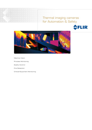
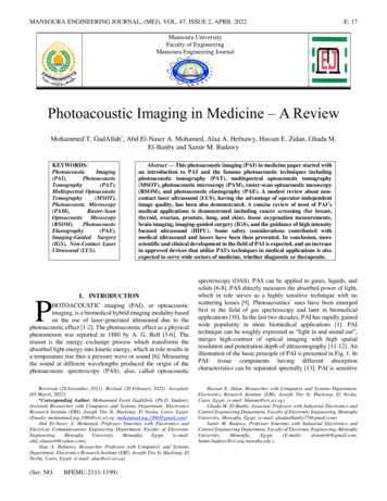
MANSOURA ENGINEERING JOURNAL, (MEJ), VOL. 47, ISSUE 2, APRIL 2022X-ray-based Mammography [97], Breast ultrasound (BUS)imaging [99-101], and Magnetic resonance imaging (MRI)[102, 103], are the three most famous imaging techniquesutilized for breast cancer screening. X-ray-based screeningmammography, beginning in the 1980s, helps in the earlydetection of breast cancer [97]. But it uses ionizing radiation,painful breast compression, and has poor performance in radiodense breasts. BUS imaging can be utilized as mammography’sadjunct [100, 101]. However, X-ray and BUS imaging are bothsuffer from having non-optimal sensitivity and specificity[104]. MRI could be used for breast imaging in cases ofuncertain findings in X-ray and BUS imaging [104]. However,MRI suffers from limited specificity, requires contrast agents,excludes more patients for instance who have pacemakers, orclaustrophobia, moreover it is a little obstructed in premenopausal women by the requirement to time imaging duringcertain phases of the menstruation cycle [104]. PAI, anonionizing modality, has the potential to achieve higherspecificity in the diagnosis of malignant and benign breastmasses, helping to reduce the number of false-positive scansand unnecessary biopsies of benign [104]. Over the last fewyears, there were various breast’ PAI systems have beenproposed [105-110]. Recently, Li Lin et al [111], haveintroduced a high-speed 3D Photoacoustic computedtomography (PACT) system for both preclinical and clinicalapplications, achieving an in vivo imaging depth of 4 cm in thehuman breast within a single breath-hold (SBH) of 10 s. Figure4 is illustrating a sample image of a healthy human subject’sright and left breasts in vivo taken by the 3D-PACT systemintroduced in [111].B. Thyroid cancerIn 2020, the expected number of new cases of thyroid cancerdiagnosed only in the United States (US) is estimated at 52,890.The incidence rate is about 3 times higher in women than inmen [92]. Thyroid cancer was the most rapidly increasingcancer in the US till now [92]. In 2012, there were an estimated40,000 deaths globally due to thyroid cancer [93]. The thyroidgland, normally located in the lower front of the neck, is abutterfly-shaped endocrine gland [112]. Ultrasound combinedwith fine-needle aspiration cytology (FNAC) is the primarydiagnostic tool for thyroid cancer, followed by histology [10].PAI can augment, FNAC combined ultrasound, for molecularimaging of thyroid nodules. It is because the thyroid gland issuperficial (2–3 cm deep) allowing sufficient penetration oflight [10]. Recently, more researches have been introduceddiscussing PAI’s utilization in thyroid cancer diagnosis [113115].C. Ovarian cancerIn 2020, an expected 21,750 new cases of ovarian cancerwill be diagnosed only in the United States and about 13,940women are expected to die from the disease [92]. Globally,ovarian cancer is the eighth most frequent cause of cancer deathamong women with 152,000 deaths, as stated by the 2014’sworld cancer report [93]. Ovarian cancer is rarely detected at anE: 21early stage because of its low spread in the general population[116], Bin Rao et al, have investigated the feasibility of “opticalbiopsy”, using high OR-PAM to quantify the microvasculatureof ovarian and fallopian tube tissue [116]. Through the lastdecade, there was more than one prototype PAI-based systemstudying the ability of PAI to enhance ovarian cancer diagnosis[116-119].Fig. 4. 3D-PACT of a healthy human subject’s breast in vivo (Taken andProcessed with permission from [111]). A is a processed perspectiveangiogram of the Right breast. B is the Left breast image of the samehuman subject.D. Prostate cancerGlobally, prostate cancer (PCa) is the second most commoncancer in men worldwide [76]. In 2020, the expected new casesof prostate cancer are estimated at 191,930 only in the UnitedStates, and 33,330 men are expected to die from the disease[92]. To visualize prostatic anatomy and guide needle biopsy,transrectal ultrasound (TRUS) has been routinely used since the1980s [120]. A trial in validating if ex‑vivo multispectral PAIcan characterize malignant prostate tissue, benign prostatichyperplasia (BPH), and normal human prostate tissue, has beenproposed by Dogra et al [121], concluding that multispectralPAI had the potential to differentiate between malignantprostate, BPH and normal prostate tissue. B. L. Bungart et al[122], have published their success in providing prostate biopsytargets by 1064 nm PAT and ultrasonography texture-basedfeature analysis. Through the last few years, more researcheshave been emerged concerning enhancing PAI’s applicationson the prostate [120-125].E. Lung cancerLung cancer is the most frequent cancer worldwide; it is themost common cancer in men and the third in women [93]. Asestimated in 2012: there were more than 1.8 million new casesand almost 1.6 million deaths [93]. Only in the United States:the expected new cases of lung cancer in 2020 were estimatedat 228,820, whereas the expected mortality from the diseasewas estimated at 135,720 [92]. Early detection could be a reasonfor decreasing mortality from lung cancer because mostdiagnoses occur in a late stage of cancer with a low survival rate[126]. PAI has been studied to help in lung cancer diagnosis
E: 22MOHAMMED T. GADALLAH*, ABD EL-NASER A. MOHAMED, ALAA A. HEFNAWY, HASSAN E. ZIDAN, GHADA M.EL-BANBY AND SAMIR M. BADAWYthrough more than one study during the last decade assuring itspromising role in lung cancer diagnosis [126-129].F. Skin cancerIn recent years, skin cancer cases number has risen globally[1]. Melanoma, Basal cell carcinoma, and squamous cellcarcinoma; are the three main types of skin cancer, in which thelast two types are called non-melanoma skin cancers (NMSCs)and are seldom life-threatening, whereas the first type:melanoma skin cancers are rare but aggressive and start inpigmented lesions like a mole or birthmark [1]. Nearby lymphnodes are the first site of metastasis where melanoma can spreadthroughout the body [1]. For precise melanoma prognosis,ascertaining, staging, and planning treatment; the pathologicalstatus of the sentinel lymph node is significant [130]. Initialresults using PAT by Jose et al [130], suggested that PA coulddevelop into an intraoperative imaging method for detectingmelanoma metastases in sentinel lymph nodes. Recently, moreresearches have been emerged discussing PAI’s utilization inskin cancer imaging [131-135].VI. PAI IN TISSUE OXYGENATION MEASUREMENTSOxygen was named by Joseph Priestley and AntoineLavoisier who had isolated it. Carl Scheele had discoveredoxygen in the late eighteenth century. Living cells obtainoxygen through air inhaled into the respiratory system fromwhere it is absorbed into the bloodstream. In red blood cells(RBCs), oxygen is bound to hemoglobin. In some tissues suchas muscle, oxygen is also stored bound to myoglobin [136].William G. K., Peter J. R., and Gregg L. S. have been awardedthe 2016 Albert L. basic medical research award for thediscovery and clarification of pathways by which humans andother multicellular organisms sense and respond to changes inoxygen availability [137]. The Nobel prize for Physiology orMedicine for 2019 had been given to the same three men fordiscoveries on the mechanisms by which animals cells respondto changes in oxygen levels [136]. To facilitate bloodoxygenation, a specialized oxygen-binding molecule(hemoglobin) is needed since oxygen does not dissolve readilyin the plasma because plasma is 93% water [138]. Tissueoxygenation can be imaged by PA based on the oxygendependent light absorption characteristics of hemoglobin [139].Molar extinction coefficient (cm-1/M) spectra curves for HBO2and HB are shown in Fig. 5. Where: M is referring to the molarconcentration in moles/liter. Where HbO2 (red): refers toOxygenated Hemoglobin, and HB (blue): refers toDeoxygenated Hemoglobin.Oxygen saturation (sO2) can be described by equation (1)taken from [138]; Where THb: refers to Total HemoglobinConcentration.Blood oxygenation measurements by PAT are an importantapplication been widely carried out in PA studies of tumorhypoxia, brain functions, cancer therapy, and wound healing[140]. PAM has been able to image sO2 in the microvasculatureof biological tissues [141]. PAI can deduce the spatialallocation of sO2 in blood, and be co-registered withultrasonography images of the surrounding anatomy [142]. sO2can be expected from the partial pressure of blood’s oxygen(pO2) based on a standard dissociation curve shown in [142].Real-time assessment of tissue hypoxia in vivo can be achievedby combining PA and high-frequency ultrasound [143]. Inblood flow measurements, PAI has an efficient role [144-146].VII. PAI IN BRAIN IMAGINGThe absorption spectra of deoxyhemoglobin (Hb) andoxyhemoglobin (HbO2) enabled PAT to provide label-freefunctional brain images of oxygen saturation (sO2) and totalhemoglobin concentration (THb) [13]. For improving thevisibility of the neurovascular structures, optical contrastagents, like nanoparticles and organic dyes can be used [13].Different PAI brain imaging techniques have emerged throughthe last few years [13, 147]. Visualizing Alzheimer’s disease ofa mouse’s brain has been reported by Park SJ et al [148],applying the MSOT technique utilizing an optical imagingprobe (CDnir7: Compound of Designation near-infrared 7).Recently, more researches have been emerged discussingdifferent types of PAI schemes, techniques, and applications inbrain imaging [148, 149-162].VIII. PAI IN IMAGING-GUIDED SURGERY (IGS)Imaging-guided surgery (IGS) is a branch of the concept ofImage-guided therapy (IGT) which includes any intervention orsurgery that utilizes improved imaging for monitoring,localizing, controlling, and targeting procedures [163]. Due toPAI’s ability in noninvasive diagnosis of various types oftissues including b
MANSOURA ENGINEERING JOURNAL, (MEJ), VOL. 47, ISSUE 2, APRIL 2022 E: 17 Mansoura University Faculty of Engineering Mansoura Engineering Journal (Ser. NO. BFEMU-2111-1199) I. INTRODUCTION which in role serves as a highly sensitive technique with no