Transcription
PALSStudy Guide2016Bulletin:New resuscitation science and American Heart Association treatment guidelines were releasedOctober 28, 2015!The new AHA Handbook of Emergency Cardiac Care (ECC) contains these 2016 Guidelines and is requiredstudy for this course. The 2016 PALS Provider Manual is not yet available. This study guide will provide you withadditional study information.Website: www.heart.org/eccstudent Keyword: pals15 (Pretest)www.phsinstitute.com (study info. For class for rhythm review)What is required to successfully complete PALS? C o m p l e t e d PALS Pre-test is required for admission to the course. S c o r e 84% on the multiple-choice post-test.It is a timed test and you may be allowed to use your ECC Handbook. Y o u must be able to demonstrate: The PALS rapid cardiopulmonary assessment Effective infant and child CPR using an AED on a child Safe defibrillation with a manual defibrillator maintaining an open airway Confirmation of effective ventilation addressing vascular access stating rhythm appropriate drugs, route and dose Consideration of treatable causesWhat happens if I do not do well in the course?The Course Director or Instructor will first “remediate” (tutor) you and you may be allowed to continue in the course. If itis decided you need more time to study, you will be placed into the next course.Where do I start? CPR/AED: You will be tested with no coaching. If you cann ot perform these skills well withoutcoaching, you can/may be directed to take the course at another time. Know p. 7 of this study guide well. Arrhythmias: Before you come be sure you can identify: Sinus Rhythm (SR), Sinus Bradycardia (SB),Sinus Tachycardia (ST), Supraventricular Tachycardia (SVT), Ventricular Tachycardia (VT), Ventricular Fibrillation(VF), Torsades de Pointes, Pulseless Electrical Activity (PEA) and Asystole.
You will need to know:*Respiratory RateAgeInfantToddlerPreschoolerSchool-age childAdolescentHeart RateRate3022201812-Age53372825201- 12 months12 months - 2 years2 – 5 years5 - 10 years10-15 yearsSleeping -Awake9090805850205180140118100-ECC Handbook p. 77* Hypotension by Systolic Blood Pressure (SBP)Age 1 month1 month – 1 year1 – 10 years10 years SBP607070 (2 x age in years)90Hypotension signs of poor perfusion Decompensated shock.ECC Handbook p. 77* Treat Possible Causes.5 Hs5 TsH ypoxiaH ypo-volemiaH ypo-thermiaHypo /hyper kalemiaHydro gen ion (acidosis)Hydro- GlycemiaT amponadeT ension pneumothoraxT oxins – poisons, drugsT hrombosis – coronary (AMI)T hrombosis – pulmonary (PE)T raumaSpacing separations may help as a memory aid.Rapid Cardiopulmonary Assessment and AlgorithmsThis is a systematic head-to-toe assessment used to identify infants and children in respiratory distress and failure,shock and pulseless arrest. Algorithms are “menus” that guide you through recommended treatment interventions.Know the following assessment because it begins all PALS case scenarios. The information you gather duringthe assessment will determine which algorithm you choose for the patient’s treatment. After each intervention youwill reassess the patient again using the head-to-toe assessment.
‹Start with child’s general appearance:Is the level of consciousness:Is the overall color: goodIs the muscle tone: goodA awake V responds to verbal P responds to pain U unresponsiveor bad?or floppy?‹Then assess CABs: (stop and give immediate support when needed, then continue with assessment)Circulation: Is central pulse presentIs the rate normalIs the rhythm regularIs the QRS narrowAirway:or absent?or too slowor irregular?or wide?or too fast?Open and hold with head tilt-chin liftBreathing: Is it presentIs the rate normalIs the pattern regularIs the depth normalIs there nasal flaringIs there stridoror absent?or too slowor irregularor shallowor sternal retractionsor gruntingor too fast?or gasping?or deep?or accessory muscle use?or wheezing?‹Next look at perfusion:Is the central pulse versus peripheral pulse strength equalIs skin color, pattern and temperature normalIs capillary refill normalIs the liver edge palpated at the costal margin (normal or dry)or unequal?or abnormal?or abnormal (greater than 2 seconds)?or below the costal margin (fluid overload)?‹And check:Is systolic BP acceptable for age (normal or compensated)Is urine output adequate for: infants and children (1– 2cc/kg/hr)or hypotensive?or adolescents (30cc/hr)?‹Now classify the physiologic status:Stable:needs little support; reassess frequentlyUnstable:needs immediate support and interventionRespiratory distress: increased rate, effort and noise of breathing; requires much energyRespiratory failure: slow or absent rate, weak or no effort and is very quietCompensated shock: SBP is acceptable but perfusion is poor: central vs. peripheral pulse strength is unequalperipheral color is poor and skin is coolcapillary refill is prolongedDecompensated shock: Systolic hypotension with poor or absent pulses, poor color, weak compensatory effort.
‹Apply the appropriate treatment algorithm: Bradycardia with a PulseTachycardia with Adequate PerfusionTachycardia with Poor PerfusionPulseless Arrest: VF/VT and Asystole/PEAAdvanced AirwayA cuffed or uncuffed Endotracheal Tube (ET) may be used on Infants and children.To estimate tube size:ECC Handbook p. 94 4) 4(Age in years 4) 3.5(Age in years 2) 12Uncuffed (Age in yearsExample: (4 yearsCuffed Example: (4 yearsDepth Example: (4 years 4) 4) 2) 1 4 5 1 3.5 4.5 2 12 14Immediately confirm tube placement by clinical assessment and a device: Clinical assessment: Look for bilateral chest rise.Listen for breath sounds over stomach and the 4 lung fields (left and right anterior and midaxillary).Look for water vapor in the tube (if seen this is helpful but not definitive). Devices: End-Tidal CO2 Detector (ETD): if weight 2 kgƒ Attaches between the ET and Ambu bag; give 6 breaths with the Ambu bag:Litmus paper center should change color with each inhalation and each exhalation.-Original color on inhalation Color change on exhalation OkayCO2!!O2 is being inhaled: expected.Tube is in trachea.-Original color on exhalation Oh-OH!!Litmus paper is wet: replace ETD.Tube is not in trachea: remove ET.Cardiac output is low during CPR.Esophageal Detector (EDD): if weight 20 kg and in a perfusing rhythm* Resembles a turkey baster:Compress the bulb and attach to end of ET.Bulb inflates quickly!Tube is in the trachea.Bulb inflates poorly?Tube is in the esophagus.* No recommendation for its use in cardiac arrest. When sudden deterioration of an intubated patient occurs, immediately check:Displaced tube is not in tracheaObstruction consider secretionsPneumothorax consider chest traumaEquipment check oxygen sourceor has moved into a bronchus (right main stem most common)or kinking of the tubeor barotraumasor non-compliant lung diseaseand Ambu bagand ventilator
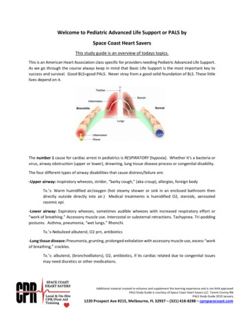
PALS DrugsIn Arrest:Epinephrine: catecholamineECC Handbook p. 92Increases heart rate, peripheral vascular resistance and cardiac output; during CPR increases myocardial and cerebral blood flow.IV/IO: 0.01 mg/kg of 1:10 000 solution (equals 0.1 mL/kg of the 1:10 000 solution); repeat q. 3–5 minET:0.1 mg/kg of 1:1000 solution (equals 0.1 mL/kg of the 1:1000 solution); repeat q. 3–5 minAnti-arrhythmic Drugs:Amiodarone: atrial and ventricular antiarrhythmicECC Handbook p. 89Slows AV nodal and ventricular conduction, increases the QT interval and may cause vasodilation.Refractory VF/PVT:IV/IO: 5 mg/kg bolus (may repeat up to 2 times)Perfusing VT:IV/IO: 5 mg/kg over 20-60 minPerfusing SVT: IV/IO: 5 mg/kg over 20-60 minMax:15 mg/kg per 24 hours – Max single dose 300mgCaution:hypotension, Torsade; half-life is up to 40 daysLidocaine: ventricular antiarrhythmic to consider when amiodarone is unavailableECC Handbook p. 94Decreases ventricular automaticity, conduction and repolarization.VF/PVT:IV/IO: 1 mg/kg bolus repeat 15 minÆET: 2 -3 mg/kgPerfusing VT:IV/IO: 1 mg/kg bolus repeat 15 minInfusion:20-50 mcg/kg/minCaution:neuro toxicity seizuresMagnesium: ventricular antiarrhythmic for Torsade and hypomagnesemiaECC Handbook p. 94Shortens ventricular depolarization and repolarization (decreases the QT interval).IV/IO:25-50 mg/kg over 10–20 min; give faster in TorsadeMax:2 gmCaution: hypotension, bradycardiaProcainamide: atrial and ventricular antiarrhythmic to consider for perfusing rhythmsECC Handbook p. 96Slows conduction speed and prolongs ventricular de- and repolarization (increases the QT interval).Perfusing recurrent VT:IV/IO: 15 mg/kg infused over 30–60 minRecurrent SVT:IV/IO: 15 mg/kg infused over 30–60 minCaution:hypotension; use it with extreme caution with amiodarone as it can cause AV block or TorsadeIncrease heart rate:Epinephrine: drug of choice for pediatric bradycardia after oxygen and ventilationECC Handbook p. 80Increases heart rate, peripheral vascular resistance and cardiac output; during CPR increases myocardial and cerebral blood flow.IV/IO: 0.01 mg/kg of 1:10 000 solution (equals 0.1 mL/kg of the 1:10 000 solution); repeat q. 3–5 minET:0.1 mg/kg of 1:1000 solution (equals 0.1 mL/kg of the 1:1000 solution); repeat q. 3–5 minAtropine: vagolytic to consider after oxygen, ventilation and epinephrineECC Handbook p. 87Blocks vagal input therefore increases SA node activity and improves AV conduction.IV/IO:0.02 mg/kg; (max dose 0.5mg)Caution:do not give less than 0.1 mg or may worsen the bradycardia2010 (New): Atropine is not recommended for routine use inthe management of PEA/asystole and has been removed fromthe PALS Cardiac Arrest Algorithm. The treatment of PEA/asystole is now consistent in the PALS
Decrease heart rate:Adenosine: drug of choice for symptomatic SVT & Wide Complex Monomorphic VTSee ECC Handbook p. 88 forinjection techniqueBlocks AV node conduction for a few seconds to interrupt AV node re-entry.IV/IO: first dose:0.1 mg/kgmax: 6 mgsecond dose:0.2 mg/kgmax: 12 mgCaution: transient AV block or asystole; has very short half-lifeIncrease blood pressure:Dobutamine: synthetic catecholamineECC Handbook p. 92Increases force of contraction and heart rate; causes mild peripheral dilation; may be used to treat shock.IV/IO infusion:2- 20 mcg/kg/min infusionCaution:tachycardiaDopamine: catecholamineECC Handbook p. 92May be used to treat shock; effects are dose dependent.Low dose:increases force of contraction and cardiac output.Moderate:increases peripheral vascular resistance, BP and cardiac output.High dose:higher increase in peripheral vascular resistance, BP, cardiac work and oxygen demand.IV/IO infusion:2–20 :ECC Handbook p. 93Increases blood glucose in hypoglycemia; prevents hypoglycemia when insulin is used to treat hyperkalemia.IV/IO:0.5–1 g/kg; this equals: 2–4 mL/kg of D25 or 5–10 mL/kg of D10 or 10–20 mL/kg of D5Caution: maximum recommended concentration should not exceed D25%; hyperglycemia may worsen neuro outcomeNaloxone: opiate antagonistECC Handbook p. 95Reverses respiratory depression effects of narcotics. 5 yr or 20 kg: IV/IO: 0.1 mg/kg 5 yr or 20 kg: IV/IO: up to 2 mgCaution:half-life is usually less than the half-life of narcotic, so repeat dosing is often required;ÆET dose can be given but is not preferred; can also give IM or SQ.Sodium bicarbonate: pH buffer for prolonged arrest, hyperkalemia, tricyclic overdose: ECC Handbook p. 97Increases blood pH helping to correct metabolic acidosis.IV/IO:1mEq/kg slow bolus; give only after effective ventilation is establishedCaution: causes other drugs to precipitate so flush IV tubing before and afterET drug administration: distribution is unpredictable as is the resulting blood level of the drug; if there is no IV/IO access,give the drug down the ET and flush with 5 mL NS then give 5 ventilations to disperse the drug.
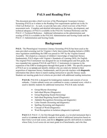
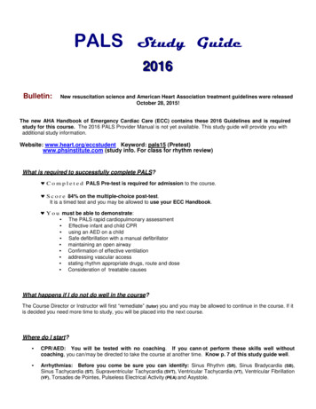
2015 (Modification of Previous Recommendation):For ease of placement and education, the anterior-lateral pad position is a reasonable defaultelectrode placement. Anyof 3 alternative pad positions (anterior-posterior, anterior-left infrascapular, andanterior-right infrascapular) may beconsidered on the basis of individual patient characteristics.Placement of AED electrode pads on the victim’ s bare chest inany of the 4 pad positions is reasonablefor defibrillation.2015 : Continuous quantitative waveform capnographyis now recommended for intubated patients throughout theperiarrest period. When quantitative waveform capnographyis used for adults, applications now include recommendationsfor confirming tracheal tube placement and for monitoring CPRquality and detecting ROSC based on end-tidal carbon dioxideCapnography to monitor effectiveness of resuscitation efforts. PETCO2 should read 35 to 40mm Hh inindividual of ROSC, High Quality CPR is confirmed by a Capnography read of 10mm Hg on the vertical axisover time. This patient is intubated and receiving CPR. Note that the ventilation rate is approximately 8 to 10breaths per minute. Chest compressions are given continuously at a rate of slightly faster than 100/min but arenot visible with this tracing.
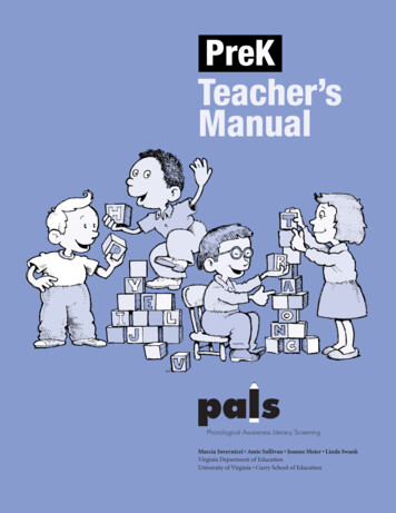
Child and Infant CPRChild CPR 1. Tap and ask: Are you OK?o If inadequate: Send someone to call 911/call cod blue and bring an AED (AEDs are approved for children 0 –until puberty).C. Check Brachial or femoral pulse for no more than 10 seconds. If pulse is felt, give 12-20 breaths per minute (one every 3-5 seconds). If pulse not definitely felt, give 30 compressions in center of chest on low half of theSternum. Compress 2” depth of chest wall with one or two hands. (at least 1/3 of the depth of the chest One cycle of CPR is 30 compressions and 2 breaths. Give 5 cycles of CPR; minimize interruptions (about 2 minutes).A. Open the airway with the head-tilt/chin lift. give 2 breaths over 1 second each. Each breath should make the chest rise.4. When an AED arrives: After 5 cycles of CPR, turn it on and follow AED’s voice prompts.
PALS Study Guide 220011666 Bulletin: New resuscitation science and American Heart Association treatment guidelines were released October 28, 2015! The new AHA Handbook of Emergency Cardiac Care (ECC) contains these 2016 Guidelines and is required study for this course. The 2016 PALS Provider Manual is not yet available. This study guide will provide you with additional study information .