Transcription
12/29/2012818.766.1111ACLS123.COMPALSSTUDYGUIDEC R IT IC A L C A R E T R A IN IN G C E N T E R C O P Y R IG H T 2 0 1 2
Course OverviewThis study guide is an outline of content that will be taught in the American Heart Association Accredited PediatricAdvance Life Support (PALS) Course. It is intended to summarize important content, but since all PALS contentcannot possibly be absorbed in a class given every two years, it is expected that the student will have the 2010Updated ECC Handbook readily available for review as a reference. The student is also required to have the AHAPALS Textbook available for reference and study for more in depth content. AgendaoooooWelcome, Introduction, OverviewVideo ReviewBLS ReviewSimulation Base Scenarios§ PALS Algorithms§ Rapid Cardiopulmonary Assessment§ Skills Stations§ Skills EvaluationWritten EvaluationEvidence Based UpdatesApproximately every 5 years the AHA updates the guidelines for CPR and Emergency Cardiovascular Care. Theseupdates are necessary to ensure that all AHA courses contain the best information and recommendations that can besupported by current scientific evidence experts from outside the United States and outside the AHA. The guidelineswere then classified as to the strength of evidence that supports the recommendation.ObjectivesUpon the completion of this PALS course the participant will be able to: Identify lethal rhythmsDescribe Rapid Cardiopulmonary Assessment and use it as a guide while working through scenariosVerbalize treatment algorithms for each of the following lethal rhythms:o Pulseless arresto Bradycardiao TachycardiaVerbalize steps to assess and treat shockPerform skills in 4 required skill stationso Bag-Mas Ventilation and Advance Airwayo Arrhythmia recognition and Management, Cardioversion and Defibrillationo Vascular Accesso BLSBLS Review(Primary Survey Approach to ECC)C–A–BC circulationA AirwayB BreathingD DefibrillationRescue Techniques – CAB and DUnresponsiveness: After determining that the scene is safe, check to see if victim is responsive and breathingnormally. If the infant or child victim is unresponsive and NOT breathing normally, send someone to activate theemergency response system (EMS) – phone 911 and get the AED.IF IN THE HOSPITAL, CALL THE CODE!!
If alone the rescuer calls out for “HELP” immediately for infants and children and begins C-A-B CPR and then phone911 after two minutes of rescue support. The goal of “phone fast” approach is to deliver oxygen quickly because themost common cause of cardiac arrest in infants and children is a severe airway breathing problems, respiratoryarrest, or shock.Exception: for sudden, witnessed collapse of child or infant, active EMS immediately after verifying that victim isunresponsive.Circulation: Check for pulse for 5 – 10 seconds.Push Hard. Push Fast. Allow for full chest recoil. Minimize interruptions. Avoid Hyperventilation The best location for performing a pulse check for a child is the carotid artery of the neck. On an infant upto the age of one year, check the brachial pulseYou should start cycles of chest compression and breathing when the victim is unresponsive, is notbreathing adequately, and does not have a pulseThe compression to ventilation ratio is 30:2Proper compression technique requires the right rate and depth of compression, as well as full chest recoil.Take your weight off your hands and allow the chest to come back to its normal position. Full chest recoilmaximizes the return of blood to the heart after each compression.The rate of performing chest compression for a victim of any age (adult, child and infant) is at a rate of atleast 100 compressions per minute.Compressions on the child, two hands are placed in the center of the chest between the nipples on the lowerhalf of the sternum.Compressions on an infant are performed by using the two finger technique (pressing two fingers along thesternum, just below the nipple line, and the fingers of the hand wrap around the back and press in with eachcompression)Compression depth is about 2 inches on a child and about 1 ½ inches on an infant.Rotation of 2 – man CPR is every 2 minutes (5 cycles of 30:2) or after 5 cycles of 15:2 for two person CPR oninfant and children.Minimize interruptions in chest compression will increase the victim’s chance of survival.Airway: Open the Airway The head tilt-chin lift is the best way to open unresponsive victim’s airway when you do NOT suspectcervical spine injury.The jaw-thrust with cervical spine immobilization is used for opening airway without tilting the head ormoving the neck if a neck injury is suspected (this includes drowning victims) – after two unsuccessfulattempts, use head tilt-chin lift.Breathing: Given two breaths To give breaths, pinch the victim’s nose closed, or for an infant place your mouth over the infant’s nose andmouth, and given 1 breath (blow for 1 second), watch for the chest to rise. If the chest does not rise, make asecond attempt to open the airway with a health tilt-chin lift. Then give 1 breath (blow over 1 second) andwatch for the chest to rise. Of course, if using mask barrier device or bag mask ventilation, there is no needto pinch the nose. Only provide enough air to see the chest rise and fall. If using a bag mask, there is noneed to compress the bag completely.DO NOT over-inflate the lungs. The positive pressure in the chest that is created by rescue breaths willdecrease venous return to the heart. This limits the refilling of the heart, so it will reduce cardiac outputcreated by subsequent chest compressions.Some victims may continue to demonstrate agonal or gasping breaths for several minutes after a cardiacarrest, but these breaths are too slow or too shallow and will not maintain oxygenation. If there is a pulse,perform rescue breathing.
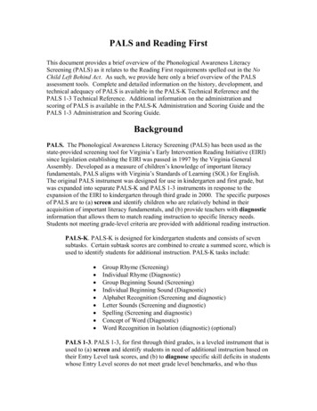
Defibrillation: Attach the Automated External Defibrillator (AED) The probably of successful defibrillation diminishes rapidly over time. Immediate CPR and defibrillationwithin no more than 3 to 5 minutes given a person in sudden cardiac arrest the best chance of survival.The AED is used on an adults, children and infants.If pediatric pads are unavailable, it is acceptable to use adult pads on an infant in cardiac arrestAdult or Child victim: place one pad on the victim’s upper right chest just below the collar bone and to theright of the sternum and the other pad on the left side and below the nipple, being careful that the pads donot touch. If the infant or child is small and the pads would touch, place the pads in an anterior/posteriorposition.Steps for defibrillation are: Power on the AED & Attach pads, clear the victim and allow the AED to analyzethe rhythm – make sure not to touch the victim during the analyze phase, clear the victim and deliver shock,if advised.Make sure to clear the victim before shocking so that you and others helping do not get shocked.If not shock is advised, leave the AED pads on the victim and continue CPR, beginning with compressions.CPR alone may not save the life of sudden cardiac arrest victim. Early defibrillation is needed.Primary Assessment: ABCDEA – Airway Look for movement of the chest or abdomenListen for air movement and breath soundsStatusDescriptionAirway is open and unobstructed for normalbreathingAirway is obstructed but can be maintained bysimple measures (eg, head tilt-chin lift)Airway is obstructed and cannot be maintainedwithout advanced interventions (eg, intubation)ClearMaintainableNot MaintainableB – Breathing Respiratory RateRespiratory EffortChest expansion and air movementLung and airway soundsOxygen saturation by pulse oximetryA consistent respiratory rate of less than 10 or more than 60 breaths/min in a child of any age is abnormal and suggeststhe presence of a potentially serious problem.AgeBreaths/minInfant ( 1 year)30 to 60Toddler (1 - 3 years)24 to 40Preschooler (4 - 5 years)22 to 34School Age (6 to 12 years)18 to 30Adolescent (13 - 18 years)12 to 16
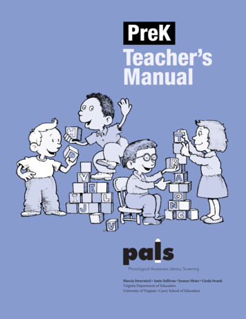
Head Bobbing – often indicate that the child has increased risk for deterioration Caused by the use of neck muscles to assist breathingMost frequently seen in infants and can be a sign of respiratory failurePulse Oximetry Readings A child may be in respiratory distress yet maintain normal oxygen saturation by increasing respiratory rateand effort, especially if supplementary oxygen is administered.C – Circulation Heart rate and rhythmPulses (both peripheral and central)Capillary refill timeSkin color and temperatureBlood o1901302yearsto10years60to14080 2795to13147to8545to85
geSystolicBloodAgePressure(mmHg)TermNeonates(0- ‐28days) 60Infants(1to12months) 70Children1to10years(5thblood 70 (ageinyearspressurepercentile)x2)Children 10years 90D – DisabilityDecrease level of consciousnessLoss of muscular toneGeneralized seizuresPupil dilation rmalextensioninresponsetopain)NoneNoneNone3
E – Exposure Undress the seriously ill or injured child as necessary to perform a focused physical examinationMaintain cervical spine precaution when turning any child with a suspected neck or spine injury.Look for evidence of trauma such as bleeding, burns, or unusual markings that suggest nonaccidentaltraumaManagement of Respiratory EmergenciesRespiratory Problems can be categorized into two categories based upon severity Respiratory Distress: tachypnea, increased respiratory effort, grunting, stridor, wheezing, seesawing or“abdominal” breathing, head bobbingRespiratory Failure: bradypnea, periodic apnea, falling heart rate/bradycardia, diminished air movement,low oxyhemoglobin saturation, stupor, coma, poor muscle tone, cyanosisRespiratory Problems are categorized into four categories based upon type: Evaluate by observing symmetric chestexpansion and by listening for bilateral breath sounds. Breath sounds should be auscultated over the anterior andposterior chest wall and in the axillary areas. Listen for intensity and pitch of sounds.Upper Airway obstruction:oooCroup, anaphylaxis and foreign body airway obstruction are common causesInspiratory StridorTreatment – treat croup with modified oxygen and nebulized epinephrine, corticosteroids treatanaphylaxis with IM epinephrine or auto injector, albuterol, antihistamines, corticosteroids, aspirationforeign body by allowing position of comfort and specialty consultation.Lower Airway obstruction:oooAsthma and bronchiolitis are common causes.Expiratory wheezesTreatment – treat bronchiolitis with nasal suctioning and bronchodilator, treat asthma withalbuterol, corticosteroids, SQ epinephrine, magnesium sulfate, terbutalineLung Tissue (Parenchymal) diseaseooPneumonia/Pneumonitis and pulmonary edema are common causesTreatment – treat pneumonia/pneumonitis with albuterol and antibiotics, treat pulmonary edemawith ventilator and vasoactive support, and consider a diureticDisordered control of ventilation:oooIncrease ICP, Poisoning/overdose, and neuromuscular disease are common causesIrregular breathing pattern (“funny breathing”)Treatment – treat increased ICP by avoiding hypoxemia, hypercarbia, and hyperthermia, treatpoisoning/overdose with antidote and call poison control center, treat neuromuscular disease withventilator support.Respiratory DistressoooooooTachypneaIncreased respiratory efforts (eg, nasal flaring, retractions)Inadequate respiratory effort (eg, hypoventilation or bradypnea)Abnormal airway sounds (eg, stridor, wheezing, grunting)TachycardiaPale, cool skinChanges in level of consciousnessRespiratory Failure
ooooooooMarked tachypnea (early)Bradypnea, apnea (late)Increased, decreased, or no respiratory effortsPoor or absent distal air movementTachycardia (early)Bradycardia (late)CyanosisStupor, coma (late)Airway DevicesNasopharyngeal Airway – semi – consciousoooChoose size based upon the diameter of the nostril (a 12F or 3mm will generally fit a fullterm infant)For proper length, measure from the nose to the earA shortened E.T. tube may be usedOral pharyngeal Airway – unconsciousChoose correct size by measuring from the corner of the mouth to the angle of the jaw.Insert while using a tongue depressor to hold the tongue on the floor of the mouthIt is still necessary to keep the head and neck in the sniffing position after the oralpharyngeal airway is in placeoDo not suction for more than 10 seconds at a timeLaryngeal Mask Airway (LMA) – recommended if provider is inexperienced with E.T. tubeoooEndotracheal Tube – usually the ideal airway in hospitalized patientsoThe E.T. tube is placed using a l
Advance Life Support (PALS) Course. It is intended to summarize important content, but since all PALS content cannot possibly be absorbed in a class given every two years, it is expected that the student will have the 2010 Updated ECC Handbook readily available for review as a reference. The student is also required to have the AHA PALS Textbook available for reference and study for more in .