Transcription
Test SummaryANA Screen, IFA, Reflex Titer/Pattern,Reflex Mplx 11 Ab Cascade With IdentRA Test Code: 94954Specimen Requirements: 4 mL room-temperatureserum (red-top tube [no gel]); 2 mL minimumCPT Codes*: 86038, 86431, 86200, 83520. If the ANA IFAtest is positive, reflex testing for titer and pattern (CPT code86039) and 5 antibodies will be performed: dsDNA (CPTcode 86225), Sm/RNP (CPT code 86235), RNP (CPT code86235), Sm (CPT code 86235), chromatin (CPT code 86235).If all 5 are negative, 4 additional antibodies will be tested:SSA (CPT code 86235), SSB (CPT code 86235), Scl-70 (CPTcode 86235), Jo-1 (CPT code 86235). If all 4 are negative, 2additional antibodies will be tested: ribosomal P (CPT code83516) and centromere B (CPT code 86235). Reflex tests areperformed at additional charge.CLINICAL USE Evaluate suspected autoimmune rheumatic diseasesCLINICAL BACKGROUNDAutoimmune rheumatic diseases are conditions in whichthe immune system attacks the joints and certain systems.They are often difficult to diagnose, as their symptoms canbe vague, vary from patient to patient, and often overlap.Laboratory testing can provide useful information, butno single test provides a definitive diagnosis for any onerheumatic disease.Diagnosis is most often based on a compilation of symptomsand signs, including clinical information and laboratory testresults. Testing for antinuclear antibodies (ANAs) using animmunofluorescence assay (IFA) is a good first approach forlaboratory evaluation of patients suspected of having certainautoimmune rheumatic diseases.ANAs, a group of autoantibodies directed against diversenuclear and cytoplasmic antigens, are associated with severalautoimmune rheumatic diseases (Table 1). These includesystemic lupus erythematosus (SLE), systemic sclerosis (SSc),and mixed connective tissue disease (MCTD). Although variousplatforms can be used to detect ANAs, an IFA with HEp-2 cellsremains the gold standard1 because of its high sensitivity forseveral of the autoimmune rheumatic diseases.2 The highsensitivity stems from the large number of autoantigens (upto 150) in HEp-2 cells.The diagnostic value of ANA testing varies with the specificclinical condition (Table 1). For example, positive ANA resultsare required for diagnosis of drug-associated lupus andMCTD. Testing is also recommended when SLE or SSc aresuspected. ANA testing may also be helpful in evaluatingSjögren syndrome and polymyositis/dermatomyositis.Individuals with rheumatoid arthritis (RA) or other rheumatoiddisorders may be positive or negative for ANAs.Knowing the ANA titer can be helpful in interpreting positiveANA results. A titer of at least 1:40 is considered positive,although most patients with autoimmune disease will havehigher levels. Low-positive titers (eg, 1:40) are not uncommonin healthy individuals.6 For example, about 20% to 30%of healthy individuals have positive titers of 1:40, but thisprevalence drops to 15% for titers of 1:80 and 5% for titers 1:160.4 Nevertheless, using a low threshold of 1:40 canincrease sensitivity for SLE, systemic sclerosis, and Sjögrensyndrome.6For patients with positive ANA screening results, nuclear andcytoplasmic antibody fluorescence patterns can be used toevaluate the likelihood of certain autoimmune diseases.7,8These patterns may inform the differential diagnosis,although they may not be specific for individual antibodies ordiseases. For example, a homogenous nuclear pattern may beassociated with SLE, drug-induced SLE/vasculitis, or juvenileidiopathic arthritis (JIA), while a diffuse cytoplasmic patterncould be consistent with SLE or an inflammatory myopathy.1Given the range of conditions associated with positive ANA IFAresults, positive results are not highly specific for any singlecondition. Tests for individual antibodies may offer greaterspecificity9,10 and are often needed to help establish thediagnosis.4,7 The combination of the highly sensitive ANA IFAscreen, followed by specific autoantibody testing when the IFAis positive, can help maximize sensitivity as well as specificity.Testing tiers of specific antibodies following a positive ANAIFA result, as depicted in the Figure, can help speed theevaluation. Table 2 summarizes the sensitivity of individualantibodies across disorders.The ANA Screen, IFA, Reflex Titer/Pattern, Reflex Mplx 11 AbCascade with IdentRA includes ANA IFA and a tiered cascade
Test Summaryof specific autoantibodies (Figure). The “IdentRA” componentsof the test include widely used laboratory markers for RA—rheumatoid factor (RF) and cyclic citrullinated peptide (CCP)antibodies11—as well as serum 14-3-3η protein, a biomarkerfor diagnosis of RA.26 Used in conjunction with RF and CCPantibody testing, 14-3-3η improves sensitivity for diagnosisof early and established RA (Table 3).26 In 21% to 67% ofseronegative RA patients, 14-3-3η was found to be positive.Since a positive result for any of these RA markers facilitatestimelier diagnosis of ANA-negative RA, simultaneous testingcan enable prompt initiation of disease-modifying therapybefore significant joint erosion develops. Although RA isnot generally associated with a high prevalence of ANA,occasional patients may present with features of RA and SLE(“rhupus”) and be positive for ANA and IdentRA analytes. RAcan be present as a comorbid condition in patients with otherautoimmune rheumatic diseases, and also in patients withsuspected rheumatic diseases who test negative for ANAs.METHOD CCP antibody and 14-3-3η testing performed usingELISA-based assays RF testing performed with immunonephelometry ANA screening conducted using an IFA performed withHEp-2 cells–– Negative ANA IFA results at the 1:40 dilution terminatethe reflex cascade–– Positive ANA IFA results at the 1:40 dilution promptINDIVIDUALS SUITABLE FOR TESTING Individuals with signs and symptoms associated withreflex to Reporting of the corresponding antibodyfluorescence pattern Titer is determined by serial dilution until thepattern cannot be observed, or to a dilution of1:1280; specimens may be titrated to endpointupon request The 3-tier autoantibody reflex cascade (Figure) isperformed using a multiplex immunoassayautoimmune disease(s)Table 1. Utility of Antinuclear Antibody IFA Test by -associatedlupus Positive ANA part of the diagnostic criteria ANA useful for symptomatic people who are taking a drug associated with drug-induced lupusMixed connectivetissue disease(MCTD) Positive ANA part of the diagnostic criteriaANA recommended when clinical suspicion of MCTDFollow-up with RNP antibody recommended to confirm diagnosis ANA sensitivity 93%, specificity 57%Best initial test when clinical suspicion of SLE is strongSLE unlikely if ANA negativeSpecific antibody tests recommended as follow-up to positive ANAAutoimmunehepatitisSystemic lupuserythematosus (SLE)Systemic sclerosis(SSc)Sjögren syndrome Polymyositis/dermatomyositisPositive ANA part of diagnostic criteriaPositive ANA often seen in patients with diverse liver disease; does not exclude otherhepatic diseasesANA sensitivity 85%, specificity 54%ANA recommended when clinical suspicion of SScIf negative, consider other fibrosing illnesses (eg, eosinophilic fasciitis, linear scleroderma)Sensitivity 48%, specificity 52% (if ANA titer 320, sensitivity 72.8% [95% CI, 67.5–77.7%]and specificity 80.4% [95% CI, 76.9–84.0])Can help clarify whether an underlying connective tissue disease exists when Sjögrensyndrome suspected ANA sensitivity 61%, specificity 63% Positive ANA provides weak evidence of disease even when combined with clinical suspicion Must consider other connective tissue diseases (SLE or overlap syndrome) regardless ofANA statusANA, antinuclear antibody test; RNP, ribonucleoprotein.a The American College of Rheumatology Ad Hoc Committee on Immunologic Testing Guidelines 3
Table 2. Autoantibody Prevalence (%) in Rheumatic and Related Diseasesa LEANA9310048856170NACENP-B3-127c 227d 266Chromatin37-73 8012148dsDNA57-620-811-20810-43 27e 2 2177 2 2 222-48 8012148Scl-702-37f 216d 2Sm20-3084010Sm/RNP3054-94949SS-A/Ro33-5213 802342SS-B/Lag13-27 2 805 2Jo-1Rib PRNP9-30ANA, antinuclear antibody; CENP-B, centromere B; dsDNA, double-stranded DNA; Jo-1, histidyl-tRNA synthetase; NA, not available; MCTD,mixed connective tissue disease; Rib P, ribosomal P; RNP, ribonucleoprotein; Scl-70, scleroderma (topoisomerase 1); SLE, systemic lupuserythematosus; Sm, Smith; Sm/RNP, Smith/ribonucleoprotein; and SS-A, SS-B, Sjögren antibodies A and B.a Highlighted antibodies represent diagnostic criteria for the disease. Note that antibodies whose presence is a diagnostic criterion do notalways correspond to those with the highest prevalence in that disease.b CREST syndrome is a variant of systemic sclerosis defined by the presence of calcinosis cutis, Raynaud phenomenon, esophagealdysmotility, sclerodactyly, and telangiectasia. Also known as limited cutaneous scleroderma.c Typically in cases with features of polymyositis.d The presence of scleroderma-related antibodies (centromere, Scl-70, or RNA polymerase III antibodies) is not necessary or sufficient fordiagnosis, but is useful for classification in the absence of diagnostic clinical findings (“clear skin thickening of the fingers extending proximalto the metacarpophalangeal joints”24).e Especially in cases with features of muscle inflammation.f Especially in cases with features of systemic sclerosis.g SS-B is no longer included in consensus classification criteria for primary Sjögren syndrome.25Table 3. Sensitivity and Specificity of RF, CCP Antibody, and 14-3-3η for Detecting RA26Early RAa (n 99)MarkersEstablished RAa (n 57858485CCP antibody5999799914-3-3η649377937284888478789078RF and CCPantibodybRF, CCP antibody, and 14-3-3ηbEarly rheumatoid arthritis (RA) indicates disease history of 6 months or less. Established RA indicates disease history of 6 months.RF, rheumatoid factor; CCP, cyclic citrullinated peptide.a Comparison with healthy controls.b Results considered positive for RA if any of the biomarkers are positive.
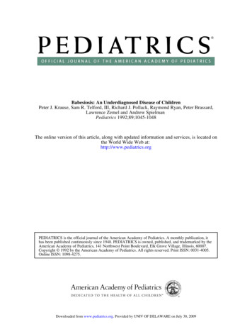
Test SummaryFigure. ANA Screen, IFA, Reflex Titer/Pattern, Reflex Multiplex 11-Ab Cascade, With IdentRA (test code 94954)Patient with symptoms suggestive of autoimmune rheumatic diseaseANA screen, IFA, reflex titer/pattern, reflexmultiplex 11-Ab cascade, with IdentRAIdentRA (14-3-3ƞ, RF, CCP IgG)ANA screen by IFANegativePositiveNegativeRA diseaseless likelyConsider RAAutoimmunedisease less likelyNegativeTier 2SS-A, SS-B, Scl-70, and Jo-1 antibodiesPositiveNegativeTier 3Centromere B and ribosomal P ychiatricSLECentromereB -RibosomalP- yositisSS-A --SS-B --Scl-70- a-Jo-1-- a Detection of RNA polymerase III and centromere antibodiesalso contribute to classification of systemic sclerosismay
PositiveTiter and patternTier 1dsDNA, chromatin, Sm, Sm/RNP, and RNP antibodiesAny positiveAntibody testSystemic LupusErythematosusMixed ConnectiveTissue DiseasedsDNA (high specificity)-Chromatin (high sensitivity)-Sm (high specificity)-Sm/RNP RNP If ANA IFA is positive, and no positive specific antibodies are detected, correlate with clinical findings and consider otherautoimmune diseases and tests.Sub-panels that can be ordered separately include test code 91472 (dark green fill ), test code 249 (blue fill), and test code 16814 (blue fill pluspurple fill).The acronym CREST refers to a syndrome defined by presence of calcinosis cutis, Raynaud phenomenon, esophageal dysmotility,sclerodactyly, and telangiectasia. CCP indicates cyclic citrullinated peptide; dsDNA, DNA; IFA, immunofluorescence assay; Jo-1, histidyl-tRNAsynthetase; RA, rheumatoid arthritis; RF, rheumatoid factor; Sm/RNP, Smith/ribonucleoprotein; SS-A and -B, Sjögren syndrome A and B; andScl-70, systemic sclerosis (topoisomerase I).This figure was developed by Quest Diagnostics based in part on references 2,9, and 11-14. It is provided for informational purposes only and isnot intended as medical advice. A physician’s test selection and interpretation, diagnosis, and patient management decisions should be basedon his/her education, clinical expertise, and assessment of the patient.
Test Summary Aside from ANA titer and pattern, panel components maybe ordered separately (Table 4). Reflex tests are performed at additional charge and areassociated with an additional CPT code(s).REFERENCE RANGESANA IFA Screen: NegativeANA Titer: Negative: 1:40 Low antibody level:1:40-1:80 Elevated antibody level: 1:80RF: 14 IU/mLCCP antibody: Negative: 20 Units Weak positive:20-39 Units Moderate positive:40-59 Units Strong positive:14-3-3η: 59 Units 0.2 ng/mLINTERPRETIVE INFORMATIONA positive ANA result in conjunction with clinical suspicionsuggests, but does not necessarily confirm, the presence ofan autoimmune disease. Positive results are not uncommonin healthy individuals (particularly as they age) and thosewith certain infectious diseases or cancer.12 In cases withstrong clinical suspicion, specific antibody testing may beappropriate even if the ANA IFA result is negative.A positive ANA IFA result with a positive result for 1 or more ofthe specific antibodies may suggest the presence of a certainautoimmune disease (Table 2). If the ANA is positive but the 11antibodies tested in the cascade antibody tests are negative,the patient may still have an autoimmune disease other thanthose typically associated with the antibodies tested. Testsfor other autoimmune diseases may be considered if clinicallyindicated; these include autoimmune hepatitis, primarybiliary cholangitis, autoimmune thyroiditis, Addison’s disease,pernicious anemia, autoimmune neuropathies, vasculitis,celiac disease, and bullous disease.A negative ANA IFA result suggests the absence of manyautoimmune diseases, but does not rule them out. Thus,Table 4. Individual Tests Included in the ANA Screen, IFA, Reflex Titer/Pattern, Reflex Mplx 11-AbCascade With IdentRA Test CodeTest NameCPT CodePatterna249ANA Screen, IFA, with Reflex to Titer and16814ANA Screen, IFA, Reflex Titer/Pattern, and Reflex to Multiplex 11 Ab Cascadea (Figure)8603886038255DNA (ds) Antibody8622538567Sm/RNP Antibody8623519887RNP Antibody8623537923Sm Antibody8623534088Chromatin (Nucleosomal) Antibody8623538568Sjögren's Antibody (SS-A)8623538569Sjögren's Antibody (SS-B)862354942Scleroderma Antibody (Scl-70)862355810Jo-1 Antibody8623534283Ribosomal P Antibody8351616088Centromere B Antibody862354418Rheumatoid Factor8643111173Cyclic Citrullinated Peptide (CCP) Antibody (IgG)862009145514-3-3 eta Proteinb83520a Reflex tests are performed at additional charge and are associated with an additional CPT code(s).b This test was developed and its analytical performance characteristics have been determined by Quest Diagnostics. It has not been clearedor approved by the U.S. Food and Drug Administration. The FDA has determined that such clearance or approval is not necessary. This assayhas been validated pursuant to the CLIA regulations and is used for clinical purposes.
specific autoantibody testing may be considered for patientswith negative ANA IFA results if clinical suspicion is high (eg,Jo-1 for suspected polymyositis/dermatomyositis, SS-A forsuspected Sjögren syndrome).A positive result on the RF, CCP antibody, or 14-3-3η testsgenerally supports a diagnosis of early or establishedRA, or potential subclinical RA (Table 3). In ANA-negativepatients, positive results are consistent with early RA.27 Anegative result on all 3 tests is consistent with absence ofRA (sensitivity 78%), although early RA cannot be ruled out(Table 3). If clinically indicated, a negative test result may alsobe followed up with tests for other autoimmune diseases.References1. Agmon-Levin N, Damoiseaux J, Kallenberg C, et al. Internationalrecommendations for the assessment of autoantibodies tocellular antigens referred to as anti-nuclear antibodies. AnnRheum Dis. 2014;73:17-23.9. Satoh M, Chan EK, Sobel ES, et al. Clinical implication ofautoantibodies in patients with systemic rheumatic diseases.Expert Rev Clin Immunol. 2007;3:721-738.10. Kumar Y, Bhatia A, Minz RW. Antinuclear antibodies and theirdetection methods in diagnosis of connective tissue diseases: ajourney revisited. Diagn Pathol. 2009;4:1.11. Aletaha D, Neogi T, Silman AJ, et al. 2010 rheumatoid arthritisclassification criteria: an American College of Rheumatology/European League Against Rheumatism collaborative initiative.Arthritis Rheum. 2010;62:2569-2581.12. Petri M, Orbai AM, Alarcon GS, et al. Derivation and validationof the Systemic Lupus International Collaborating Clinicsclassification criteria for systemic lupus erythematosus. ArthritisRheum. 2012;64:2677-2686.13. Cappelli S, Bellando Randone S, Martinovic D, et al. “To be or not tobe,” ten years after: evidence for mixed connective tissue diseaseas a distinct entity. Semin Arthritis Rheum. 2012;41:589-598.14. Stinton LM, Fritzler MJ. A clinical approach to autoantibodytesting in systemic autoimmune rheumatic disorders. AutoimmunRev. 2007;7:77-84.2. American College of Rheumatology. Methodology of testing forantinuclear antibodies (position statement). n%20Statement.pdf.Published January 2009. Updated August 2015. AccessedDecember 5, 2017.15. Hoffman IE, Peene I, Meheus L, et al. Specific antinuclearantibodies are associated with clinical features in systemic lupuserythematosus. Ann Rheum Dis. 2004;63:1155-1158.3. Solomon DH, Kavanaugh AJ, Schur PH, et al. Evidence-basedguidelines for the use of immunologic tests: antinuclear antibodytesting. Arthritis Rheum. 2002;47:434-444.17. Colglazier CL, Sutej PG. Laboratory testing in the rheumaticdiseases: a practical review. South Med J. 2005;98:185-191.4. Kavanaugh A, Tomar R, Reveille J, et al. Guidelines for clinicaluse of the antinuclear antibody test and tests for specificautoantibodies to nuclear antigens. American College ofPathologists. Arch Pathol Lab Med. 2000;124:71-81.5. Shiboski SC, Shiboski CH, Criswell L, et al. American College ofRheumatology classification criteria for Sjögren’s syndrome:a data-driven, expert consensus approach in the Sjogren’sInternational Collaborative Clinical Alliance cohort. Arthritis CareRes (Hoboken). 2012;64:475-487.6. Tozzoli R, Bizzaro N, Tonutti E, et al. Guidelines for the laboratoryuse of autoantibody tests in the diagnosis and monitoring ofautoimmune rheumatic diseases. Am J Clin Pathol. 2002;117:316324.7. Satoh M, Vazquez-Del Mercado M, Chan EK. Clinical interpretationof antinuclear antibody tests in systemic rheumatic diseases.Mod Rheumatol. 2009;19:219-228.8. Chan EK, Damoiseaux J, Carballo OG, et al. Report of the firstinternational consensus on standardized nomenclature ofantinuclear antibody HEp-2 cell patterns 2014-2015. FrontImmunol. 2015;6:412.16. Cervera R, Vinas O, Ramos-Casals M, et al. Anti-chromatinantibodies in systemic lupus erythematosus: a useful marker forlupus nephropathy. Ann Rheum Dis. 2003;62:431-434.18. Scholz J, Grossmann K, Knutter I, et al. Second generationanalysis of antinuclear antibody (ANA) by combination ofscreening and confirmatory testing. Clin Chem Lab Med.2015;53:1991-2002.19. Moder KG, Wener MH, Weisman MH, et al. Measurement ofantinuclear antibodies by multiplex immunoassay: a prospective,multicenter clinical evaluation. J Rheumatol. 2007;34:978-986.20. Binder SR, Genovese MC, Merrill JT, et al. Computer-assistedpattern recognition of autoantibody results. Clin Diagn LabImmunol. 2005;12:1353-1357.21. Mouthon L, Dunogue B, Guillevin L. Diagnosis and classificationof eosinophilic granulomatosis with polyangiitis (formerly namedChurg-Strauss syndrome). J Autoimmun. 2014;48-49:99-103.22. Tur BS, Suldur N, Ataman S, et al. Anti-neutrophil cytoplasmicantibodies in patients with rheumatoid arthritis: clinical,biological, and radiological correlations. Joint Bone Spine.2004;71:198-202.23. van Paassen P, Damoiseaux J, Tervaert JW. Laboratory assessmentin musculoskeletal disorders. Best Pract Res Clin Rheumatol.2003;17:475-494.
Test Summary24. van den Hoogen F, Khanna D, Fransen J, et al. 2013 classificationcriteria for systemic sclerosis: an American College ofRheumatology/European League Against Rheumatismcollaborative initiative. Arthritis Rheum. 2013;65:2737-2747.26. Maksymowych WP, Naides SJ, Bykerk V, et al. Serum 14-33eta is a novel marker that complements current serologicalmeasurements to enhance detection of patients with rheumatoidarthritis. J Rheumatol. 2014;41:2104-2113.25. Shiboski CH, Shiboski SC, Seror R, et al. 2016 American Collegeof Rheumatology/European League Against Rheumatismclassification criteria for primary Sjogren’s syndrome: aconsensus and data-driven methodology involving threeinternational patient cohorts. Ann Rheum Dis. 2017;76:9-16.27. Singh JA, Saag KG, Bridges SL Jr, et al. 2015 American College ofRheumatology guideline for the treatment of rheumatoid arthritis.Arthritis Rheumatol. 2016;68:1-26.* The CPT codes provided are based on AMA guidelines and are for informational purposes only. CPT coding is the sole responsibility of thebilling party. Please direct any questions regarding coding to the payer being billed.This test was developed and its analytical performance characteristics have been determined by Quest Diagnostics. It has not been clearedor approved by the U.S. Food and Drug Administration. This assay has been validated pursuant to the CLIA regulations and is used for clinicalpurposes.QuestDiagnostics.comQuest, Quest Diagnostics, any associated logos, and all associated Quest Diagnostics registered or unregistered trademarks are the property of Quest Diagnostics.All third-party marks— and —are the property of their respective owners. 2018 Quest Diagnostics Incorporated. All rights reserved. TS7392 01/2018
multiplex 11-Ab cascade, with IdentRA Sub-panels that can be ordered separately include test code 91472 (dark green fill ), test code 249 (blue fill), and test code 16814 (blue fill plus purple fill).