Transcription
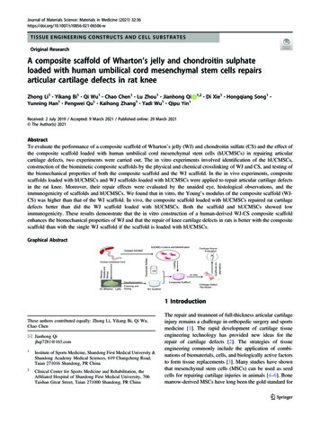
Journal of Materials Science: Materials in Medicine (2021) SUE ENGINEERING CONSTRUCTS AND CELL SUBSTRATESOriginal ResearchA composite scaffold of Wharton’s jelly and chondroitin sulphateloaded with human umbilical cord mesenchymal stem cells repairsarticular cartilage defects in rat kneeZhong Li1 Yikang Bi1 Qi Wu1 Chao Chen1 Lu Zhou1 Jianhong QiYunning Han1 Pengwei Qu1 Kaihong Zhang1 Yadi Wu1 Qipu Yin1 1,2 Di Xie1 Hongqiang Song1 1234567890();,:1234567890();,:Received: 2 July 2019 / Accepted: 9 March 2021 / Published online: 29 March 2021 The Author(s) 2021AbstractTo evaluate the performance of a composite scaffold of Wharton’s jelly (WJ) and chondroitin sulfate (CS) and the effect ofthe composite scaffold loaded with human umbilical cord mesenchymal stem cells (hUCMSCs) in repairing articularcartilage defects, two experiments were carried out. The in vitro experiments involved identification of the hUCMSCs,construction of the biomimetic composite scaffolds by the physical and chemical crosslinking of WJ and CS, and testing ofthe biomechanical properties of both the composite scaffold and the WJ scaffold. In the in vivo experiments, compositescaffolds loaded with hUCMSCs and WJ scaffolds loaded with hUCMSCs were applied to repair articular cartilage defectsin the rat knee. Moreover, their repair effects were evaluated by the unaided eye, histological observations, and theimmunogenicity of scaffolds and hUCMSCs. We found that in vitro, the Young’s modulus of the composite scaffold (WJCS) was higher than that of the WJ scaffold. In vivo, the composite scaffold loaded with hUCMSCs repaired rat cartilagedefects better than did the WJ scaffold loaded with hUCMSCs. Both the scaffold and hUCMSCs showed lowimmunogenicity. These results demonstrate that the in vitro construction of a human-derived WJ-CS composite scaffoldenhances the biomechanical properties of WJ and that the repair of knee cartilage defects in rats is better with the compositescaffold than with the single WJ scaffold if the scaffold is loaded with hUCMSCs.Graphical Abstract1 IntroductionThese authors contributed equally: Zhong Li, Yikang Bi, Qi Wu,Chao Chen* Jianhong Qijhqi7281@163.com1Institute of Sports Medicine, Shandong First Medical University &Shandong Academy Medical Sciences, 619 Changcheng Road,Taian 271016 Shandong, PR China2Clinical Center for Sports Medicine and Rehabilitation, theAffiliated Hospital of Shandong First Medical University, 706Taishan Great Street, Taian 271000 Shandong, PR ChinaThe repair and treatment of full-thickness articular cartilageinjury remains a challenge in orthopedic surgery and sportsmedicine [1]. The rapid development of cartilage tissueengineering technology has provided new ideas for therepair of cartilage defects [2]. The strategies of tissueengineering commonly include the application of combinations of biomaterials, cells, and biologically active factorsto form tissue replacements [3]. Many studies have shownthat mesenchymal stem cells (MSCs) can be used as seedcells for repairing cartilage injuries in animals [4–6]. Bonemarrow-derived MSCs have long been the gold standard for
36Page 2 of 11bone and cartilage tissue engineering [7]. However, bonemarrow MSCs face problems such as the low yield ofsingle-pumped stem cells, difficulty in culture expansion,donor infection, and differentiation potential inverselyproportional to age [8] and limited use. In contrast, umbilical cord (UC)-derived MSCs have desirable characteristics. First, the UC, which is discarded at birth, provides aninexhaustible source of stem cells for therapy. Furthermore,human UC mesenchymal stem cells (hUCMSCs) havefaster proliferation rates and greater expansion capabilitythan adult MSCs and possess broad multipotency withoutinducing teratomas [9, 10]. The efficacy of hUCMSCs incartilage repair has been demonstrated in animal studies[8, 11, 12], and hUCMSCs are thus prospective cell candidates for tissue-engineered cartilage.The construction of engineered tissues is often based onthe structures of scaffold biomaterials that support thefunction and growth of new tissue. Both the material andmanufacturing process must be considered to ensure that themechanical and physiological properties of the de novotissue closely match those of the native tissue [13].Increasing levels of complexity in repairing tissues or organsare generally consistent with a concomitant increase in thecomplexity of the associated tissue engineering approach[3]. In recent years, Wharton’s jelly (WJ) has been found tohave components similar to those of articular cartilage andcontain some chondrogenic growth factors, such as insulinlike growth factor I and transforming growth factor-β. Studies have shown that cartilage tissue engineering scaffoldsprepared with WJ can effectively repair articular cartilagedefects [14, 15, 17]. Thus, WJ has become a promising rawmaterial for tissue-engineered cartilage [16–18]. However,its physical properties, such as modulus of elasticity, usuallydo not satisfy the requirements of cartilage repair. Theseproperties may be modified by introducing intermolecularcrosslinks [19]. Chondroitin sulfate (CS), with a relativemolecular mass ranging from 10,000 Da to 50,000 Da, is anacidic polysaccharide composed of disaccharide units ofglucuronic acid and aminohexose. CS distributed in variousanimal tissues has been demonstrated to possess variousbiological activities, such as adjusting immunity and fatmetabolism, preventing infection, and reducing the risk ofheart diseases [20]. CS has broad application prospects inthe fields of medicine, food and cosmetics, and the use of CSin the preparation of bioengineered scaffolds can improvethe physical characteristics of the scaffold material [21, 22].According to the requirements of tissue-engineeredscaffolds, does the scaffold composite prepared by crosslinking WJ with CS have greater biomechanical performance? Does the composite scaffold containing WJ and CShave a better repair effect in vivo than the WJ scaffoldalone? No relevant reports that answer these questions havebeen published. In this study, a novel composite scaffoldJournal of Materials Science: Materials in Medicine (2021) 32:36was prepared with a WJ scaffold, its performance was testedin vitro, and the cartilage defect of a rat knee was repairedin vivo with different scaffolds loaded with hUCMSCs. Therepair effect was assessed by gross appearance and histological grading scores. This study provides an experimentalbasis for the clinical feasibility of tissue-engineeredcartilage.2 Methods and materials2.1 Isolation, culture and identification of hUCMSCsin vitroAll experimental protocols were approved by the MedicalEthics Committee of Shandong First Medical University &Shandong Academy Medical Sciences (No. 201758).Human UCs were collected aseptically from normal fullterm births from the Maternity Department at the AffiliatedHospital of Shandong First Medical University & ShandongAcademy Medical Sciences after informed consent wasobtained. The cords were rinsed twice with phosphatebuffered saline (PBS) (Solarbio, Beijing, China), and thecord blood was cleaned in penicillin and streptomycin. Thewashed cords were cut into 1.0–2.0 mm sized pieces andsuspended in Dulbecco’s modified Eagle’s medium (Gibco,Life Technologies, USA) containing 10% fetal bovineserum (BI, Israel), 5% horse serum (Gibco, USA), penicillinand streptomycin. The cord pieces were then incubated at37 C in a humidified atmosphere with 5% CO2 undernormoxic conditions. The nonadherent cells were thenwashed off. The medium was changed every 3 days. Whenwell-developed colonies of fibroblast-like cells appearedafter 10 days, the cultures were trypsinized (without dilution) and transferred into a new vessel for further expansion;the medium was replaced every 3 days. To detect the typicalmarkers of different passages of hUCMSCs, thirdgeneration hUCMSCs were selected.Immunofluorescence double staining was used to identify the phenotype of the hUCMSCs. A suspension of thirdgeneration hUCMSCs was prepared after digestion with acell digestion solution in 0.25% trypsin (Beijing SoleiChemical Co., Beijing, China) and added dropwise into sixwell plates. The cells were incubated at 37 C in 5% CO2for 6 h. The samples were fixed in paraformaldehyde for10 min, permeabilized with Triton X-100 (Amresco, USA)for 5 min and then incubated with 1% bovine serum albumin (BI Company, Israel) for 1 h. Anti-CD44 at a 1:1000dilution and anti-CD90 at a 1:200 dilution (Abcam, UK)were added, and the moisture of the solutions was preservedduring the overnight incubation at 4 C. The cells wereincubated in PBS for 50 min before the addition of thesecondary antibody (DAKO, Denmark). Then, after they
Journal of Materials Science: Materials in Medicine (2021) 32:36were washed in PBS, the cells were stained with 4′,6-diamidino-2-phenylindole (DAPI) for 5 min and then observedunder a fluorescence microscope (Nikon D90, Japan).2.2 Construction of WJ scaffolds in vitroThe UC tissues were cut open from the center under sterileconditions, and then the outer tissue and vascular tissuewere peeled away. The remaining adhesive tissues (WJ)were rinsed with sterile distilled water for 5 min; this procedure was repeated 3 times. The tissues were subsequentlysterilized in 3% H2O2 for 30 min and washed with steriledistilled water three times for 30 min each time. The jellywas placed in a grinder with three volumes of sterile distilled water and repeatedly crushed into a homogenate at 5 C. Then, after adding five volumes of sterile distilledwater, the homogenate was frozen at 20 C and thenthawed at room temperature. This freeze-thaw cycle wasrepeated four or five times. The homogenate was centrifuged using various centrifugation steps (Beckman Allegra X-22R, USA) for 20 min at 3000 rpm, after which thesupernatant was removed from the homogenate. Then, thesupernatant was centrifuged for 20 min at 6000 rpm andseparated again by gradient centrifugation for 30 min at7000 rpm. Afterward, it was separated again by gradientcentrifugation for 30 min at 10,000 rpm. The final supernatant was discarded, and the precipitate was used as theWharton’s jelly extracellular matrix (WJECM). TheWJECM was placed in a container in which the upperportion of the liquid was in contact with the temperaturegradient to guide the formation of crystals in the solutionalong the vertical temperature gradient. The mold and WJscaffolds were prefrozen for approximately 16 h in avacuum freeze dryer (Yonghao, 2XZ-2B, Linhai, China),after which the scaffolds were stored at 4 C.2.3 Composite scaffold construction in vitroThe WJ scaffold was irradiated under 258 nm ultraviolet lightand crosslinked for 8 h. For chemical crosslinking, the scaffoldswere soaked for 30 min in a 40% ethanol solution supplemented with 50 mmol/L 2-morpholinoethanesulphonic (MES)acid and then soaked for 4 h in CS solution containing 40%ethanol, 24 mmol/L 1-(3-dimethylaminopropyl)-3-ethylcarbodiimide hydrochloride, 5.0 mmol/L N-hydroxysuccinimide and50 mmol/L MES. The CS fraction was divided into threegroups. In group A, the CS quality fraction was 1% (WJ 1%CS); in group B, the CS quality fraction was 2% (WJ 2%CS); and in group C, the CS quality fraction was 3% (WJ 3% CS). The scaffolds were washed repeatedly in PBS, placedin a 80 C freezer for 30 min and then placed into a vacuumfreeze dryer, where they were dried again. After disinfection byultraviolet rays, the samples were stored at 4 C, and thePage 3 of 11 36composite scaffold structure was observed using scanningelectron microscopy, hematoxylin and eosin (H&E) stainingand toluidine blue (TB) staining; subsequently, the porosity,density, water absorption, degradation rate, and physicalproperties of the scaffolds were examined.2.4 Biomechanical testing of the scaffoldsThe WJ scaffold and the 1% CS, 2% CS, and 3% CScomposite scaffolds were shaped into cylinders (n 5) witha diameter of 15.0 mm and a height of 6.0 mm. An Instron3343 biomechanical testing instrument (Instron, 3343,USA) was used to evaluate the biomechanical performanceof the scaffolds in axial compression. First, the scaffoldswere placed in PBS to remain hydrated. After themechanical testing software was opened, the compressionmethod was selected, the mechanical sensors were connected, the preload was set to 0.05 N, and the scaffolds wereplaced between two rigid plates to ensure contact withthe stent surface; the testing speed was 0.05 mm/s, and themeasurement was stopped at a maximum load of 50 N. Themagnitude of the reasonable force was determined according to the compression state. All tests were conductedwithin this range of reasonable forces. The experimentaldata in the test were automatically collected, and Young’smodulus was calculated from the slope of the stress–straincurve. Each experiment was performed three times, and theresults were averaged.2.5 Cartilage defect in a rat model and implantationin vivoThe animal experiments were conducted according to theAnimal Care and Use Committee of Taishan MedicalUniversity (No. 2017041). Male Sprague-Dawley (SD) rats(six weeks old) were selected for the cartilage defect modeland subjected to anesthesia with an intraperitoneal injectionof 0.1 ml/kg of 1% pentobarbital sodium. The rat kneejoints were opened using a medial parapatellar approach. Afull-thickness cylindrical cartilage defect with a diameter of2 mm and a depth of 2 mm was introduced with a cornealtrephine on the groove of the femur in both legs. Processingof the rat defect was divided into three groups: untreatedcontrol group (cartilage defect was not treated with a scaffold or hUCMSCs) (n 10); WJ with hUCMSCs group(cartilage defect was treated with the WJ scaffold andhUCMSCs) (n 10); and WJ CS with hUCMSCs (cartilage defect was treated with the WJ CS scaffold andhUCMSCs) (n 10). After processing the different ratdefects, the incision in the patella was closed by suturing,and an intramuscular injection of penicillin was given toprevent infection. During the postoperative period, thegeneral condition of the animals was observed, and any sign
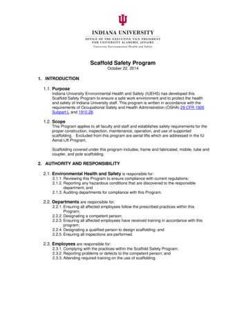
36Page 4 of 11Journal of Materials Science: Materials in Medicine (2021) 32:36Fig. 1 Isolation, culture and identification of hUCMSCs. A hUCMSCswere isolated from WJ and cultured for 7 days. Scale bar 500 μm.B hUCMSCs were cultured and differentiated into chondrocytes for21 days and then stained with alkaline blue. The cells around the cellmass that appear blue and round are chondrocytes rich in acidmucopolysaccharides, as shown in (1). Scale bar 200 μm. (2)represents a portion of the enlarged box in (1). Scale bar 100 μm.C (1) CD90 is shown by green fluorescence; (2) CD44 by red fluorescence; (3) the nucleus by blue fluorescence; (4) merge. Scale bar 100 μmof knee infection was noted. At 4 and 12 weeks, the animalswere anesthetized with sodium pentobarbital and sacrificed;the joint cavity was opened, the knee joint was removed,and the repair state of the cartilage defects was observedfollowed by gross observations. Finally, histological analysis of the samples was performed.2.8 Cell-derived detection of repaired cartilagein vivo2.6 Gross observations in vivoThe surface texture, defect filling/size, and graft-recipientcartilage integration of the samples were grossly examinedto evaluate the macroscopic structure of the repair tissue onthe cartilage defects. The gross score was rated using a scaleof 0–9, where high scores indicated degeneration and/orpoor repair of the cartilage defects [23]. The scores of twoblinded observers were averaged.2.7 Histological staining and scoring in vivoThe samples of repair tissue were fixed in 10% neutralbuffered formalin, decalcified in 10% EDTA, and paraffinembedded for histological analysis. Section 5 μm thick werecut, deparaffinized, and then stained with H&E, TB, safranin O and fast green according to standard procedures. Allhistological staining results were scored according to thehistological scoring protocol described by Wakitani et al.[24]. The score ranged from 0–14 points, with 0 denotingcomplete regeneration and 14 denoting no regeneration.Thirty pathology images were selected, independentlyscored by three observers, and then averaged to increase theaccuracy.Human-specific ribonucleoprotein immunohistochemicalstaining (Chemicon, Temecula, CA, USA) was performedto determine the source of the hUCMSCs in the repair tissuein the cartilage defects of the three groups in vivo. Theprocedure described by Liu et al. [8] was followed [11, 12].2.9 Measurement of interleukin 6 (IL-6) in ratvenous blood in vivoVenous blood was obtained from rats of each group at 2 days,1 week, 2 weeks, and 4 weeks after the operation and compared with that from nonoperative rats. The level of IL-6 wasmeasured by enzyme-linked immunosorbent assay (ELISA).Venous blood samples were collected, and ethylenediaminetetraacetic acid disodium salt was added as an anticoagulant.After mixing for 10–20 min, the supernatant fluid was collected and centrifuged for 20 min (2000–3000 rpm/min). Thereagents, samples, and standards in the ELISA kit (LangtonBiotechnology Co., Ltd., Shanghai, China) for rat IL-6 wereprepared according to the manufacturer’s requirements. Thestandards were prepared, and the biotin-labeled secondaryantibody and ELISA reagents were added to the samples andincubated at 37 C for 60 min. After the samples were washed5 times, a color indicator was added and developed for 10 minat 37 C, after which a stop solution was added. Within10 min, the enzyme-labeled plate was placed into a standardmicroplate reader (Biotek, ELx800, Vermont, USA), and theabsorbance was read.
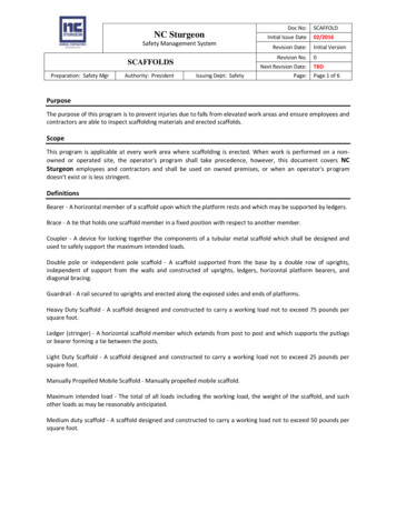
Journal of Materials Science: Materials in Medicine (2021) 32:36Page 5 of 11 36Fig. 2 Scaffold test results. A (1) Gross observation of the scaffold: alacunary, cylindrical and cavernous porous structure. Scale bar 5 mm. (2) The WJ scaffolds were stained with H&E, and the lowmagnification image shows multiple holes and a lattice-like networkwith a three-dimensional structure. Scale bar 100 μm. (3) Scaffoldwith TB staining showing the secretion of proteoglycans. Scale bar 100 μm. B (1) Longitudinal electron microscopy image of a scaffold,with the microtubules in an oriented structure. Scale bar 500 μm. (2)The three-dimensional porous structure of the composite scaffold canbe observed by electron microscopy. Scale bar 100 μm. (3) Scalebar 50 μm. C The Young’s modulus of the 2% composite group wasthe highest, followed by the 1% composite group, the 3% compositegroup and the WJ scaffold group; significant differences were foundbetween the groups (**P 0.01)2.10 Statistical analysisSPSS 19.0 and GraphPad Prism 6 statistical analysis software were used for the statistical analysis and plottinganalysis. All experimental data are expressed as the mean standard deviation. The scores, contents of proteoglycan,acid mucopolysaccharide, and collagen as well as themechanical properties and IL-6 ELISA data for each groupwere compared using variance analysis, and the least significant difference method was used to compare any pairwise difference; a difference of P 0.05 was consideredstatistically significant.microscope, the cells appeared polygonal and spindleshaped, and cell proliferation was robust. After 2 weeks ofculture, the cells were spindle shaped and arranged randomly (Fig. 1A). The hUCMSCs were cultured for 21 days.Next, 5-μm-thick paraffin sections were prepared andstained with alkaline blue; the blue color represents theacidic mucopolysaccharide of cartilage (Fig. 1B). ThehUCMSCs were subjected to double immunofluorescencestaining to detect CD44 and CD90 expression;CD44 staining is shown by red fluorescence, whileCD90 staining is shown by green fluorescence; bothshowed positive results (Fig. 1C).3 Results3.2 Gross observation, histological observation, andphysical properties of the scaffolds in vitro3.1 Characterization and surface markers ofhUCMSCs in vitroThe hUCMSCs were isolated from the WJ of UC tissue.After 7 days in primary culture, the adherent cells of thehUCMSCs could be observed. Under an invertedA white, cylindrical, sponge-like porous structure wasobserved in the scaffolds. The three-dimensional structureof the scaffold was macroscopically visible (Fig. 2A(1)),and H&E staining revealed that the scaffold had a porous,lattice-like three-dimensional structure (Fig. 2A(2)). Positive TB staining indicated proteoglycan secretion in the
36Page 6 of 11Table 1 Physical performance ofthe scaffolds in each groupJournal of Materials Science: Materials in Medicine (2021) 32:36GroupPorosity (%)Density (μg/mm3)Water absorption rate (%)Degradation rate (%)WJ91.13 2.1275.13 6.221147 85.7120.69 1.02WJ 1% CS93.33 1.1182.33 3.791287 72.3921.34 1.73WJ 2% CS92.33 0.7683.40 2.921237 56.0421.24 1.01WJ 3% CS90.83 1.4679.11 3.131036 72.8420.22 2.02cartilage ECM (Fig. 2A(3)). Microscopically, the scaffoldexhibited a three-dimensional porous structure with voids.Under a scanning electron microscope, the compositescaffold with a 2% mass fraction of CS combined with WJshowed a three-dimensional porous structure. The scaffoldswere directional and arranged in parallel, and the intersticeswere interpenetrated. The microtubules were also orientedin a structure (Fig. 2B(1)–(3)). After the physical propertieswere determined, the porosities of the WJ scaffold and thecomposite scaffold were both found to be greater than 90%,and other physical features, such as density, water absorption and degradation rate, met the requirements of tissueengineered scaffolds (Table 1).3.3 Biomechanical properties of the scaffoldsin vitroThe axial compression biomechanical test showed that theYoung’s modulus of the composite scaffold composed ofdifferent concentrations of CS was higher than that of theWJ scaffold (P 0.05) (Fig. 2C). The Young’s modulus ofthe WJ 2% CS composite scaffold was higher than that ofeither the WJ 1% CS or WJ 3% CS composite scaffold.The biomechanical performance of the WJ 2% CS wasthe highest of all the composite scaffolds (Fig. 2C).3.4 Gross observation in vivoAt 12 weeks after surgery, the cartilage defects of theuntreated control group were basically filled, but the repairsite was still rough, and integration with the surroundingnormal cartilage was poor. The repair surface of the rat kneein the WJ with hUCMSCs group was less smooth but waswell integrated into the surrounding normal tissue. In theWJ CS with hUCMSCs group, the color of the repairtissue was close to that of the normal cartilage tissue, andthe flat repair surface was integrated well into the surrounding tissue (Fig. 3(1)). Twelve weeks after surgery, thecartilage repair scores of the three groups were evaluatedbased on the surface features, degree of filling defect, andintegration of the host grafts. The total scores of theuntreated control, WJ with hUCMSCs and WJ CS withhUCMSCs groups were 8.80 0.45, 5.40 1.14, and4.00 0.71, respectively. The P value indicates that thecartilage repair performance of the WJ CS withhUCMSCs group was the best, followed by that of the WJwith hUCMSCs group, and the repair performance of theuntreated control group was the worst (Table 2).3.5 Histological observation and score in vivo3.5.1 H&E stainingAt 4 weeks after surgery, the repaired tissue of theuntreated control group was mainly fibrous tissue, andnumerous fusiform fibrillar cells were observed. Therepaired tissue had obvious fissures, and the wound surface was uneven. The WJ with hUCMSCs group contained a mixture of fibroblasts and chondrocytes, thechondrocytes were unevenly distributed, and the woundsurface was smooth and flat. The repaired tissue of theWJ CS with hUCMSCs group was mainly hyaline cartilage, and many chondrocytes were observed. Thearrangement of the cells in the repaired tissue was similarto that in normal cartilage tissue. The repaired tissueintegrated well with the surrounding normal tissues;however, the wound surface was still uneven. At12 weeks, the repaired tissue of the untreated controlgroup was still fibrous tissue, the repair tissue of the WJwith hUCMSCs group was a mixture of fibrous tissue andhyaline cartilage, and the repaired tissue of the WJ CSwith hUCMSCs group was hyaline cartilage. Thearrangement of the chondrocytes in the WJ CS withhUCMSCs group was very similar to that in normalarticular cartilage. The repaired tissue integrated well withthe surrounding normal cartilage tissues, and the woundsurface was smooth and complete (Fig. 3(2)).3.5.2 TB stainingAt 4 weeks after surgery, the untreated control group wasnegative for TB staining, the WJ with hUCMSCs group wasweakly positive in a small area, and the WJ CS withhUCMSCs group was strongly positive. At 12 weekspostsurgery, the repaired tissue in the untreated controlgroup was still negative for TB staining, that in the WJ withhUCMSCs group was partially positive, and that in the WJ CS with hUCMSCs group was strongly positive and very
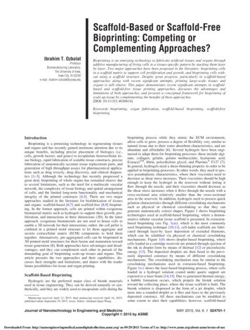
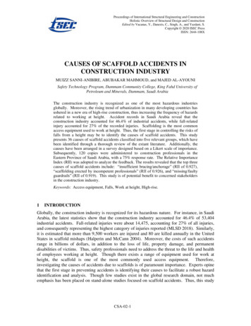
Journal of Materials Science: Materials in Medicine (2021) 32:36Page 7 of 11 36Fig. 3 Gross observation and histological analysis of the cartilage defectrepair. (1) Gross observation of the defect site in each group at 12 weeksafter surgery. The lower figures are partially enlarged views of the upperfigures. The defects of the untreated control group were basically filled, butthe repair surface was still rough and poorly integrated with the surrounding normal cartilage. The repair surface of the WJ with hUCMSCsgroup was less smooth but integrated well with the surrounding normaltissue. The color of the repair tissue was very similar to that of the normalcartilage tissue in the WJ CS with hUCMSCs group, and the flat repairsurface integrated well with the surrounding normal tissue. (2) The imagesof cartilage repair defects include H&E (2), TB (3), safranin O (4) and typeII collagen immunohistochemical staining (5). WJ CS with hUCMSCsgroup: all stains in the WJ CS with hUCMSCs group were positive. Therepaired tissue was hyaline cartilage, and the arrangement of the chondrocytes was very similar to that in normal cartilage tissue. The repairedtissue integrated well with the surrounding normal tissue, and the woundwas smooth and complete. The cartilage content in the extracellular matrixwas very similar to that of the surrounding normal cartilage tissue; the typeII collagen content of the repaired cartilage was similar to that of thenormal cartilage. WJ with hUCMSCs group: the H&E, TB, safranin O andtype II collagen immunohistochemical staining results were weakly positive; the repaired tissue was a mixture of fibrous tissue and hyaline cartilage, and the wound was not smooth; the cartilage content in theextracellular matrix was lower than that of the WJ CS with hUCMSCsgroup; and the type II collagen content of the repaired tissue was low.Untreated control group: the H&E, TB, safranin O and type II collagenimmunohistochemical staining results were weakly positive; the mainrepair tissue was fibrous tissue; the structure of the hyaline cartilage wasdifficult to detect in the defect site; the reconstructed tissue was poorlyintegrated with the surrounding normal tissue, and the wound was irregular; and the chondrocyte content in the extracellular matrix was thelowest. Scale bar 100 μm3.5.3 Type II collagen immunohistochemical stainingPositive immunohistochemical staining for type II collagen is indicated by a specific brownish-yellow color. At4 weeks after surgery, the repaired tissue of the untreatedcontrol group was negative for type II collagen, that of theWJ with hUCMSCs group was weakly positive for type IIcollagen, and that of the WJ CS with hUCMSCs groupwas positive and similar to that of the surrounding normaltissue. At 12 weeks after surgery, the immunohistochemical staining of the repaired tissue of the untreatedcontrol group was still negative for type II collagen, thatof the WJ with hUCMSCs group was weakly positive,and that of the WJ CS with hUCMSCs group wasstrongly positive and similar to that of the surroundingnormal tissue (Fig. 3(5)).3.5.4 Histological scoresimilar to the surrounding normal cartilage tissue(Fig. 3(3)). The results of the safranin O staining weresimilar to those of the TB staining (Fig. 3(4)).According to the rules of the Wakitani law histology score,the lower the score is, the better the effect. The histologicalscores of the WJ CS with hUCMSCs group at 4 and12 weeks were lower than those of the WJ with hUCMSCsand untreated control groups (P 0.05). The histologicalscores of the WJ CS with hUCMSCs groups were lowestat each time point (P 0.05) (Fig. 4C).
36Page 8 of 11Table 2 Comparison of grossobservation scores among thethree groups (χ s)Journal of Materials Science: Materials in Medicine (2021) 32:36Groupn Characteristics of thecartilage surfaceFillingdefect degreeHostgraft fusionTotal scoreUntreated control5 2.80 0.453.00 0.003.00 0.008.80 0.45WJ withhUCMSCs5 2.00 0.71a1.00 0.00a2.40 0.55a5.40 1.14aWJ CS withhUCMSCs5 0.60 0.55ab2.20 0.45ab1.20 0.45ab4.00 0.71abF value18.60076.00025.20045.700P value 0.001 0.001 0.001 0.001aCompared with the untreated control group, P 0.05Compared with the WJ with hUCMSCs group, P 0.05bFig. 4 Immunogenicity and histological scoring in each in vivotransplantation group. A Immunohistochemical staining for an antihuman nucleoprotein at 12 weeks after surgery. The repaired cartilagein the untreated control group was stained blue, which indicates thatthe staining results were negative (1). The cartilage in the WJ withhUCMSCs and WJ CS with hUCMSCs groups was stained brown,which indicates positive staining ((2) and (3)). Scale bar 100 μm.B The histological scores of the WJ CS with hUCMSCs group at 4and 12 weeks were lower than those of the WJ with hUCMS
heart diseases [20]. CS has broad application prospects in the fields of medicine,food andcosmetics, and the use of CS in the preparation of bioengineered scaffolds can improve the physical characteristics of the scaffold material [21, 22]. According to the requirements of tissue-engineered scaffolds, does the scaffold composite prepared by cross-