Transcription
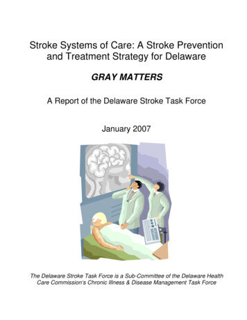
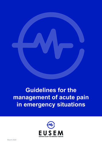
How-I-do-itAcute MR Stroke Protocol in Six MinutesKambiz Nael; Rihan Khan; Kevin Johnson; Diego MartinUniversity of Arizona, Department of Medical Imaging, Tucson, AZ, USABackgroundStroke is a common and serious disorder, with an annual incidenceof approximately 795,000. Based onAmerican Heart Association statisticsupdate in 2010, approximately610,000 of these are first attacks,and 185,000 are recurrent attacks. Onaverage, every 40 seconds, someonein the United States has a stroke withan estimated mortality rate of 5.5%,claiming approximately 1 of every18 deaths in the United States [1].Neuroimaging plays a central role inthe evaluation of patients with acuteischemic stroke (AIS). With improvedtechnology over the last decade, imag ing now provides information beyondthe mere presence or absence of intra 92-year-old man with sudden onset of right-sided weakness and aphasia presented to ouremergency department after receiving IV-tPA at an outside institution. The acute stroke protocolwas performed after 9 hours from the onset in our institution and selective images are shown.1A1B1A Serial aligned DWI, ADC, EPI-FLAIR and EPI-GRE imagesare shown. There is acute infarction of the left MCAdistribution involving the left operculum and insula.Small focus of hemorrhagic conversion is presentwithin the area of infarction seen on both EPI-FLAIRand EPI-GRE images.1B Aligned DSC-Tmax, DSC-CBF and DSC-CBV images are shown.DSC maps show a heterogeneous pattern of perfusion deficitcontaining a small perfusion defect in the region of hemorrhageand predominant luxury perfusion along the left MCA territoryseen on Tmax and CBF maps.44MAGNETOM Flash 5/2013 www.siemens.com/magnetom-world
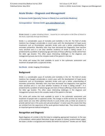
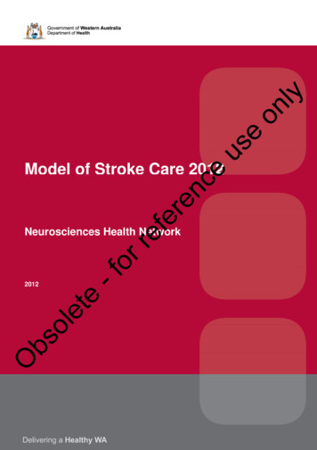
How-I-do-itcranial hemorrhage including tissueviability, site of occlusion, and collat eral status. While computed tomo graphy (CT) is the most widely avail able and faster imaging modality,some comprehensive stroke centersfavor streamlined MR protocols overCT in the acute stroke setting dueto the higher specificity and superiortissue characterization afforded byMRI. The success of CT in initial eval uation of AIS is due, in part, to fastacquisition time, widespread avail ability and ease of interpretation inthe emergency setting. The introduc tion of multi-slice technology hasdramatically increased the speed andsimplicity of CT techniques and hasset a high standard for alternativeimaging techniques. A comprehen sive CT stroke algorithm includingparenchymal imaging (non-contrasthead CT), CT angiography (CTA), andperfusion/penumbral imaging by CTperfusion can now be acquired andprocessed in less than 10 minutes[5, 6].MRI has been demonstrated to bemore sensitive for the detection ofacute ischemia and more specific fordelineation of infarction core volumewhen compared to CT [7, 8]. How ever, due to longer acquisition timeand limited availability; it has beenmainly used in large institutionsand comprehensive stroke centers.A comprehensive MR protocol includ ing parenchymal imaging, MRA andMR perfusion can now be obtainedin the order of 20 minutes as demon strated in several clinical trials [9–13].If MRI is to compete with CT forevaluation of acute stroke, there isneed for further improvements inacquisition speed.In this article we describe our modi fied acute stroke MRI protocol thatcan be obtained in approximately6 minutes rivaling that of any com prehensive acute stroke CT protocol.We describe the technical aspectsand review a few clinical examplesbased on our preliminary results.1C1C Coronal MIP from CE-MRA of the entire supra-aortic arteries and cropped volume-rendered reconstruction of the intracranial arteries show no evidence of hemodynamic significant arterialstenosis nor occlusion involving the proximal arteries. Note the high diagnostic image qualityof the CE-MRA images which are obtained after administration of 8 ml of contrast.MAGNETOM Flash 5/2013 www.siemens.com/magnetom-world45
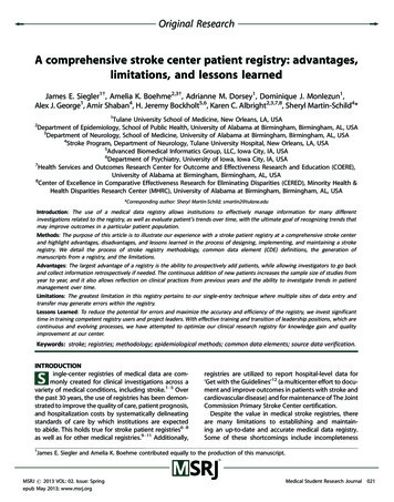
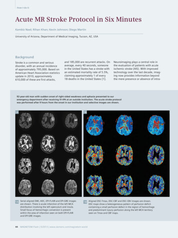
How-I-do-itTechnical considerationA comprehensive MR stroke protocolhas three essential components:1) Parenchymal imaging that identi fies the presence and size of an irre versible infarcted core and deter mines the presence of hemorrhage;2) MR angiogram to determine thepresence of proximal arterial occlu sion and/or intravascular thrombusthat can be treated with thrombolysisor thrombectomy;3) Pwerfusion imaging to determinethe presence of hypoperfused tissueat risk for subsequent infarction ifadequate perfusion is not restored.Below we describe each of thesecomponents in detail and explainhow recent technical advances canbe used to enhance the performanceof the different aspects of acutestroke imaging.1. Parenchymal imagingThis encompasses three parts:1) DWI (diffusion-weighted imaging)that can detect ischemic tissue withinminutes of its occurrence and hasemerged as the most sensitive andspecific imaging technique for acuteischemia, far beyond NECT or anyother type of MRI sequences [14].2) FLAIR that helps to age the infarc tion and permits the detection ofsubtle subarachnoid hemorrhage;3) GRE to detect parenchymal hemor rhage with comparable accuracy forthe acute intraparenchymal hemor rhage to CT [15].Both FLAIR and GRE images havebeen used to detect intraarterial clotwith variable sensitivity and specificity[16, 17].Introduction of fast imaging tech niques such as parallel acquisition [18]and EPI [19, 20] has significantlyenhanced the performance of MRimaging in terms of acquisition speed.The main advantage of EPI, as in thecase of DWI imaging, is rapid acquisi tion time, which is made possible byrapid gradient switching which per mits the acquisition of all frequencyand phase encoding steps during a sin gle pulse cycle. The addition of parallelimaging can further enhance theacquisition speed and may also serveto mitigate the geometric distortionand susceptibility artifacts commonly68-year-old man with left sided weakness and altered level of consciousness of unknown onset.2A2B2A Serial aligned DWI, ADC, EPI-FLAIR and EPI-GREimages are shown. There is acute right hemisphericinfarction involving both the ACA and MCA territories.The EPI-FLAIR images demonstrate correspondinghyperintensity suggestive of completed infarction.There is associated mass effect. No hemorrhage isidentified on corresponding EPI-GRE images.2B Aligned DSC-Tmax, DSC-CBF and DSC-CBVimages are shown. There is a matchedperfusion defect with the region of infarction.46MAGNETOM Flash 5/2013 www.siemens.com/magnetom-world
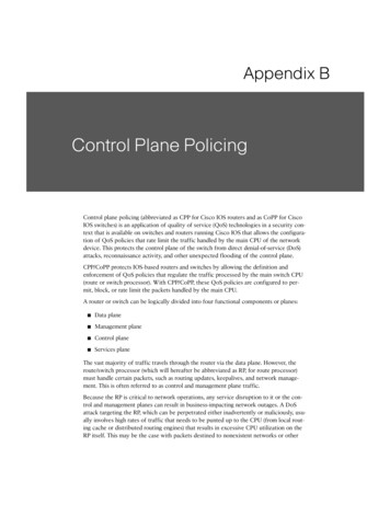
How-I-do-itassociated with long echo-trainsequences such as EPI [21, 22]. If theirpotential is realized, the applicationof EPI and parallel imaging techniquesto the FLAIR and GRE sequences canresult in reduction of image acquisitiontime of the entire brain to less thana minute, a three-fold reduction inscan time over conventional imaging[23, 24].2. MR AngiogramAn important aspect of the workupof patients with AIS is the imaging ofboth the intracranial and extracranialvasculature. Precise imaging of thevascular tree is required during the initial assessment of patients withacute stroke to accurately detect thesite of arterial disease, which in turncan be crucial in determining the2C2C Coronal MIP from CE-MRA of theentire supra-aortic arteries showscomplete occlusion of the rightcervical ICA shortly after the origin.There is some reconstitution offlow signal at the supracliniod ICAlikely via collaterals.type of acute therapy they are given.Intravenous thrombolysis has beenshown to be more effective in smalldistal vessels than in the large vessels[25]. Larger vessel occlusion may bemore effectively treated with intraarterial thrombolysis or clot retrievaldevices while associated with fewercomplications [26, 27]. In addition,MRA of the extracranial circulation(neck arteries) is essential to estab lish the mechanism of ischemia andto prevent subsequent episodes.Extracranial tandem stenoses withplaque involving the carotid or verte bral arteries can be the source of disease that triggers an acute stroke.Time-of-flight MRA (TOF-MRA) hasbeen traditionally used in routinestroke protocols to evaluate thestatus of neck and brain arteries.Despite its promising results [28],TOF-MRA has significant disadvan tages including spin saturation andphase dispersion due to slow or tur bulent flow [29, 30]. This can resultin overestimation of arterial stenosisand increase false positive rates, usually due to slow flow distal toa subocclusive thrombus or clot.Most importantly the acquisitiontime u sually is long, typically lasting5–7 minutes.The general consensus is thatcontrast-enhanced MR angiography (CE-MRA) provides more accurateimaging of extracranial vessel mor phology and of the degree of steno sis than TOF-MRA techniques [31–33].However, CE-MRA has not beenwidely incorporated into acute strokeprotocols for several reasons. First,CE-MRA has lower spatial resolutionrelative to TOF-MRA, since the com peting requirements of coverage andacquisition speed generally force acompromise in spatial resolution for CE-MRA [34]. A second potential limi tation to incorporation of CE-MRAinto clinical stroke protocols is relatedto the requirement of an extra con trast dose, which would be in addi tion to the intravenous contrastbolus normally utilized for perfusionimaging. With introduction of highperformance MR scanners and recentadvances in fast imaging tools suchas parallel acquisition (GRAPPA) [18],high matrices can now be spread outover a large field-of-view encompass ing the entire head and neck, result ing in acquisitions with submillimetervoxel sizes and acquisition times onthe order of 20 seconds [35, 36].3. MR PerfusionMR perfusion imaging has been usedbroadly in the identification of poten tially salvageable tissue to determinethe best treatment strategy in patientswith acute ischemic stroke. Althoughthe concept of perfusion-diffusionmismatch remains controversial[37, 38], it has been used with some success to identify patients who mayrespond favorably to revasculariza tion therapies in several clinical trials[12, 13, 39].Faster image acquisition combinedwith higher signal-to-noise ratio(SNR) resulting from the use of gado linium contrast agents has helpeddynamic susceptibility contrast (DSC)perfusion become a more robust andwidely accepted technique in com parison to arterial spin labeling (ASL)to identify the presence of perfusionabnormalities in patients with AIS.A refined MR stroke protocol thatcan combine both CE-MRA and DSCperfusion with improved acquisitiontime and diagnostic image qualityas previously suggested [47, 48] mayhave important therapeutic andprognostic implications in the man agement of patients with acutestroke. Higher inherent SNR of highermagnetic fields such as 3T withimproved multi-coil technology hasresulted in acquisition of low doseCE-MRA of the supra-aortic arterieswith contrast dose as low as 8 ml[40, 41]. A modified 2-phase contrastinjection scheme [46] can be usedto perform both CE-MRA and DSCperfusion imaging, without the needfor additional contrast. The influenceof contrast dose reduction on DSCperfusion has been evaluated byseveral investigators [42, 43] andcontrast dose as low as 0.05 mmol/kghas been used to perform DSC perfu sion with promising results [44, 45].Advances in MR technology includinghardware and software, faster gradi ent performance of MR scanners,improved sequence design and fastimaging tools such as EPI and parallelMAGNETOM Flash 5/2013 www.siemens.com/magnetom-world47
How-I-do-itCE-MRA and DSC perfusion. The clini cal i ndications for using this acuteMR stroke protocol are patients withacute ( 9 hours) presentation fromthe onset of symptoms, unknownonset of symptoms, NIHSS 4, oraphasia. Table 1 shows the sequenceparameters of our acquisitionprotocol.acquisition have promised the potential for a fast but comprehen sive MR stroke protocol that can beperformed in approximately 6 min utes rivaling those of CT protocols.Next we review our stroke protocolin terms of image acquisition andsequence parameters and showsome of the clinical examples thatwere performed at our institution.How we do itAt our institution, absent contraindi cation, MR is the default imagingmodality for AIS. An MR safety ques tionnaire is administered, and MRcompatible ECG leads are placed inthe emergency department aspatients are being evaluated by theneurology team. Patients are thenplaced onto an MR compatible tableand wheeled to the MR magnet forrapid imaging.We use both 3T and 1.5T MR scanners(MAGNETOM Skyra and MAGNETOMAera, Siemens Healthcare, Erlangen,Germany), with 3T the default scannerfor acute stroke imaging when avail able. For signal reception, a combina tion of a 16-element array coil [head(n 12), neck (n 4)] will be used.The coil design allows for applicationof parallel acquisition in both thephase and slice encoding directions.Our 6-minute MR imaging protocolconsist of DWI, EPI-FLAIR, EPI-GRE,A modified 2-phase contrast injectionscheme [46] is used to perform bothCE-MRA and DSC perfusion imaging,without the need for additional con trast. To accomplish this, the totalvolume of 20 ml of gadolinium (Multihance, Bracco Diagnostics Inc.,Princeton, NJ, USA) that is used rou tinely for MR perfusion is diluted withnormal saline to a total 50 ml vol ume. Using a timing bolus, a total of3 ml of contrast solution (1.2 ml ofgadolinium) is injected at 1.5 ml/s todetermine the transit time from thearm vein to the cervical carotid arter ies. Then, a total of 22 ml contrastsolution (8.8 ml of gadolinium) isinjected at the same flow rate as thetiming injection for the CE-MRAacquisition. A centric ordering k-spaceis used for CE-MRA to minimize intra cranial venous contamination. Sub sequently, the remaining 25 ml of contrast solution (10 ml of gadolin ium) is injected at 5 ml/s for the MRperfusion scan which is performedat the end.Image analysisFollowing data acquisition, CE-MRAimage processing is performed on thescanner console with standard com mercial software using a maximumintensity projection (MIP) algorithm.All of the reconstructed data, as wellas the source images are available onthe workstation for image analysis. Perfusion analysis will be performedoff-line on a dedicated FDA-approvedworkstation (Olea-sphere, Olea MedicalSA, France). The arterial input functionis selected automatically and multi parametric perfusion maps includingtime-to-peak (TTP), time-to-maximum(Tmax) cerebral blood flow (CBF) andcerebral blood volume (CBV) are thencalculated using a block-circulant singular value decomposition tech nique [49].Our initial results using the describedstroke MR protocol have been promis ing. We have scanned more than 600patients with ASI since January 2013.More than 97% of our studies havebeen rated with diagnostic image qual ity. The EPI-FLAIR sequence has beenused in parallel to conventional FLAIRin a subset of patients with compara ble qualitative and quantitative results[24]. In a study of 52 patients withAIS, the mean SD of the signal inten sity ratios on EPI-FLAIR and FLAIR forDWI positive lesions were 1.28 0.16and 1.25 0.17 respectively with sig Table 1: Imaging protocolDWIEPI-FLAIREPI-GRECE-MRADSCTR (ms)460010000 (TI:2500)18603.361450TE6588481.2422FA 220340220Slices (n thickness)30 430 440 3120 0.830 4Bandwidth (Hz/pixel)125014889645901502Parallel acquisition (GRAPPA)33–33Acquisition time58 sec52 sec56 sec20 sec1 min and 30 sec48MAGNETOM Flash 5/2013 www.siemens.com/magnetom-world
How-I-do-itnificant correlation (r 0.899, z value 8.677, p 0.0001). The EPI-GREsequence has been also used in paral lel to conventional GRE in a subsetof patients with comparable resultsin terms of detection of hemorrhage(Fig. 1) and blood clot in proximalarteries.The combination of CE-MRA andDSC has been successfully tested inour institution [48] with diagnosticimage quality. In a cohort of 30patients with acute stroke, the speci ficity of CE-MRA for detection of arterial stenosis 50% was 97% com pared to 89% for TOF-MRA when com pared to DSA as the standard of refer ence [48]. DSC perfusion imaging withreduced contrast dose is feasible withcomparable quantitative and qualita tive results to a full-dose control group[48]. Importantly, the presence ofcontrast in the circulating blood of the CE-MRA half-dose group does not neg atively impact the image quality northe quantitative analysis of perfusiondata when compared to the controlfull-dose group.ConclusionDescribed multimodal MR protocolis feasible for evaluation of patientswith acute ischemic stroke with totalacquisition time of 6 minutes rivalingthat of the multimodal CT protocol.References1 Lloyd-Jones D, Adams RJ, Brown TM,et al. Heart disease and stroke statistics– 2010 update: a report from theAmerican Heart Association. Circulation.Feb 23 2010;121(7):e46-e215.2 Saver JL. Time is brain-quantified.Stroke. Jan 2006;37(1):263-266.3 Michel P, Bogousslavsky J. Penumbra isbrain: no excuse not to perfuse. AnnNeurol. Nov 2005;58(5):661-663.4 Gonzalez RG. Imaging-guided acuteischemic stroke therapy: From “timeis brain” to “physiology is brain”. AJNR.American journal of neuroradiology.Apr 2006;27(4):728-735.5 Zhu G, Michel P, Aghaebrahim A, et al.Computed tomography workup ofpatients suspected of acute ischemicstroke: perfusion computed tomographyadds value compared with clinicalevaluation, noncontrast computedtomography, and computed tomographyangiogram in terms of predictingoutcome. Stroke; a journal of cerebralcirculation. Apr 2013;44(4):1049-1055.6 Schaefer PW, Roccatagliata L, Ledezma C,et al. First-pass quantitative CT perfusionidentifies thresholds for salvageablepenumbra in acute stroke patients treatedwith intra-arterial therapy. AJNR.American journal of neuroradiology.Jan 2006;27(1):20-25.7 Jauch EC, Saver JL, Adams HP, Jr., et al.Guidelines for the early managementof patients with acute ischemic stroke:a guideline for healthcare professionalsfrom the American Heart Association/American Stroke Association. Stroke.Mar 2013;44(3):870-947.8 Chalela JA, Kidwell CS, Nentwich LM,et al. Magnetic resonance imaging andcomputed tomography in emergencyassessment of patients with suspectedacute stroke: a prospective comparison.Lancet. Jan 272007;369(9558):293-298.9 Kang DW, Chalela JA, Dunn W, Warach S,Investigators NI-SSC. MRI screeningbefore standard tissue plasminogenactivator therapy is feasible and safe.Stroke. Sep 2005;36(9):1939-1943.10 Hjort N, Butcher K, Davis SM, et al.Magnetic resonance imaging criteria forthrombolysis in acute cerebral infarct.Stroke. Feb 2005;36(2):388-397.11 Schellinger PD, Jansen O, Fiebach JB,Hacke W, Sartor K. A standardized MRIstroke protocol: comparison with CT inhyperacute intracerebral hemorrhage.Stroke; a journal of cerebral circulation.Apr 1999;30(4):765-768.12 Albers GW, Thijs VN, Wechsler L, et al.Magnetic resonance imaging profilespredict clinical response to early reper fusion: the diffusion and perfusionimaging evaluation for understandingstroke evolution (DEFUSE) study. AnnNeurol. Nov 2006;60(5):508-517.13 Davis SM, Donnan GA, Parsons MW, et al.Effects of alteplase beyond 3 h afterstroke in the Echoplanar ImagingThrombolytic Evaluation Trial (EPITHET):a placebo-controlled randomised trial.Lancet neurology. Apr 2008;7(4):299-309.14 Fiebach JB, Schellinger PD, Jansen O,et al. CT and diffusion-weighted MRimaging in randomized order: diffusionweighted imaging results in higheraccuracy and lower interrater variabilityin the diagnosis of hyperacute ischemicstroke. Stroke; a journal of cerebralcirculation. Sep 2002;33(9):2206-2210.15 Kidwell CS, Chalela JA, Saver JL, et al.Comparison of MRI and CT for detectionof acute intracerebral hemorrhage. JAMA :the journal of the American Medical Association. Oct 20 2004;292(15):1823-1830.16 Flacke S, Urbach H, Keller E, et al. Middlecerebral artery (MCA) susceptibility signat susceptibility-based perfusion MRimaging: clinical importance andcomparison with hyperdense MCA sign atCT. Radiology. May 2000;215(2):476-482.17 Assouline E, Benziane K, Reizine D, et al.Intra-arterial thrombus visualized on T2*gradient echo imaging in acute ischemicstroke. Cerebrovasc Dis. 2005;20(1):6-11.18 Griswold MA, Jakob PM, Heidemann RM,et al. Generalized autocalibrating partiallyparallel acquisitions (GRAPPA). Magneticresonance in medicine : official journal ofthe Society of Magnetic Resonance inMedicine / Society of Magnetic Resonancein Medicine. Jun 2002;47(6):1202-1210.19 Mansfield P. Real-time echo-planar imagingby NMR. Br Med Bull. Apr 1984;40(2):187-190.20 DeLaPaz RL. Echo-planar imaging. Radiographics. Sep 1994;14(5):1045-1058.21 Pruessmann KP. Parallel imaging at highfield strength: synergies and jointpotential. Topics in magnetic resonanceimaging : TMRI. Aug 2004;15(4):237-244.22 Wiesinger F, Van de Moortele PF, AdrianyG, De Zanche N, Ugurbil K, PruessmannKP. Potential and feasibility of parallel MRIat high field. NMR in biomedicine.May 2006;19(3):368-378.23 Kinoshita T, Okudera T, Tamura H,Ogawa T, Hatazawa J. Assessment oflacunar hemorrhage associated withhypertensive stroke by echo-planargradient-echo T2*-weighted MRI.Stroke. Jul 2000;31(7): 1646-1650.24 Meshksar A, Khan R, Carmody R, Nael K.The Role of Echo-planar Fluid-AttenuatedInversion Recovery (EPI-FLAIR) in AcuteStroke Setting: A Feasibility Study.Paper presented at: ASNR; May 22, 2013,2013; San Diego, CA.25 del Zoppo GJ, Poeck K, Pessin MS, et al.Recombinant tissue plasminogen activatorin acute thrombotic and embolic stroke.Ann Neurol. Jul 1992;32(1):78-86.26 Furlan A, Higashida R, Wechsler L, et al.Intra-arterial prourokinase for acuteischemic stroke. The PROACT II study:a randomized controlled trial. Prolyse inAcute Cerebral Thromboembolism.JAMA : the journal of the AmericanMedical Association. Dec 1 1999;282(21):2003-2011.MAGNETOM Flash 5/2013 www.siemens.com/magnetom-world49
How-I-do-it27 Becker KJ, Brott TG. Approval of the MERCIclot retriever: a critical view. Stroke;a journal of cerebral circulation. Feb 2005;36(2):400-403.28 Yucel EK, Anderson CM, Edelman RR, et al.AHA scientific statement. Magneticresonance angiography : update on appli cations for extracranial arteries. Circulation. Nov 30 1999;100(22):2284-2301.29 Isoda H, Takehara Y, Isogai S, et al. MRAof intracranial aneurysm models:a comparison of contrast-enhanced threedimensional MRA with time-of-flight MRA.J Comput Assist Tomogr. Mar-Apr 2000;24(2):308-315.30 Lin W, Tkach JA, Haacke EM, Masaryk TJ.Intracranial MR angiography: applicationof magnetization transfer contrast and fatsaturation to short gradient-echo,velocity-compensated sequences.Radiology. Mar 1993;186(3):753-761.31 Somford DM, Nederkoorn PJ, Rutgers DR,Kappelle LJ, Mali WP, van der Grond J.Proximal and distal hyperattenuatingmiddle cerebral artery signs at CT:different prognostic implications.Radiology. Jun 2002;223(3):667-671.32 Cosottini M, Pingitore A, Puglioli M, et al.Contrast-enhanced three-dimensionalmagnetic resonance angiography ofatherosclerotic internal carotid stenosisas the noninvasive imaging modality inrevascularization decision making. Stroke;a journal of cerebral circulation. Mar 2003;34(3):660-664.33 Huston J, 3rd, Fain SB, Wald JT, et al.Carotid artery: elliptic centric contrastenhanced MR angiography compared withconventional angiography. Radiology.Jan 2001;218(1):138-143.34 Fellner C, Lang W, Janka R, Wutke R,Bautz W, Fellner FA. Magnetic resonanceangiography of the carotid arteries usingthree different techniques: accuracycompared with intraarterial x-ray angiog raphy and endarterectomy specimens.Journal of magnetic resonance imaging :JMRI. Apr 2005;21(4):424-431.35 Nael K, Villablanca JP, Pope WB, McNamaraTO, Laub G, Finn JP. Supraaortic arteries:contrast-enhanced MR angiography at 3.0T-highly accelerated parallel acquisitionfor improved spatial resolution over anextended field of view. Radiology.Feb 2007;242(2):600-609.36 Phan T, Huston J, 3rd, Bernstein MA,Riederer SJ, Brown RD, Jr. Contrastenhanced magnetic resonance angiog raphy of the cervical vessels: experiencewith 422 patients. Stroke; a journal ofcerebral circulation. Oct 2001;32(10):2282-2286.5037 Hacke W, Furlan AJ, Al-Rawi Y, et al. Intra venous desmoteplase in patients withacute ischaemic stroke selected by MRIperfusion-diffusion weighted imagingor perfusion CT (DIAS-2): a prospective,randomised, double-blind, placebocontrolled study. Lancet neurology.Feb 2009;8(2):141-150.38 Kidwell CS, Jahan R, Gornbein J, et al.A trial of imaging selection and endovas cular treatment for ischemic stroke.The New England journal of medicine.Mar 7 2013;368(10):914-923.39 Lansberg MG, Straka M, Kemp S, et al.MRI profile and response to endovas cular reperfusion after stroke (DEFUSE2): a prospective cohort study. Lancetneurology. Oct 2012;11(10):860-867.40 Tomasian A, Salamon N, Lohan DG,Jalili M, Villablanca JP, Finn JP. Supra aortic arteries: contrast material dosereduction at 3.0-T high-spatial-resolutionMR angiography--feasibility study.Radiology. Dec 2008;249(3):980-990.41 Nael K, Moriarty JM, Finn JP. Low doseCE-MRA. Eur J Radiol. Oct 2011;80(1):2-8.42 Heiland S, Reith W, Forsting M, Sartor K.How do concentration and dosage ofthe contrast agent affect the signalchange in perfusion-weighted magneticresonance imaging? A computersimulation. Magnetic resonanceimaging. Jul 2001;19(6):813-820.43 Alger JR, Schaewe TJ, Lai TC, et al.Contrast agent dose effects in cerebraldynamic susceptibility contrast magneticresonance perfusion imaging. Journalof magnetic resonance imaging: JMRI.Jan 2009;29(1):52-64.44 Manka C, Traber F, Gieseke J, Schild HH,Kuhl CK. Three-dimensional dynamicsusceptibility-weighted perfusion MRimaging at 3.0 T: feasibility and contrastagent dose. Radiology. Mar 2005;234(3):869-877.45 Alger JR, Schaewe TJ, Liebeskind DS,Saver JL, Kidwell CS. On the feasibilityof reduced dose Dynamic SusceptibilityContrast perfusion MRI for stroke. Paperpresented at: Intl. Soc. Mag. Reson.Med; 7-13 May 2011, 2011; Montréal,Québec, Canada.46 Habibi R, Krishnam MS, Lohan DG, et al.High-spatial-resolution lower extremityMR angiography at 3.0 T: contrast agentdose comparison study. Radiology.Aug 2008;248(2):680-692.47 Ryu CW, Lee DH, Kim HS, et al. Acqui sition of MR perfusion images andcontrast-enhanced MR angiography inacute ischaemic stroke patients: whichprocedure should be done first? TheBritish journal of radiology. Dec 2006;79(948):962-967.MAGNETOM Flash 5/2013 www.siemens.com/magnetom-world48 Nael K, Pirastehfar M, Villablanca JP,Salamon N. Addition of a Low-doseContrast Enhanced MRA at 3.0T in theAssessment of Acute Stroke: A MoreEfficient and Accurate Stroke Protocol.Paper presented at: ASNR 50th AnnualMeeting; April, 2012; New York, NY.49 Wu O, Ostergaard L, Weisskoff RM,Benner T, Rosen BR, Sorensen AG. Tracerarrival timing-insensitive technique forestimating flow in MR perfusion-weightedimaging using singular value decompo sition with a block-circulant deconvo lution matrix. Magnetic resonance inmedicine : official journal of the Societyof Magnetic Resonance in Medicine /Society of Magnetic Resonance inMedicine. Jul 2003;50(1):164-174.ContactKambiz Nael, M.D.Assistant Professor of RadiologyDirector of Neuroradiology MRIUniversity of ArizonaMedical CenterDepartment of Medical Imaging,Neuroradiology Section1501 N. Campbell,PO Box 245067Tucson, AZ 85724-5067USAPhone: 1 520-626-2138Fax: 1 520-626-7093kambiz@radiology.arizona.edu
ent performance of MR scanners, improved sequence design and fast imaging tools such as EPI and parallel 2C Coronal MIP from CE MRA of the entire supra aortic arteries shows complete occlusion of the right cervical ICA shortly after the origin. There is some reconstitution of flow signal at the supracliniod ICA likely via collaterals. 2C How .