Transcription
Scottish Universities Medical Journal, 2014Published Online: May 2014Vol 3 (Issue 1): PP. 18-27.Smith, G. (2014). Acute Stroke - Diagnosis and ManagementAcute Stroke – Diagnosis and ManagementDr Gemma Smith (Specialty Trainee in Elderly Care and Stroke Medicine)Correspondence - Gemma Smith: gem.eaton@gmail.comABSTRACTStroke (noun): a sudden disabling attack caused by an interruption in the flow of blood tothe brain, especially through thrombosis.Stroke is a considerable cause of mortality and morbidity in the UK. The field of strokemedicine has changed considerably in recent years with the development of hyper-acutetreatments such as thrombolysis, specialist stroke units and a better understanding ofsecondary prevention. Mortality rates may have decreased but diagnostics have becomemore sensitive and it is not clear whether incidence of stroke is falling overall. It ispredominantly a problem of advancing age and many of those suffering a stroke will be fromthe older age bracket. This often raises interesting challenges in the diagnosis andmanagement process due to the complex needs of the patient in the bed.This article will review the tools available to assist in the systematic assessment andtreatment of people with a suspected stroke.Key Words: stroke; imaging; thrombolysisBackgroundStroke is a considerable cause of mortality and morbidity in the UK. The field of strokemedicine has changed considerably in recent years with the development of hyper-acutetreatments such as thrombolysis, specialist stroke units and a better understanding ofsecondary prevention. Mortality rates may have decreased2 but diagnostics have becomemore sensitive and it is not clear whether incidence of stroke is falling overall3,4. It ispredominantly a problem of advancing age and many of those suffering a stroke will be fromthe older age bracket. This often raises interesting challenges in the diagnosis andmanagement process due to the complex needs of the patient in the bed.This article will review the tools available to assist in the systematic assessment andtreatment of people with a suspected stroke. The change in stroke services has meant thatacute events are now often admitted directly to acute stroke units as these are proven toprovide optimal care in the acute and sub-acute phases5. In some cases this means that theyare omitted from the experience of the acute general medical take. They remain however,an important general medical emergency.Recognition and DiagnosisRapid diagnosis of a stroke is the first step to instigating appropriate treatment. In the caseof thrombolysis, where potent fibrinolytic drugs are given to restore cerebral blood supply,the faster a stroke is recognised and treated, the better the outcome6. Strokes are not “black
and white” clinical entities however and many “mimics” have similar symptoms that canlead to diagnostic uncertainties.The symptoms of a stroke are due to the acute interruption of the blood supply to an area ofthe brain. This can be through blockage of a blood vessel (infarcts) or haemorrhage. Infarctscan be caused by emboli, usually from thrombus in the carotid arteries or left atrium, orfrom in-situ clot formation. In both haemorrhagic strokes and infarcts the onset is sudden.The vascular territory involved and the presence of associated symptoms may assist in thediagnosis. Anterior circulation – weakness, sensory deficits, dysphasia (expressive andreceptive), visual field defects, dyspraxia and higher cortical dysfunction,contralateral signs.Posterior circulation – visual field defects, ataxia and vertigo, inco-ordination, cranialnerve deficits, ipsilateral signs.To allow rapid identification of these stroke patterns and to assist in the exclusion of otherpossible diagnoses, the following screening tools have been developed.F.A.S.T.This stands for Face, Arm, Speech, Time and has been the subject of a national televisionawareness campaign. It is used by paramedics and emergency department triage staff toscreen for stroke symptoms and can be up to 81% sensitive7. It prompts assessment forfacial asymmetry, arm weakness, slurred or disordered speech and then rapid transfer to theappropriate acute care setting for further assessment. FAST is not infallible and isparticularly prone to missing posterior circulation events.R.O.S.I.E.R8.This tool is for Recognition of Stroke in the Emergency Room. It was developed to helpemergency department staff assess possible stroke patients and provides some tools forscreening out mimics such as hypoglycaemia, seizures and syncope. It has a sensitivity of93% and specificity of 83%8. See Table 1.Table 1 – ROSIER scoreIs the blood glucose above 3.5mmol? If not, treat and re-assess.1. Has there been loss of consciousness or syncope? Yes (-1) No (0)2. Has there been seizure activity? Yes (-1) No (0)3. Is there NEW ACUTE onset (or on awakening from sleep) of: Asymmetrical face weakness Yes ( 1) No (0) Asymmetrical arm weakness Yes ( 1) No (0) Asymmetrical leg weaknessYes ( 1) No (0) Speech disturbanceYes ( 1) No (0) Visual field defectYes ( 1) No (0)Paramedic teams trained in the use of FAST screening will often refer patients directly toacute stroke units. Use of the ROSIER scale in emergency departments and medicaladmissions units allows self-presenting patients or those admitted via GPs to be assessed
rapidly so that appropriate admission pathways and investigations can be instigatedpromptly.Following assessment with screening tools, patients with suspected strokes should have athorough history taken and a comprehensive neurological examination. This may need to besimultaneous with the ordering of investigations such as CT imaging if the patient may be acandidate for thrombolysis as will be discussed later.History and ExaminationTiming is a crucial point when taking a patient’s history of a suspected stroke. The onset ofsymptoms is usually sudden, with all deficits occurring together, as opposed to a “marching”or progressive deficit. The time of onset is vital, especially when considering patients forthrombolysis that must be initiated within 4.5 hours of symptom onset9. It is necessary toclearly determine when the patient was last well. Often patients wake up with symptomshowever they may have got up in the night or spoken to their partner in the early hours ofthe morning and the time they were last well can still be identified.It is useful when reviewing neurological symptoms to consider them as positive or negative.Positive symptoms involve gaining a quality such as extra movements or shaking, addedsensations such as prickling or burning and extra visual signs such as flashing lights. Negativesymptoms describe the loss of a normal function such a weakness, loss of sensation, loss ofcomprehensible and useful speech, loss of vision (full or partial) or incoordination. Strokesgenerally produce negative symptoms. The presence of positive symptoms can sway theassessing physician towards a diagnosis of a mimic. The negative symptom of lossconsciousness is rarely a feature of stroke (Table 2).Table 2- Positive and negative features on examination and stroke mimicsPositive symptomFlashing lights and colours in vision.Shaking or jerking limbs.Tingling / prickling sensation.Stroke MimicMigraineSeizures (focal or generalized).Radiculopathy, herpes zoster infection.Negative symptomLoss of consciousnessStroke MimicSyncope, seizures.Associated symptoms mainly have a role in identifying mimics or increasing the suspicion ofan intra-cerebral haemorrhage. Haemorrhagic strokes may present with headache, nauseaand vomiting, which are otherwise uncommon in strokes but may also be present inmigraine. Confusion and agitation can occur with intra-cranial haemorrhages or non-strokediagnoses such as infection or hypoglycaemia. A medication history is also important,particularly if patients are taking antiplatelet or anticoagulant therapy.When considering the diagnosis of a stroke it is useful to look at the vascular risk factors ofthe patient. This provides a guide to the probability of a stroke but also as a target for futuresecondary prevention. Hypertension, diabetes, smoking, hyperlipidaemia, family history,male gender, age and other vasculopathies are potent indicators of stroke risk howeverstrokes are still possible in those without these factors.Each patient with a suspected stroke should have a thorough neurological examinationincluding assessment of motor and sensory modalities and cerebellar function. Pronator
drift is an excellent sign of subtle motor weakness. Higher cortical function such as speechand praxis should be assessed. Speech quality, fluency and word finding skills can beassessed throughout taking the history. Providing simultaneous stimuli to bilateral visualfields or sensory pathways can identify inattention. The inability to identify when both sidesare stimulated suggests cortical dysfunction. Cranial nerve examinations should differentiatebetween upper and lower motor neuron facial weakness and should include an assessmentof visual fields. Neurological examination findings can be applied to the National Institute forHealth Stroke Scale (NIHSS) to give a way of communicating and monitoring stroke severity.The application of this scale in a reproducible way requires training. The examinationfindings will also allow the stroke to be classified as per the Bamford Classification10. Thisgives some guide of mortality and morbidity with a total anterior circulation stroke carryinga higher chance of both.Total anterior circulation stroke (TACS) – All three of the following: 1.Unilateral weakness(and/or sensory deficit) of face, arm and leg 2. Homonymous hemianopia 3. Higher cerebraldysfunction (dysphasia, visuospatial disorder).Partial anterior circulation stroke (PACS) – Two of the following: 1.Unilateral weakness(and/or sensory deficit) of face, arm and leg 2. Homonymous hemianopia 3. Higher cerebraldysfunction (dysphasia, visuospatial disorder).Lacunar stroke (LACS) – One of the following: 1. Unilateral weakness (and/or sensory deficit)of face and arm, arm and leg or all three. 2. Pure sensory stroke. 3.Ataxic hemiparesis.Posterior circulation stroke (PoCS) – One of the following: 1. Cerebellar or brainstemsyndromes 2. Loss of consciousness3. Isolated homonymous hemianopiaPlease note that both TACS and PACS involve the anterior and middle cerebral arteries (ACAand MCA), whilst the PoCS involves the posterior circulation.Transient Ischaemic AttackTIAs can present with symptoms in either of the vascular territories described above butresolve entirely within 24 hours and usually in less than one hour. Amaurosis fugax,described as a curtain coming over the vision in one eye and resolving rapidly is also a formof TIA. TIAs can generally be managed as an outpatient and therefore should rarely be seenon medical admission units having been assessed and referred from GPs or emergencydepartments.Following a TIA, the risk of progression to a full stroke within the next 7 days can bepredicted by the use of the ABCD2 score (Table 3).Table 3 – ABCD2 scoreA – age 60(1 point)B – blood pressure 140/90 (1 point)C – clinical picture:Unilateral weakness (2 points)Speech disturbance(1 point)D – Duration 60 minutes(2 points)1-59 minutes(1 point)D – Diabetes(1 point)Total out of max. 7
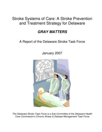
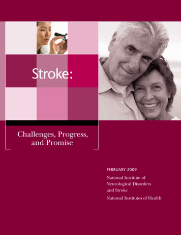
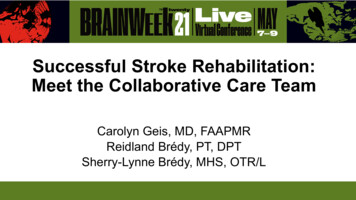
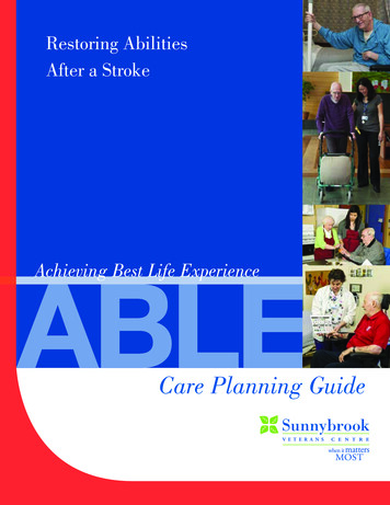
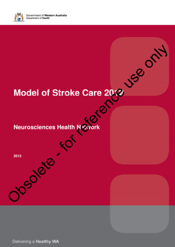
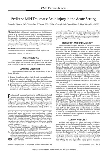
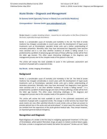
Once diagnosed with a TIA, with particular emphasis that all symptoms should have resolvedentirely, patients can be commenced on an antiplatelet agent. An ABCD2 score of 4 or lesscan be referred for assessment in a TIA clinic. The aim of this service is to identify modifiablerisk factors, in particular carotid stenosis that may be amenable to surgical intervention, andatrial fibrillation for which oral anticoagulants may be appropriate. TIA clinics are often runfive or even seven days a week to allow rapid access. A score greater than 4 carries a verysignificant risk of progressing to a stroke and these cases should be assessed within 24 hourswith carotid imaging, as should patients presenting with crescendo TIAs (more than oneepisode in a week). In centres without seven-day TIA clinics this may require admission tohospital.InvestigationsOnce a stroke is suspected clinically, the appropriate investigation should be pursuedurgently to allow optimal treatment.Laboratory tests.A blood glucose level is necessary to exclude hypoglycaemia as a stroke mimic and this canusual be done as a bedside finger prick test. Blood tests should be sent for full blood countand biochemistry. A coagulation screen should be sent particularly if a bleed is suspected, ifthe patient is anti-coagulated or if thrombolysis is being considered. In the days after astroke, thyroid function tests, lipid profile and ESR are also useful investigations.Imaging and their common appearances.CT remains the mainstay in acute radiological investigation in suspected stroke. This shouldhappen urgently, within 1 hour, in cases being considered for thrombolysis and those caseswith a low Glasgow come score (GCS), signs of meningism or a high suspicion of intracerebral bleed11. All strokes should have imaging within 24 hours of presenation11,12.The role of CT in the immediate phase is mainly to exclude the presence of intracranialhaemorrhage. Intra-cerebral, subarachnoid, subdural and extradural bleeds have acharacteristic appearance on CT. Intra-cerebral haemorrhages are generally rounded, wellcircumscribed lesions within the brain parenchyma (Figure 1). They may have surroundingoedema or extend into the ventricles.Infarcted cerebral tissue appears unchanged on CT scans in the first few hours. The changesseen later develop at a range of 2-3 Hounsfield units per hour. The human eye can onlydetect a difference in contrast of over 6 Hounsfield units so a clinically apparent stroke willonly become radiological apparent after a few hours.There are patterns on CT that can indicate cerebral infarction, particularly when applied withthe suspected vascular territory. There may be a loss of differentiation between grey andwhite matter. When this occurs in the insular region it is referred to as the insular ribbonsign. Loss of differentiation may also occur between structures such as the basal ganglia andinternal capsule. There may be sulcal effacement due to underlying oedema. Clot may bevisible in the proximal middle cerebral artery, “dense MCA” sign (Figure 2) or as a “dot sign”if a more distal branch has been occluded. In the posterior circulation, a hyper-dense basilarartery may be visible (Figure 3) and a high suspicion of impending occlusion is necessary inpatients presenting with nausea and vertigo and posterior circulation signs. Cerebellar andbrainstem strokes are poorly imaged on CT and may require MRI if the diagnosis is in doubt.
Figure 1: CT of an intra-cranial bleedFigure 2: Dense MCA SignFigure 3: Hyper-denseHyperbasilar artery
fection9.html (Fig 3) www.bjcardio.co.uk(Fig2)ww.med-MRI has become a useful tool in stroke diagnosis in recent years. This is primarily due to theuse of diffusion weighted imaging (DWI). Acute infarcts will appear bright on DWI for up tofourteen days. The detailed images produced with MRI, as well as the superior imaging ofthe posterior intra-cranial structures makes this modality useful when a PoCS is suspected orif the diagnosis or vascular territory is in doubt. Other MRI modalities such as gradient echocan identify microhaemorrhages.The availability of MRI, particularly out of hours, means that it does not have a place in thehyper-acute phases of stroke management and is unlikely to replace CT in the near futurehowever it remains a useful adjunct.Imaging of the cerebral blood supply, particularly at a carotid level, can assist in identifyingthe pathophysiology of embolic strokes. Plaque formation causing stenosis in the internalcarotid arteries leads to thrombus formation that then embolises to more distal cerebralvessels. This imaging can be done by Doppler measurements in the acute phase after strokeso decisions can be made about carotid endarterectomy. The use of CT angiography isbecoming increasingly popular as new hyper-acute treatments including intra-arterialthrombolysis and mechanical clot retrieval are investigated. These interventions rely onearly and detailed imaging of the cerebral blood supply. It is also useful in cases of suspectedcarotid or vertebral dissection. CT angiography is often not routinely available out of normalworking hours and its use is at the discretion of stroke physicians and radiologists.Cardiac investigations.The aim of cardiac investigations in stroke patients is to identify atrial fibrillation as a sourceof cardiac emboli. In the acute phase, this can be done with a bedside 12 lead ECG. In time,patients with embolic strokes will need further cardiac investigations to guide secondaryprevention measures.Treatment and ManagementIn the acute phase, there are several important areas of stroke care to consider includingtreatment of the acute event and prevention of complications. Haemorrhagic strokes mayneed discussion with the neurosurgeons.Apart from thrombolysis or antiplatelet measures, the management of these cases has manysimilarities to cerebral infarcts and is discussed below. For cerebral infarcts, specifictreatment options need to be considered.Acute Treatment OptionsThrombolysis.All stroke patients presenting with a suspected cerebral infarction and a time of onset within4.5 hours should be considered for thrombolysis. This requires the urgent exclusion ofhaemorrhage and discussion with a stroke consultant. Thrombolysis has been shown toimprove functional outcome though it has no effect on mortality. It involves the giving of anintravenous fibrinolytic agent (recombinant tissue plasminogen activator).Thrombolysis is licensed for adults of all ages and benefit has been confirmed in the elderlybeyond the age of 80 years9. Treatment is most effective when the “door to needle” time is
shortest. The primary risk of treatment is haemorrhage, either intra- or extra-cranial. Askingabout contraindications can minimise this risk.Contraindications include a time of onset greater than 4.5 hours prior to treatment, bloodpressure 180mmHg systolic or 110mmHg diastolic, blood glucose 22 or 2.8mmol,abnormal clotting or known anti-coagulation, thrombocytopenia, established infarct on CT,history of haemorrhage, recent surgery, previous strokes in patients with diabetes, seizureactivity at any time in the presentation and pre-existing significant disability. Somecontraindications are relative where others absolute. Some may be modifiable, such asblood pressure. The issue of pre-existing disability relates to the aim of thrombolysis toimprove the chances of good functional recovery, this requires a reasonable premorbid levelof function.Stroke physicians will review each case on an individual basis and balance the risks andbenefits. The counselling undertaken before thrombolysis can be complex and emotional forpatients and relatives. After thrombolysis patients require intensive monitoring in aspecialist unit and repeat scan after 24 hours to exclude haemorrhagic transformation.Following this they can be started on high dose aspirin.Antiplatelet and anticoagulant therapy.For those patients not having thrombolysis but in whom a haemorrhage has been excluded,the mainstay of treatment is high dose aspirin (300mg daily). This is continued for fourteendays after the initial event with the aim of reducing the risk of a further embolic event. Longterm antiplatelet therapy is with clopidogrel.Anticoagulant therapy is usually omitted for fourteen days post-stroke. In those onanticoagulants at the time of infarction the decision to continue or stop may be taken by thestroke consultant and depends on the infarct size and risks involved.Blood pressure control.Blood pressure levels often rise around the time of an acute stroke. The risk of allowingblood pressure to rise is that intra-cerebral haemorrhage may expand and cerebral infarctsmay develop haemorrhagic transformation. Lowering blood pressure in the acute phaseafter a stroke is contentious as it may be that the rise seen is a physiological response tomaintain cerebral perfusion. Ideally blood pressures should be kept below 180mmHg butadvice should be taken from a stroke specialist.Nutrition and hydration.Many stroke patients will have an unsafe swallow in the early phases after the event. A wardbased swallow assessment should be completed as soon as possible. In those with an unsafeswallow, medication should be reviewed and either suspended or given via an alternativeroute (rectal, intravenous or topically). Intravenous fluids can be given and artificial feedingshould be considered within the first 24 hours by a nasogastric tube. Aspiration pneumoniais a common complication of stroke and a low threshold for treatment should bemaintained.Thrombo-embolism prophylaxis.Thrombo-embolic events are an important cause of death following stroke. Routineprophylaxis with low molecular weight heparin carries risks. It is contra-indicated in acutehaemorrhagic stroke and can increase the risk of haemorrhagic transformation in cerebralinfarction. Graduated compression stockings have been shown to cause more harm than
benefit including pressure damage in stroke patients and are now contraindicated13. Recentresearch suggests the use of intermittent pneumatic compression stockings can be used toreduce thrombo-embolic risk14 and the case for using low molecular weight heparin can bediscussed on a case-by-case basis dependent on the individual risk.Review of patients with acute strokes.All stroke patients should be transferred to the acute stroke unit as soon as possible afteradmission. They should be seen within the next 24 hours by a stroke consultant or associatespecialist and within 72 hours by the multidisciplinary team therapists.The Next StepThe aim of all management in stroke is two fold, to restore as much function as possible tothe individual and to reduce the chance of future similar events. The former relies onrehabilitation and the skills of the multidisciplinary team. The latter has grown from anevidence base that can then be tailored to each individual patient’s needs. These issues willbe reviewed in the next article on stroke ford English Dictionary sh/strokeLee S et al. UK stroke incidence, mortality and cardiovascular risk management 1999–2008:time-trend analysis from the General Practice Research Database. BMJ Open 2011;1:e000269doi:10.1136/bmjopen-2011-000269Wolf P A et al. Secular trends in stroke incidence and mortality. The Framingham Study.Stroke.1992; 23: 1551-1555Brown RD et al. Stroke incidence, prevalence, and survival: secular trends in Rochester,Minnesota, through 1989. Stroke; a Journal of Cerebral Circulation [1996, 27(3):373-380]Indredavik B et al. Benefit of a Stroke Unit: A randomized controlled trial Stroke1991;20:1026-1031.Hack W et al. Association of outcome with early stroke treatment: pooled analysis ofATLANTIS, ECASS, and NINDS rt-PA stroke trials. Lancet. 2004;363:768–774.Whiteley WN et al. Clinical Scores for the identification of stroke and transient ischaemicattack in the emergency department: a cross-sectional study. Neurol Neurosurg Psychiatry.2011 Sep;82(9):1006-10. doi: 10.1136/jnnp.2010.235010. Epub 2011 Mar 14.Nor AM et al. The Recognition of Stroke in the Emergency Room (ROSIER) scale:development and validation of a stroke recognition instrument. Lancet Neurol. 2005Nov;4(11):727-34.P Sandercock et al. The benefits and harms of intravenous thrombolysis with recombinanttissue plasminogen activator within 6 h of acute ischaemic stroke (the third internationalstroke trial [IST-3]): a randomised controlled trial. Lancet. 2012 June 23; 379(9834): 2352–2363.Pittock SJ et al. The Oxfordshire Community Stroke Project classification: correlation withimaging, associated complications, and prediction of outcome in acute ischemic stroke. JStroke Cerebrovasc Dis. 2003 ckRefGuide/pdf/English CG68: Stroke.http://www.sign.ac.uk/pdf/qrg108.pdf Guideline 108: Management of patients with strokeor TIA: assessment, investigation, immediate management and secondary preventionThe CLOTS trial collaboration. Effectiveness of thigh-length graduated compression stockingsto reduce the risk of deep vein thrombosis after stroke (CLOTS trial 1): a multicentre,randomised controlled trial. The Lancet, Volume 373, Issue 9679, Pages 1958 - 1965, 6 June2009The CLOTS trial collaboration. Effectiveness of intermittent pneumatic compression inreduction of risk of deep vein thrombosis in patients who have had a stroke (CLOTS 3): a
multicentre randomised controlled trial. The Lancet, Volume 382, Issue 9891, Pages 516 524, 10 August 2013
assessing physician towards a diagnosis of a mimic. The negative symptom of loss consciousness is rarely a feature of stroke (Table 2). Table 2- Positive and negative features on examination and stroke mimics . Positive symptom Stroke Mimic . Flashing lights and colours in vision. Migraine Shaking or jerking limbs.