Transcription
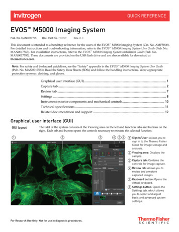
EVOS M5000 Imaging SystemPub. No. MAN0017765Doc. Part No. 710209Rev. B.0This document is intended as a benchtop reference for the users of the EVOS M5000 Imaging System (Cat. No. AMF5000).For detailed instructions and troubleshooting information, refer to the EVOS M5000 Imaging System User Guide (Pub. No.MAN0017563). For installation instructions, refer to the EVOS M5000 Imaging System Installation Guide (Pub. No.MAN0017783). These documents are provided on the USB flash drive and are also available for download atthermofisher.com.Note: For safety and biohazard guidelines, see the “Safety” appendix in the EVOS M5000 Imaging System User Guide(Pub. No. MAN0017563). Read the Safety Data Sheets (SDSs) and follow the handling instructions. Wear appropriateprotective eyewear, clothing, and gloves.Graphical user interface (GUI). 1Capture tab . 2Review tab . 7Settings . 8Instrument exterior components and mechanical controls . 10Technical specifications . 11Related documentation and support . 12Graphical user interface (GUI)GUI layoutThe GUI of the system consists of the Viewing area on the left and function tabs and buttons on theright. Each tab and button opens the controls necessary to execute the selected function.Sign In/User: Allows you tosign in to the Thermo FisherCloud for image storage andanalysis.Viewing area: Displays thesample.Capture tab: Contains thecontrols for image capture.Review tab: Allows you toreview and annotatecaptured images.Keyboard button: Opens thevirtual keyboard.Settings button: Opens theSettings tab, which allowsyou to select and adjustbasic and advanced systemsettings.For Research Use Only. Not for use in diagnostic procedures.
Capture tabCapture tabSign In/User: Allows you to sign in to and sign out of your Thermo Fisher Cloud account. You canchoose to save your captured images in your Cloud account for image storage (in addition to localstorage) and subsequent analysis with the EVOS Image Analysis application.Viewing area: Displays the sample.Zoom slider: Zooms in and out of the Viewing area. The zoom range is100% to 1000%.Active objective and phase ring: Displaysthe active objective and phase ringinformation.Channel: Selects the light source from theinstalled LED light cubes (fluorescent channels) orfrom the condenser (transmitted light).RGB Trans option outputs a color image frominterlaced RGB images in the Live mode. Select a channel to turn on the excitation light and enter the instrument in the Livemode. In the Live mode, you can use the brightness and focus controls. Click the selected Channel button again to turn off the excitation light and exit the Livemode. The current channel remains selected as indicated by the blue line underneaththe channel button.Brightness mode: Toggles between Simple and Actual modes for brightness controls. Simple mode allows you to control light intensity as a single Light parameter. Actual mode allows you to adjust Brightness (i.e., LED intensity), Exposure, and Gainparameters individually.2EVOS M5000 Imaging System Quick Reference Guide
Brightness controls: Adjust the brightness for the selected channel.SimpleActualCurrent Z (µm): Displays the current focus position in µm along the Z-axis.To change the Z-axis focus position, enter the desired value.Autofocus (Coarse and Fine): Runs the coarse and fine autofocusalgorithms in the selected channel.Coarse and Fine focus sliders: Allow you to adjust the focus in the selectedchannel when in the Live mode.Using the Autofocus Maximum and Autofocus Minimum controls (bluetriangles on the Coarse focus slider), you can limit the autofocus algorithmto the selected region along the Z-axis.Saved Settings: Allows you to save and edit Z-axis focus positionsand brightness settings for future use.Press thebutton to save the current settings.Lock Z: Allows you to lock the Z-axis offsets, which specify the optimal focus position ineach channel relative to the focus position in other channels. When locked, movement of the Z-position in one channel changes the Z-position in all channels,preserving their relative Z-positions. When unlocked, adjusting the Z-position only affects the currently selected channel.Enable focus knob: Enables the manual focus knobs.Capture: Captures an image using the current capture settings and stores it in theimage cache of the channel in which it was captured.You can capture multiple channels by selecting thecorresponding channel checkboxes.Thumbnails: Display the most recently capturedimage stored in the memory buffer for thechannel.EVOS M5000 Imaging System Quick Reference Guide3
Display settings and analysis tools: Allow you tochange image display settings for the Viewing area,and analyze and annotate captured images.To open a tool, click the corresponding button. Click the button again to close it.Image Display Settings: Opens the Image Display Settings tool, which allows you to adjustimage display parameters (Brightness, Constrast, Gamma Correction) for theselected channels.Grid/Grid Settings: Display Grid button switches the display ofthe grid in the Viewing area on and off.Grid Settings allows you to select the grid size.Scale Bar/Scale Bar Settings: Display Scale Bar button switches thedisplay of the scale bar in the Viewing area on and off.Scale Bar Settings allows you to select scale bar color and to display orhide end bars.Histogram: Opens the IntensityHistogram window, which displaysthe Pixel count vs. Intensity plot,where Intensity is the number ofphotons detected by the camerasensor.Measurements and Annotations:Allows you to draw regions of interest(rectangle, ellipse, polygon, line, orfree-form) on the captured image andmeasure dimensions, area, orperimeter of the drawn region.Show Cell Count: Allows you to perform cell counts using the Auto Count or Manual Counttools. Auto Count: Allows you define auto count parameters by selecting representative targetobjects, then count the objects by intensity, area, and circularity. Manual Count: Allows you to manually mark items onscreen using up to six separatelabels and keep a running tally of the counts with percentages for each label. Confluence: Allows you to select up to 5 reference objects each for target (i.e., cells) andbackground in your image to automatically calculate the percentage confluence of yourculture.4EVOS M5000 Imaging System Quick Reference Guide
Auto CountManual CountConfluenceToggle pseudo color: Allows you to display images in pseudocolor or in grayscale in theViewing area. By default, color display is on.Automate: Allows you to display the controls for the Time Lapse and Z-Stacktools, and the EVOS Onstage Incubator.Time Lapse: Opens the Time Lapse tool, which allows you tocreate and run time lapse routines to capture images atgiven intervals over a time period based on yourspecifications.EVOS M5000 Imaging System Quick Reference Guide5
Z Stack: Opens the Z Stack tool, which allows you to capturemultiple images along the Z-axis based on yourspecifications. You can use the images captured with the ZStack tool to generate a Z-Stack Projection.Incubator: Opens the Incubator Controls, which allow you tocontrol the EVOS Onstage Incubator and monitor its statusduring your experiments.Save: Opens the Save dialog, which allows you to select asave location, set save options, and enable the Quick Saveoption.6EVOS M5000 Imaging System Quick Reference Guide
Review tabReview tabViewing area: Displays the captured image currently selected from the list of image files.Folder: Displays the location of thecurrent folder or image.Up: Goes up one folder from the current folder.Refresh: Refreshes the list or grid of images in the current folder.Grid view and List view buttons: Display the folders or saved images in grid or list view.Folder/Image display size: Increase and decrease the display size of the folders orimage files.Browse: Allows you to navigate to the foldercontaining your saved images and to select theimage to display in the viewing area. Currentlyselected folder or image is indicated with a boxaround it.Image properties: Displays the metadata for theselected image file.Display settings and analysis tools: Allow you tochange image display settings in the Viewing area,and analyze and annotate captured images.Export: Allows you to export the currently selected folder or image to a storagedevice.Save: Saves the currently opened image.EVOS M5000 Imaging System Quick Reference Guide7
SettingsSettings tabObjectives: Allows you to assign and unassignobjectives on the objective turret, and tocalibrate objective magnification.Visuals: Allows you to calibrate color channellighting (White Balance Calibration) and toassign saturated pixel colors in theFluorescence, Transmitted, and RGBTransmitted channels.General: Allows you to define saving options forTIFF files and Saved Settings, and to reversethe focus wheel action.8EVOS M5000 Imaging System Quick Reference Guide
Filter Cubes: Allows you to add or removeEVOS LED light cubes from the instrumentand to assign pseudocolors to specificchannels.Network: Allows you to connect to a Wi-Finetwork and to map network drives.Service: Displays the EVOS M5000 hardwareand software information, allows you to updatethe EVOS M5000 firmware and software fromthe Thermo Fisher Cloud or using a USB flashdrive, and to copy the error logs.Done: Closes the Settings tabs and returns to the previously opened tab.EVOS M5000 Imaging System Quick Reference Guide9
Instrument exterior components and mechanical controlsFront viewLCD monitorCondenser slider slotCondenser headMechanical X-Y stageStage X-axis positioning knobStage Y-axis positioning knobObjective selection wheelFocusing knobVessel holderObjectivePhase annuli selectorRear viewLCD monitorMechanical X-Y stageFocusing knobsPower switch4-pin power input port (12 VDC, 5 A)Display port (video output)Network portUSB-A 2.0 ports (4 )USB 3.0 port (1 )Stage X-axis positioning knobStage Y-axis positioning knob** Not visible in this perspective.10EVOS M5000 Imaging System Quick Reference Guide
Technical specificationsNote: Technical specifications of the EVOS M5000 Imaging System are subject to change without notice. For the latestproduct information, see the product page ensions (W H D): 46 59 46 cm (18 23 18 in)Weight: 16 kg (35 lbs.)Footprint: Approximately 92 cm 92 cm (36 in 36 in)Operating temperature: 4 –32 C (40 –90 F)Operating humidity: 90%, non-condensingOperating power: 100–240 VAC, 1.8 AFrequency: 50–60 HzElectrical input: 12 VDC, 5 AHardwareOptics: Infinity‐corrected optical system; RMS‐threaded objectives with 45 mm parfocal distance.Illumination: Adjustable intensity LED ( 50,000-hour life per light cube).Light cubes (not included): 5 position chamber for 4 fluorescence cubes plus brightfield. Broadselection standard and specialty light cubes.Contrast methods: Fluorescence and transmitted light (brightfield & phase contrast).Objective turret: 5-position; front-mounted control.Objectives (not included): Wide selection of high‐quality LWD and coverslip‐corrected objectives.Condenser: 60-mm long working distance condenser, 4 position turret with a clear aperture and 3phase annuli.Stage: Manual X-Y scanning stage. Travel range of 120 mm 80 mm with sub‐micron resolution,drop‐in inserts to receive vessel holders and lock down holders to fix sample in place during longscans.Focus mechanism: Automated focus mechanism with sub‐micron resolution.LCD display: 18.5-inch high-resolution LCD display (1920 1080 pixel resolution)Camera: High-sensitivity monochrome CMOS camera (2048 1536 pixel resolution, 3.2 Megapixels)Captured images: 16‐bit monochrome (12‐bit dynamic range) TIFF or PNG; 8-bit per RGB channelTIF, PNG, BMP, or JPG.Output ports: 4-pin power input port (12 VDC, 5 A), 1 USB 3.0, 4 USB 2.0Networking capability: Connection through Windows/SMB network via an Ethernet cableconnection or wirelessly using the supplied Wi-Fi adaptor.Power supply: AC adaptor with country‐specific power cords.EVOS M5000 Imaging System Quick Reference Guide11
Related documentation and supportRelateddocumentationThe publication numbers in this section are for the latest product versions available at the timeof publication. For documentation supporting newer product versions, go tothermofisher.com/support.DocumentPub. No.EVOS M5000 Imaging System User GuideMAN0017563EVOS M5000 Imaging System Installation GuideMAN0017783 Customer andtechnical supportVisit thermofisher.com/support for the latest in services and support, including: Worldwide contact telephone numbers Product support, including:---Limited productwarrantyProduct FAQsSoftware, patches, and updatesTraining for many applications and instruments Order and web support Product documentation, including user guides, manuals, and protocolsLife Technologies Corporation and/or its affiliate(s) warrant their products as set forth in theLife Technologies’ General Terms and Conditions of Sale found on Life Technologies’ website -conditions.html. If you have anyquestions, please contact Life Technologies at www.thermofisher.com/support.Manufacturer: Life Technologies Corporation 22025 20TH Ave SE Bothell, WA 98021The information in this document is subject to change without notice.DISCLAIMER: TO THE EXTENT ALLOWED BY LAW, THERMO FISHER SCIENTIFIC AND/OR ITS AFFILIATE(S) WILL NOT BE LIABLE FOR SPECIAL,INCIDENTAL, INDIRECT, PUNITIVE, MULTIPLE OR CONSEQUENTIAL DAMAGES IN CONNECTION WITH OR ARISING FROM THIS DOCUMENT,INCLUDING YOUR USE OF IT.Revision history: MAN0017765RevisionDateDescriptionB.008 January 2019Add Thermo Fisher Cloud sign in and Confluence tool, and update the screenshots.A.006 August 2018New quick reference guide for the EVOS M5000 Imaging System.Trademarks: All trademarks are the property of Thermo Fisher Scientific and its subsidiaries unless otherwise specified. 2019 Thermo Fisher Scientific Inc. All rights reserved.thermofisher.com/support thermofisher.com/askaquestionthermofisher.com08 January 2019
EVOS M5000 Imaging System . Pub. No. MAN0017765 Doc. Part No. 710209 Rev. B.0. This document is intended as a benchtop reference for the users of the EVOS M5000 Imaging System (Cat. No. AMF5000). For detailed instructions and troubleshooting information, refer to the EVOS M5000 Imaging System User Guide (Pub. No. MAN0017563).