Transcription
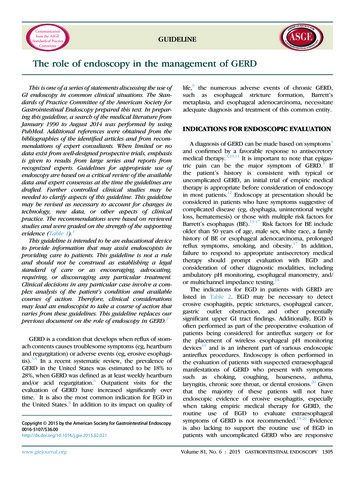
GUIDELINEThe role of endoscopy in the management of GERDThis is one of a series of statements discussing the use ofGI endoscopy in common clinical situations. The Standards of Practice Committee of the American Society forGastrointestinal Endoscopy prepared this text. In preparing this guideline, a search of the medical literature fromJanuary 1990 to August 2014 was performed by usingPubMed. Additional references were obtained from thebibliographies of the identified articles and from recommendations of expert consultants. When limited or nodata exist from well-designed prospective trials, emphasisis given to results from large series and reports fromrecognized experts. Guidelines for appropriate use ofendoscopy are based on a critical review of the availabledata and expert consensus at the time the guidelines aredrafted. Further controlled clinical studies may beneeded to clarify aspects of this guideline. This guidelinemay be revised as necessary to account for changes intechnology, new data, or other aspects of clinicalpractice. The recommendations were based on reviewedstudies and were graded on the strength of the supportingevidence (Table 1).1This guideline is intended to be an educational deviceto provide information that may assist endoscopists inproviding care to patients. This guideline is not a ruleand should not be construed as establishing a legalstandard of care or as encouraging, advocating,requiring, or discouraging any particular treatment.Clinical decisions in any particular case involve a complex analysis of the patient’s condition and availablecourses of action. Therefore, clinical considerationsmay lead an endoscopist to take a course of action thatvaries from these guidelines. This guideline replaces ourprevious document on the role of endoscopy in GERD.2GERD is a condition that develops when reflux of stomach contents causes troublesome symptoms (eg, heartburnand regurgitation) or adverse events (eg, erosive esophagitis).3-5 In a recent systematic review, the prevalence ofGERD in the United States was estimated to be 18% to28%, when GERD was defined as at least weekly heartburnand/or acid regurgitation.6 Outpatient visits for theevaluation of GERD have increased significantly overtime.7 It is also the most common indication for EGD inthe United States.8 In addition to its impact on quality ofCopyright ª 2015 by the American Society for Gastrointestinal Endoscopy0016-5107/ w.giejournal.orglife,9 the numerous adverse events of chronic GERD,such as esophageal stricture formation, Barrett’smetaplasia, and esophageal adenocarcinoma, necessitateadequate diagnosis and treatment of this common entity.INDICATIONS FOR ENDOSCOPIC EVALUATIONA diagnosis of GERD can be made based on symptoms3and confirmed by a favorable response to antisecretorymedical therapy.3,10,11 It is important to note that epigastric pain can be the major symptom of GERD.3 Ifthe patient’s history is consistent with typical oruncomplicated GERD, an initial trial of empiric medicaltherapy is appropriate before consideration of endoscopyin most patients.12 Endoscopy at presentation should beconsidered in patients who have symptoms suggestive ofcomplicated disease (eg, dysphagia, unintentional weightloss, hematemesis) or those with multiple risk factors forBarrett’s esophagus (BE).13-17 Risk factors for BE includeolder than 50 years of age, male sex, white race, a familyhistory of BE or esophageal adenocarcinoma, prolongedreflux symptoms, smoking, and obesity.17 In addition,failure to respond to appropriate antisecretory medicaltherapy should prompt evaluation with EGD andconsideration of other diagnostic modalities, includingambulatory pH monitoring, esophageal manometry, and/or multichannel impedance testing.18The indications for EGD in patients with GERD arelisted in Table 2. EGD may be necessary to detecterosive esophagitis, peptic strictures, esophageal cancer,gastric outlet obstruction, and other potentiallysignificant upper GI tract findings. Additionally, EGD isoften performed as part of the preoperative evaluation ofpatients being considered for antireflux surgery or forthe placement of wireless esophageal pH monitoringdevices19 and is an inherent part of various endoscopicantireflux procedures. Endoscopy is often performed inthe evaluation of patients with suspected extraesophagealmanifestations of GERD who present with symptomssuch as choking, coughing, hoarseness, asthma,laryngitis, chronic sore throat, or dental erosions.20 Giventhat the majority of these patients will not haveendoscopic evidence of erosive esophagitis, especiallywhen taking empiric medical therapy for GERD, theroutine use of EGD to evaluate extraesophagealsymptoms of GERD is not recommended.21-26 Evidenceis also lacking to support the routine use of EGD inpatients with uncomplicated GERD who are responsiveVolume 81, No. 6 : 2015 GASTROINTESTINAL ENDOSCOPY 1305
The role of endoscopy in the management of GERDTABLE 1. GRADE system for rating the quality of evidence forguidelinesQuality ofevidenceDefinitionSymbolHigh qualityFurther research is very unlikelyto change our confidence inthe estimate of effect.4444ModeratequalityFurther research is likely to havean important impact on ourconfidence in the estimate of effectand may change the estimate.444BLow qualityFurther research is very likelyto have an important impact onour confidence in the estimateof effect and is likely to changethe estimate.44BBVery lowqualityAny estimate of effect is veryuncertain.4BBBAdapted from Guyatt et al.1to medical therapy. There is a paucity of outcomesresearch to suggest that early or even once-in-a-lifetimeEGD has a favorable effect on the management, course,or health-related quality of life of patients with typicalsymptoms of GERD without alarm features (dysphagia,odynophagia, weight loss, bleeding, or anemia).10DIAGNOSIS AND CLASSIFICATION OF GERDINDUCED ESOPHAGEAL INFLAMMATIONPatients with reflux esophagitis have endoscopic and/orhistopathologic changes of esophageal mucosal injury andinflammation. The presence of typical findings of refluxesophagitis on EGD such as erythema, erosions, ulceration,peptic strictures, and BE is diagnostic of GERD with a specificity as high as 95%.27,28 However, at least 50% of patientswith reflux symptoms have normal esophageal endoscopicfindings (nonerosive reflux disease) or uncomplicatedGERD.3,26 In addition, dyspepsia is a diagnosis often confusedwith GERD. Furthermore, the severity of GERD symptomsdoes not correlate with the degree of underlying esophagealdamage, supporting current recommendations to initiateempiric antisecretory therapy in patients with typical GERDsymptoms in the absence of alarm features.10,29There are several classification systems for grading theendoscopic severity of erosive reflux esophagitis and associated adverse events.30 These classification systems havebeen primarily used in clinical trials to study the efficacyof medical therapy of reflux esophagitis. However, thesesystems are useful in clinical practice for documentingdisease severity. Currently, the most commonly usedsystems are the Los Angeles classification and the SavaryMiller classification (Table 3). The Los Angelesclassification has been shown to be reliable, with goodintra- and interobserver agreement when tested among1306 GASTROINTESTINAL ENDOSCOPY Volume 81, No. 6 : 2015expert and inexperienced endoscopists.13,30 When usingthis system, the severity of esophagitis has been demonstrated to correlate with the extent of esophageal acidexposure determined by 24-hour pH monitoring.31When esophagitis is encountered endoscopically, tissuesamples of the esophageal mucosa should be obtained under the following circumstances: underlying immunocompromised state, the presence of irregular or deepulceration, proximal distribution of esophagitis, the presence of an esophageal mass lesion or nodularity, bullouschanges suggestive of esophageal pemphigus vulgaris,visible changes of eosinophilic esophagitis (rings, linearfurrows, white plaques, fragile mucosa), changes of esophageal dessicans superficialis, or an irregular or malignantappearing esophageal stricture. In these situations, forcepstissue samples and/or brush cytology specimens are necessary to exclude other diagnoses, including infectious etiologies and malignancy. Tissue sampling has also beenrecommended in patients with dysphagia without evidenceof erosive esophagitis to evaluate for eosinophilic esophagitis.29 However, routine sampling of the esophagus orgastroesophageal junction in patients with heartburn anda normal findings on endoscopy are not recommended.32Historically, follow-up EGD for patients with GERD andesophagitis has been reserved for patients whose symptoms fail to respond to medical therapy, those who had severe esophagitis or an esophageal ulcer, or those whoneeded additional biopsies to clarify a diagnosis such asBE or BE-associated dysplasia because the presence oferosive esophagitis may impair the accurate histopathologic detection of BE and dysplasia.32,33 Multiple trials havedemonstrated that 8 weeks of proton pump inhibitor(PPI) treatment is adequate to achieve mucosal healingin most patients with erosive esophagitis due to GERD.34-36Upon healing of erosive esophagitis, BE can be identified inas many as 12% of these patients.32,37,38Adverse events of GERDPeptic strictures. The endoscopic evaluation andmanagement of peptic strictures is discussed in anotherASGE guideline.39Barrett’s esophagus. BE is a premalignant conditionin which the squamous epithelium of the distal esophagusis replaced by an abnormal columnar epithelium known asspecialized intestinal metaplasia.40,41 BE is found in asmany as 15% of patients undergoing EGD for GERD.42,43Recommendations regarding the role of EGD for screeningand surveillance for BE were recently published.17The value and optimal method of screening for BE remains unclear.17 Widespread screening of the entirepopulation with GERD would not be feasible given boththe high prevalence of GERD in the Western world andthe presence of many asymptomatic individualsharboring BE.44,45 However, several factors associatedwith BE may facilitate the selection of at-risk individualsfor screening EGD. These include white race, male sex,www.giejournal.org
The role of endoscopy in the management of GERDolder age (older than 50 years of age), prolonged GERDsymptoms (O5 years), a family history of BE and/or adenocarcinoma of the esophagus, nocturnal reflux symptoms,hiatal hernia, increased body mass index (BMI R25 kg/m2), tobacco use, and intra-abdominal distribution offat.10,16,17,46,47 The ASGE suggests that endoscopicscreening for BE be considered in select patients withmultiple risk factors for BE and esophageal adenocarcinoma, but patients should be informed that there is insufficient evidence to affirm that this practice prevents canceror prolongs life.17 Given the high costs of endoscopy andlimitations of using GERD symptoms to screen for BE,alternative screening methods have been sought.Endoscopy with tissue sampling is the most accuratetool for the detection and diagnosis of BE. To determinethe presence of BE endoscopically, the squamocolumnarand gastroesophageal junctions must be clearly identified.Although proximal displacement of the squamocolumnarjunction relative to the gastroesophageal junction is suggestive of BE, the endoscopic appearance of salmoncolored mucosa or an irregular Z line, either alone or incombination, is not sufficient to establish the diagnosis.40Esophageal tissue specimens should always be obtainedfor histopathologic confirmation of columnar epitheliumwhen BE is suspected. The optimal number of tissuesamples necessary to identify intestinal metaplasia is notknown, but it is generally accepted that multiple biopsyspecimens should be obtained in all areas of suspectedBE.48,49 Care should be taken to avoid obtaining specimensfrom a normal-appearing squamocolumnar junction orfrom the proximal cardia because tissue samples fromthese areas may demonstrate intestinal metaplasia and provide a false diagnosis of BE.49-51 Patients with a negativescreening EGD for BE do not need follow-up endoscopybecause only 1.8% of such patients were found to haveBE on repeat EGD performed within 5 years.37ENDOLUMINAL ANTIREFLUX PROCEDURESEndoluminal therapies for GERD have been used formore than a decade. The techniques used have included delivery of thermal energy intended to constrict the loweresophageal sphincter, intramural injection of bulking agentsto augment lower esophageal sphincter pressures, and mechanical alterations of the gastroesophageal junction tomimic results achieved with surgical fundoplication.52-55Several devices and/or techniques have been abandoneddue to a lack of efficacy or durability or for safety reasons.Currently, there are 2 endoluminal GERD therapies usedin the United States: the Stretta procedure (Mederi Therapeutics, Greenwich, Conn) and transoral incisionless fundoplication (TIF) (Endogastric Solutions, Redmond, Wash).54The Stretta procedure received initial U.S. Foodand Drug Administration approval in 2000. This techniqueuses radiofrequency energy delivery to the distalwww.giejournal.orgTABLE 2. Indications for endoscopy in patients with GERDGERD symptoms that are persistent or progressive despite appropriatemedical therapyDysphagia or odynophagiaInvoluntary weight loss O5%Evidence of GI bleeding or anemiaFinding of a mass, stricture, or ulcer on imaging studiesScreening for Barrett’s esophagus in selected patients (as clinicallyindicated)Persistent vomiting (7-10 days)Evaluation of patients before or with recurrent symptoms afterendoscopic or surgical antireflux proceduresPlacement of wireless pH monitoringesophagus and appears to reduce GERD by decreasing tissue compliance and reducing transient lower esophagealrelaxations.54 A recent meta-analysis of 18 studies involving1441 patients found significant improvement in heartburnand GERD quality of life scores after the Stretta procedure.56 However, although the esophageal acid exposure,as measured by the DeMeester score, was significantlyreduced after treatment (44.4 vs 28.5, P Z .007), it didnot normalize. In addition, no significant increase inlower esophageal sphincter pressure was observed.Adverse events were infrequent and typically minor. Thetechnique appears to durably relieve GERD symptomsfor up to 10 years in the majority of patients.57,58The TIF procedure received U.S. Food and Drug Administration approval in 2007 and has undergone several device and technique modifications since the initialapproval.54 Most studies involving TIF have been smallwith short-term follow-up. The results have been variable,with poorer results observed with earlier versions of thedevice/technique. A systematic review of TIF that included15 studies and 550 procedures found improved GERDhealth-related quality of life scores (21.9 vs 5.9, P !.0001) after the procedure.59 PPI use was discontinued in67% of patients. Limitations of the analysis of thesestudies include a lack of routine reporting of pH dataand a mean follow-up period of only 8.3 months. Majoradverse events were reported in 3.2% of patients. Mostrecently, 3 randomized trials with at least 6-month followup found that TIF was more effective than high-dose PPItherapy in eliminating troublesome regurgitation or extraesophageal symptoms of GERD.60-62Endoluminal antireflux techniques represent potentiallynew therapeutic indications for GI endoscopy. Prospectivetrials comparing these therapies with existing medical andsurgical options by using objective measures of GERD asthe primary endpoint could be useful in further definingthe clinical role of these procedures. Appropriate patientselection and endoscopist experience and training shouldbe carefully considered before pursuing these therapies.Volume 81, No. 6 : 2015 GASTROINTESTINAL ENDOSCOPY 1307
The role of endoscopy in the management of GERDTABLE 3. The Los Angeles and Savary-Miller classifications of esophagitisClassificationLos AngelesSavary-MillerGradeDescriptionAOne (or more) mucosal break no longer than 5 mm that does not extend between the tops of 2 mucosal foldsBOne (or more) mucosal break O5 mm that does not extend between the tops of 2 mucosal foldsCOne (or more) mucosal break that is continuous between the tops of R2 mucosal folds but that involves !75%of the circumferenceDOne (or more) mucosal break that involves at least 75% of the esophageal circumference1Single erosion above the gastroesophageal mucosal junction2Multiple, noncircumferential erosions above the gastroesophageal mucosal junction3Circumferential erosion above the mucosal junction4Chronic change with esophageal ulceration and associated stricture5Barrett’s esophagus with histologically confirmed intestinal differentiation within the columnar epithelium.ROLE OF ENDOSCOPY IN PEDIATRIC GERDAlthough most infant reflux is physiologic, there aresparse data regarding the prevalence of GERD in the pediatric population.6 Guidelines from the North AmericanSociety of Pediatric Gastroenterology, Hepatology andNutrition state that endoscopy is indicated in infants andchildren with GERD who fail to respond to pharmacologictherapy or as part of the initial management if symptomsof poor weight gain, unexplained anemia or fecal occultblood, recurrent pneumonia, or hematemesis exist.63Erosive esophagitis is reported less often in infants andchildren with GERD than in adults with GERD, butapproximately 25% of infants younger than 1 year of ageundergoing upper endoscopy will have histologicevidence of esophageal inflammation.64 When EGD isperformed in children with suspected GERD, tissuesampling of both normal and inflamed mucosa should beperformed to exclude other conditions such aseosinophilic esophagitis, gastritis, and celiac disease.65,66SUMMARY We recommend that uncomplicated GERD be diagnosed on the basis of typical symptoms without theuse of diagnostic testing, including EGD. 4444 We recommend EGD for patients who have symptomssuggesting complicated GERD or alarm symptoms.444B We recommend that EGD not be routinely performedsolely for the assessment of extraesophageal GERDsymptoms. 444B We recommend that endoscopic findings of refluxesophagitis be classified according to an acceptedgrading scale or described in detail. 444B We suggest that repeat EGD be performed in patientswith severe erosive esophagitis after at least an 8-weekcourse of PPI therapy to exclude underlying BE ordysplasia. 44BB1308 GASTROINTESTINAL ENDOSCOPY Volume 81, No. 6 : 2015 We recommend against obtaining tissue samples fromendoscopically normal tissue to diagnose GERD orexclude BE in adults. 444B We suggest that endoscopy be considered in patientswith multiple risk factors for Barrett’s esophagus.4BBB We recommend that tissue samples be obtained toconfirm endoscopically suspected Barrett’s esophagus.4444 We suggest that endoscopic antireflux therapy beconsidered for selected patients with uncomplicatedGERD after careful discussion with the patient regardingpotential adverse effects, benefits, and other availabletherapeutic options. 44BBDISCLOSUREDr Khashab is a consultant for and member of theadvisory board of Boston Scientific, is a consultant forOlympus American, and has received research supportfrom Cook Medical. Dr Chathadi is a consultant for Boston Scientific. Dr Muthusamy is a consultant for and hasreceived honoraria and research support from CovidienGI Solutions. Dr Fanelli is a consultant for EndoGastricSolutions, has received royalties for unrelated inventionsand product development from Cook Surgical Inc, andhas minor ownership interest in Allurion TechnologiesInc and Mozaic Medical Inc. All other authors disclosedno financial relationships relevant to this article.Abbreviations: BE, Barrett’s esophagus; PPI, proton pump inhibitor; TIF,transoral incisionless fundoplication.REFERENCES1. Guyatt GH, Oxman AD, Vist GE, et al. GRADE: an emerging consensuson rating quality of evidence and strength of recommendations. BMJ2008;336:924-6.2. Lichtenstein DR, Cash BD, Davila R, et al. Role of endoscopy in themanagement of GERD. Gastrointest Endosc 2007;66:219-24.www.giejournal.org
The role of endoscopy in the management of GERD3. Vakil N, van Zanten SV, Kahrilas P, et al. The Montreal definition andclassification of gastroesophageal reflux disease: a global evidencebased consensus. Am J Gastroenterol 2006;101:1900-20.4. DeVault KR, Castell DO. Updated guidelines for the diagnosis and treatment of gastroesophageal reflux disease. Am J Gastroenterol 2005;100:190-200.5. Jones R, Galmiche JP. Review: what do we mean by GERD?–definitionand diagnosis. Aliment Pharmacol Ther 2005;22(Suppl 1):2-10.6. El-Serag HB, Sweet S, Winchester CC, et al. Update on the epidemiology of gastro-oesophageal reflux disease: a systematic review. Gut2014;63:871-80.7. Friedenberg FK, Hanlon A, Vanar V, et al. Trends in gastroesophagealreflux disease as measured by the National Ambulatory Medical CareSurvey. Dig Dis Sci 2010;55:1911-7.8. Peery AF, Dellon ES, Lund J, et al. Burden of gastrointestinal disease inthe United States: 2012 update. Gastroenterology 2012;143:1179-87;e1-3.9. Becher A, El-Serag H. Systematic review: the association betweensymptomatic response to proton pump inhibitors and health-relatedquality of life in patients with gastro-oesophageal reflux disease.Aliment Pharmacol Ther 2011;34:618-27.10. Katz PO, Gerson LB, Vela MF. Guidelines for the diagnosis and management of gastroesophageal reflux disease. Am J Gastroenterol 2013;108:308-28.11. Numans ME, Lau J, de Wit NJ, et al. Short-term treatment withproton-pump inhibitors as a test for gastroesophageal reflux disease:a meta-analysis of diagnostic test characteristics. Ann Intern Med2004;140:518-27.12. Venables TL, Newland RD, Patel AC, et al. Omeprazole 10 milligramsonce daily, omeprazole 20 milligrams once daily, or ranitidine 150milligrams twice daily, evaluated as initial therapy for the relief ofsymptoms of gastro-oesophageal reflux disease in general practice.Scand J Gastroenterol 1997;32:965-73.13. Lundell LR, Dent J, Bennett JR, et al. Endoscopic assessment of oesophagitis: clinical and functional correlates and further validation ofthe Los Angeles classification. Gut 1999;45:172-80.14. Wo JM, Mendez C, Harrell S, et al. Clinical impact of upper endoscopyin the management of patients with gastroesophageal reflux disease.Am J Gastroenterol 2004;99:2311-6.15. Lieberman DA, Oehlke M, Helfand M. Risk factors for Barrett’s esophagus in community-based practice. GORGE consortium. Gastroenterology Outcomes Research Group in Endoscopy. Am J Gastroenterol1997;92:1293-7.16. Shaheen NJ, Weinberg DS, Denberg TD, et al. Upper endoscopy forgastroesophageal reflux disease: best practice advice from the clinicalguidelines committee of the American College of Physicians. AnnIntern Med 2012;157:808-16.17. Evans JA, Early DS, Fukami N, et al. The role of endoscopy in Barrett’sesophagus and other premalignanct conditions of the esophagus.Gastrointest Endosc 2012;76:1087-94.18. Fock KM, Talley N, Hunt R, et al. Report of the Asia-Pacific consensus onthe management of gastroesophageal reflux disease. J GastroenterolHepatol 2004;19:357-67.19. Chotiprashidi P, Liu J, Carpenter S, et al. ASGE Technology StatusEvaluation Report: wireless esophageal pH monitoring system. Gastrointest Endosc 2005;62:485-7.20. Poelmans J, Feenstra L, Demedts I, et al. The yield of upper gastrointestinal endoscopy in patients with suspected reflux-related chronicear, nose, and throat symptoms. Am J Gastroenterol 2004;99:1419-26.21. Richter JE. Extraesophageal presentations of gastroesophageal refluxdisease. Semin Gastrointest Dis 1997;8:75-89.22. El-Serag HB, Lee P, Buchner A, et al. Lansoprazole treatment of patientswith chronic idiopathic laryngitis: a placebo-controlled trial. Am JGastroenterol 2001;96:979-83.23. Koufman JA. Laryngopharyngeal reflux is different from classicgastroesophageal reflux disease. Ear Nose Throat J 2002;81(9 Suppl2):7-9.www.giejournal.org24. Koufman JA, Belafsky PC, Bach KK, et al. Prevalence of esophagitis inpatients with pH-documented laryngopharyngeal reflux. Laryngoscope 2002;112:1606-9.25. Gralnek IM, Dulai GS, Fennerty MB, et al. Esomeprazole versus otherproton pump inhibitors in erosive esophagitis: a meta-analysis ofrandomized clinical trials. Clin Gastroenterol Hepatol 2006;4:1452-8.26. Ronkainen J, Aro P, Storskrubb T, et al. High prevalence of gastroesophageal reflux symptoms and esophagitis with or withoutsymptoms in the general adult Swedish population: a Kalixanda studyreport. Scand J Gastroenterol 2005;40:275-85.27. Moayyedi P, Talley NJ. Gastro-oesophageal reflux disease. Lancet2006;367:2086-100.28. Richter JE. Diagnostic tests for gastroesophageal reflux disease. Am JMed Sci 2003;326:300-8.29. Kahrilas PJ, Shaheen NJ, Vaezi MF, et al. American GastroenterologicalAssociation Medical Position Statement on the management of gastroesophageal reflux disease. Gastroenterology 2008;35:1383-91;1391.e1-5.30. Rath HC, Timmer A, Kunkel C, et al. Comparison of interobserveragreement for different scoring systems for reflux esophagitis: impactof level of experience. Gastrointest Endosc 2004;60:44-9.31. Kahrilas PJ, Pandolfino JE. Review article: oesophageal pH monitoring–technologies, interpretation and correlation with clinical outcomes.Aliment Pharmacol Ther 2005;22(Suppl 3):2-9.32. Takubo K, Honma N, Aryal G, et al. Is there a set of histologic changesthat are invariably reflux associated? Arch Pathol Lab Med 2009;129:159-63.33. Hanna S, Rastogi A, Weston AP, et al. Detection of Barrett’s esophagusafter endoscopic healing of erosive esophagitis. Am J Gastroenterol2006;101:1416-20.34. Castell DO, Kahrilas PJ, Richter JE, et al. Esomeprazole (40 mg)compared with lansoprazole (30 mg) in the treatment of erosiveesophagitis. Am J Gastroenterol 2002;97:575-83.35. Richter JE, Kahrilas PJ, Sontag SJ, et al. Comparing lansoprazole andomeprazole in onset of heartburn relief: results of a randomized,controlled trial in erosive esophagitis patients. Am J Gastroenterol2001;96:3089-98.36. Labenz J, Armstrong D, Lauritsen K, et al. Esomeprazole 20 mg vs. pantoprazole 20 mg for maintenance therapy of healed erosive oesophagitis: results from the EXPO study. Aliment Pharmacol Ther 2005;22:803-11.37. Rodriguez S, Mattek N, Lieberman D, et al. Barrett’s esophagus onrepeat endoscopy: should we look more than once? Am J Gastroenterol 2008;103:1892-7.38. Modiano N, Gerson LB. Risk factors for the detection of Barrett’sesophagus in patients with erosive esophagitis. Gastrointest Endosc2009;69:1014-20.39. Pasha SF, Acosta RD, Chandrasekhara V, et al. The role of endoscopy inthe evaluation and management of dysphagia. Gastrointest Endosc2014;79:191-201.40. Spechler SJ. Barrett esophagus and risk of esophageal cancer: a clinicalreview. JAMA 2013;310:627-36.41. Spechler SJ, Sharma P, Souza RF, et al. American GastroenterologicalAssociation medical position statement on the management ofBarrett’s esophagus. Gastroenterology 2011;140:1084-91.42. Wani S, Sharma P. The rationale for screening and surveillance ofBarrett’s metaplasia. Best Pract Res Clin Gastroenterol 2006;20:829-42.43. Westhoff B, Brotze S, Weston A, et al. The frequency of Barrett’s esophagus in high-risk patients with chronic GERD. Gastrointest Endosc2005;61:226-31.44. Gerson LB, Groeneveld PW, Triadafilopoulos G. Cost-effectivenessmodel of endoscopic screening and surveillance in patients withgastroesophageal reflux disease. Clin Gastroenterol Hepatol 2004;2:868-79.45. Rex DK, Cummings OW, Shaw M, et al. Screening for Barrett’sesophagus in colonoscopy patients with and without heartburn.Gastroenterology 2003;125:1670-7.Volume 81, No. 6 : 2015 GASTROINTESTINAL ENDOSCOPY 1309
The role of endoscopy in the management of GERD46. Rubenstein JH, Morgenstern J, Appelman H, et al. Prediction ofBarrett’s esophagus among men. Am J Gastroenterol 2013;108:353-62.47. Kamat P, Wen S, Morris J, et al. Exploring the association betweenelevated body mass index and Barrett’s esophagus: a systematicreview and meta-analysis. Ann Thorac Surg 2009;87:655-62.48. Sharma P, McQuaid K, Dent J, et al. A critical review of the diagnosisand management of Barrett’s esophagus: the AGA Chicago Workshop.Gastroenterology 2004;127:310-30.49. Sharaf RN, Shergill AK, Odze RD, et al. Endoscopic mucosal tissuesampling. Gastrointest Endosc 2013;78:216-24.50. Spechler SJ, Zeroogian JM, Antonioli DA, et al. Prevalence of metaplasia at the gastro-oesophageal junction. Lancet 1994;344:1533-6.51. Hirota WK, Loughney TM, Lazas DJ, et al. Specialized intestinal metaplasia,dysplasia, and cancer of the esophagus and esophagogastric junction:prevalence and clinical data. Gastroenterology 1999;116:277-85.52. Louis H, Devière J. Ensocopic-endoluminal therapies. A criticalappraisal. Best Pract Res Clin Gastroenterol 2010;24:969-79.53. Yew KC, Chuah SK. Antireflux endoluminal therapies: past and present.Gastroenterol Res Pract 2013;2013:481417.54. Auyang ED, Carter P, Rauth T, et al. SAGES clinical spotlight review:endoluminal treatments for gastroesophageal reflux disease (GERD).Surg Endosc 2013;27:2658-72.55. Falk GW, Fennerty MB, Rothstein RI. AGA Institute technical review onthe use of endoscopic therapy for gastroesophageal reflux disease.Gastroenterology 2006;131:1315-36.56. Perry KA, Banerjee A, Melvin WS. Radiofrequency energy delivery tothe lower esophageal sphincter reduces esophageal acid exposureand improves GERD symptoms: a systematic review and meta-analysis.Surg Laparosc Endosc Percutan Tech 2012;22:283-8.57. Noar M, Squires P, Noar E, et al. Long-term maintenance effect ofradiofrequency energy delivery for refractory GERD: a decade later.Surg Endosc 2014;28:2323-33.58. Dughera L, Rotondano G, De Cento M, et al. Durability of Stretta radiofrequency treatment for GERD: results of an
mendations of expert consultants. When limited or no data exist from well-designed prospective trials, emphasis is given to results from large series and reports from recognized experts. Guidelines for appropriate use of endoscopy are based on a critical review of the available data and expert consensus at the time the guidelines are drafted.