Transcription
ArticlePsychedelics Promote Structural and FunctionalNeural PlasticityGraphical AbstractAuthorsCalvin Ly, Alexandra C. Greb,Lindsay P. Cameron, .,Kassandra M. Ori-McKenney,John A. Gray, David E. OlsonCorrespondencedeolson@ucdavis.eduIn BriefLy et al. demonstrate that psychedeliccompounds such as LSD, DMT, and DOIincrease dendritic arbor complexity,promote dendritic spine growth, andstimulate synapse formation. Thesecellular effects are similar to thoseproduced by the fast-actingantidepressant ketamine and highlightthe potential of psychedelics for treatingdepression and related disorders.HighlightsdSerotonergic psychedelics increase neuritogenesis,spinogenesis, and synaptogenesisdPsychedelics promote plasticity via an evolutionarilyconserved mechanismdTrkB, mTOR, and 5-HT2A signaling underlie psychedelicinduced plasticitydNoribogaine, but not ibogaine, is capable of promotingstructural neural plasticityLy et al., 2018, Cell Reports 23, 1–13June 12, 2018 ª 2018 The .022
Please cite this article in press as: Ly et al., Psychedelics Promote Structural and Functional Neural Plasticity, Cell Reports (2018), https://doi.org/10.1016/j.celrep.2018.05.022Cell ReportsArticlePsychedelics Promote Structuraland Functional Neural PlasticityCalvin Ly,1 Alexandra C. Greb,1 Lindsay P. Cameron,2 Jonathan M. Wong,2 Eden V. Barragan,2 Paige C. Wilson,3Kyle F. Burbach,4 Sina Soltanzadeh Zarandi,1 Alexander Sood,5 Michael R. Paddy,3 Whitney C. Duim,1Megan Y. Dennis,4,6,7 A. Kimberley McAllister,5,8,9 Kassandra M. Ori-McKenney,3 John A. Gray,5,8and David E. Olson1,5,6,10,*1Departmentof Chemistry, University of California, Davis, Davis, CA 95616, USAGraduate Program, University of California, Davis, Davis, CA 95618, USA3Department of Molecular and Cellular Biology, University of California, Davis, Davis, CA 95616, USA4Genome Center, University of California, Davis, Davis, CA 95616, USA5Center for Neuroscience, University of California, Davis, Davis, CA 95618, USA6Department of Biochemistry and Molecular Medicine, School of Medicine, University of California, Davis, Sacramento, CA 95817, USA7MIND Institute, University of California, Davis, Sacramento, CA 95817, USA8Department of Neurology, School of Medicine, University of California, Davis, Sacramento, CA 95817, USA9Department of Neurobiology, Physiology, and Behavior, University of California, Davis, Davis, CA 95616, USA10Lead Contact*Correspondence: p.2018.05.0222NeuroscienceSUMMARYAtrophy of neurons in the prefrontal cortex (PFC)plays a key role in the pathophysiology of depressionand related disorders. The ability to promoteboth structural and functional plasticity in the PFChas been hypothesized to underlie the fast-actingantidepressant properties of the dissociative anesthetic ketamine. Here, we report that like ketamine,serotonergic psychedelics are capable of robustlyincreasing neuritogenesis and/or spinogenesis bothin vitro and in vivo. These changes in neuronal structure are accompanied by increased synapse numberand function, as measured by fluorescence microscopy and electrophysiology. The structural changesinduced by psychedelics appear to result from stimulation of the TrkB, mTOR, and 5-HT2A signalingpathways and could possibly explain the clinicaleffectiveness of these compounds. Our results underscore the therapeutic potential of psychedelicsand importantly, identify several lead scaffolds formedicinal chemistry efforts focused on developingplasticity-promoting compounds as safe, effective,and fast-acting treatments for depression andrelated disorders.INTRODUCTIONNeuropsychiatric diseases, including mood and anxiety disorders, are some of the leading causes of disability worldwideand place an enormous economic burden on society (Gustavsson et al., 2011; Whiteford et al., 2013). Approximatelyone-third of patients will not respond to current antidepressantdrugs, and those who do will usually require at least 2–4 weeksof treatment before they experience any beneficial effects(Rush et al., 2006). Depression, post-traumatic stress disorder(PTSD), and addiction share common neural circuitry (Arnsten,2009; Russo et al., 2009; Peters et al., 2010; Russo andNestler, 2013) and have high comorbidity (Kelly and Daley,2013). A preponderance of evidence from a combination ofhuman imaging, postmortem studies, and animal models suggests that atrophy of neurons in the prefrontal cortex (PFC)plays a key role in the pathophysiology of depression andrelated disorders and is precipitated and/or exacerbated bystress (Arnsten, 2009; Autry and Monteggia, 2012; Christoffelet al., 2011; Duman and Aghajanian, 2012; Duman et al.,2016; Izquierdo et al., 2006; Pittenger and Duman, 2008;Qiao et al., 2016; Russo and Nestler, 2013). These structuralchanges, such as the retraction of neurites, loss of dendriticspines, and elimination of synapses, can potentially be counteracted by compounds capable of promoting structural andfunctional neural plasticity in the PFC (Castrén and Antila,2017; Cramer et al., 2011; Duman, 2002; Hayley and Litteljohn,2013; Kolb and Muhammad, 2014; Krystal et al., 2009;Mathew et al., 2008), providing a general solution to treatingall of these related diseases. However, only a relatively smallnumber of compounds capable of promoting plasticity in thePFC have been identified so far, each with significant drawbacks (Castrén and Antila, 2017). Of these, the dissociativeanesthetic ketamine has shown the most promise, revitalizingthe field of molecular psychiatry in recent years.Ketamine has demonstrated remarkable clinical potential as afast-acting antidepressant (Berman et al., 2000; Ionescu et al.,2016; Zarate et al., 2012), even exhibiting efficacy in treatmentresistant populations (DiazGranados et al., 2010; Murroughet al., 2013; Zarate et al., 2006). Additionally, it has shown promise for treating PTSD (Feder et al., 2014) and heroin addiction(Krupitsky et al., 2002). Animal models suggest that its therapeutic effects stem from its ability to promote the growth of dendriticspines, increase the synthesis of synaptic proteins, andstrengthen synaptic responses (Autry et al., 2011; Browne andLucki, 2013; Li et al., 2010).Cell Reports 23, 1–13, June 12, 2018 ª 2018 The Author(s). 1This is an open access article under the CC BY-NC-ND license ).CELREP 5086
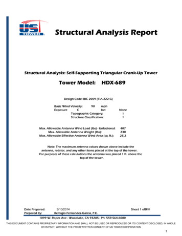
Please cite this article in press as: Ly et al., Psychedelics Promote Structural and Functional Neural Plasticity, Cell Reports (2018), https://doi.org/10.1016/j.celrep.2018.05.022Like ketamine, serotonergic psychedelics and entactogenshave demonstrated rapid and long-lasting antidepressant andanxiolytic effects in the clinic after a single dose (Bouso et al.,2008; Carhart-Harris and Goodwin, 2017; Grob et al., 2011;Mithoefer et al., 2013, 2016; Nichols et al., 2017; Sancheset al., 2016; Osório et al., 2015), including in treatment-resistantpopulations (Carhart-Harris et al., 2016, 2017; Mithoefer et al.,2011; Oehen et al., 2013; Rucker et al., 2016). In fact, therehave been numerous clinical trials in the past 30 years examiningthe therapeutic effects of these drugs (Dos Santos et al., 2016),with 3,4-methylenedioxymethamphetamine (MDMA) recentlyreceiving the ‘‘breakthrough therapy’’ designation by the Foodand Drug Administration for treating PTSD. Furthermore, classical psychedelics and entactogens produce antidepressantand anxiolytic responses in rodent behavioral tests, such asthe forced swim test (Cameron et al., 2018) and fear extinctionlearning (Cameron et al., 2018; Catlow et al., 2013; Younget al., 2015), paradigms for which ketamine has also been shownto be effective (Autry et al., 2011; Girgenti et al., 2017; Li et al.,2010). Despite the promising antidepressant, anxiolytic, andanti-addictive properties of serotonergic psychedelics, theirtherapeutic mechanism of action remains poorly understood,and concerns about safety have severely limited their clinicalusefulness.Because of the similarities between classical serotonergicpsychedelics and ketamine in both preclinical models and clinical studies, we reasoned that their therapeutic effects mightresult from a shared ability to promote structural and functionalneural plasticity in cortical neurons. Here we report that serotonergic psychedelics and entactogens from a variety of chemicalclasses (e.g., amphetamine, tryptamine, and ergoline) displayplasticity-promoting properties comparable with or greaterthan ketamine. Like ketamine, these compounds stimulatestructural plasticity by activating mammalian target of rapamycin(mTOR). To classify the growing number of compounds capableof rapidly promoting induced plasticity (Castrén and Antila,2017), we introduce the term ‘‘psychoplastogen,’’ from theGreek roots psych- (mind), -plast (molded), and -gen (producing). Our work strengthens the growing body of literature indicating that psychoplastogens capable of promoting plasticityin the PFC might have value as fast-acting antidepressantsand anxiolytics with efficacy in treatment-resistant populationsand suggests that it may be possible to use classical psychedelics as lead structures for identifying safer alternatives.RESULTSPsychedelics Promote NeuritogenesisBecause atrophy of cortical neurons is believed to be a contributing factor to the development of mood and anxiety disorders(Christoffel et al., 2011; Duman and Aghajanian, 2012), we firsttreated cultured cortical neurons with psychedelics from a variety of structural classes (Figures 1A and S1A) and measuredthe resulting changes in various morphological features. Using et al., 2006), we observed that severalSholl analysis (Ristanovicpsychedelics increased dendritic arbor complexity comparablywith ketamine, as measured by the area under the curve of theSholl plots as well as the maximum number of crossings (Figures1B–1E and S1B–S1E). This increase in arbor complexity appeared to result from large changes in both the number of dendritic branches and the total length of the arbors (Figures 1F,1H, S1F, and S1H). Psychedelics had a limited effect on the number of primary dendrites and did not alter the length of thelongest dendrite (Figures 1G, 1I, S1G, and S1I).Nearly all psychedelic compounds tested were capable ofrobustly promoting neuritogenesis, with comparable effects being produced by tryptamines (N,N-dimethyltryptamine [DMT]and psilocin), amphetamines (2,5-dimethoxy-4-iodoamphetamine [DOI] and MDMA), and ergolines (lysergic acid diethylamide [LSD]). As a positive control, we treated cells with 7,8-dihydroxyflavone (DHF), a psychoplastogen structurally dissimilarto classical psychedelics (Jang et al., 2010), and foundthat it also increased dendritic arbor complexity (Figure S2).This neurite outgrowth structural phenotype seems to onlybe induced by select compounds because serotonin andD-amphetamine, molecules that are chemically related toclassical psychedelics and entactogens, exerted minimal to noeffects on neuritogenesis (Figure S2).To establish the relative potencies and efficacies of hallucinogens and entactogens for promoting neurite outgrowth, we conducted 8-point dose-response studies (Figure S3). We defined100% and 0% efficacy as the maximum number of crossingsinduced by ketamine (10 mM) and vehicle (0.1% DMSO), respectively. We chose the 10 mM concentration of ketamine as the upper limit because this concentration of ketamine is reached in thebrain following intraperitoneal administration of an antidepressant dose in rats (Yang et al., 2018). For consistency, we usedthis same concentration when testing the effects of psychedelicsand entactogens, with DMT being the only exception. We used amaximum 90 mM concentration of DMT in our studies to moreclosely mimic the brain concentration of DMT in rats treatedwith an antidepressant dose (Cohen and Vogel, 1972; Cameronet al., 2018). In this neuritogenesis assay, ketamine’s halfmaximal effective concentration (EC50) value was 132 nM. Surprisingly, the majority of the psychedelics and entactogens wetested exhibited significantly greater potency than ketamine,with LSD being particularly potent (EC50 0.409 nM). In fact,LSD exhibited activity across 8 orders of magnitude into thelow picomolar range (Figure S3).Notably, the anti-addictive alkaloid ibogaine (Alper, 2001;Belgers et al., 2016) was the only psychedelic tested that hadabsolutely no effect (Figure S4). This was a surprising resultbecause we hypothesized that ibogaine’s long-lasting antiaddictive properties might result from its psychoplastogenicproperties. Previous work by He et al. (2005) clearly demonstrated that ibogaine increases the expression of glial cell linederived neurotrophic factor (GDNF) and that this plasticity-promoting protein is critical to ibogaine’s anti-addictive mechanismof action. Because several reports have suggested that noribogaine, a metabolite of ibogaine, might actually be the active compound in vivo (Zubaran et al., 1999; Baumann et al., 2000, 2001),we decided to test its ability to promote neuritogenesis incultured cortical neurons. Gratifyingly, noribogaine robustlyincreased dendritic arbor complexity with an EC50 value comparable with ketamine (Figure S3), providing additional evidencesuggesting that it may be the active compound in vivo.2 Cell Reports 23, 1–13, June 12, 2018CELREP 5086
Please cite this article in press as: Ly et al., Psychedelics Promote Structural and Functional Neural Plasticity, Cell Reports (2018), GJKLMNOHIFigure 1. Psychedelics Promote Neuritogenesis both In Vitro and In Vivo(A) Chemical structures of psychedelics.(B) Representative tracings of cortical neurons (DIV6) treated with compounds.(C) Sholl analysis demonstrates that psychedelics increase dendritic arbor complexity (n 39–41 neurons).(D) Area under the curve (AUC) of the Sholl plots in (C).(E) Maximum number of crossings (Nmax) of the Sholl plots in (C).(F–I) Cortical neurons treated with psychedelics display an increase in the number of branches (F), the number of primary dendrites (G), and the total length of thedendritic arbor (H) but not the length of the longest dendrite (I).(J and K) Class I neurons from Drosophila larvae treated with psychedelics during the first instar display increased branching (J) but not total length of the dendriticarbor (K) (n 3 neurons).(L) Representative images of neurons from (J) and (K).(M and N) Class I neurons from Drosophila larvae treated with psychedelics during the third instar display increased branching (M) but not total length of thedendritic arbor (N) (n 3 neurons).(O) Representative images from (M) and (N).*p 0.05, **p 0.01, ***p 0.001, ****p 0.0001, as compared to vehicle control. Scale bars, 30 mm. Data are represented as mean SEM. See alsoFigures S1–S5.To assess the in vivo effects of classical psychedelics on neuritogenesis, we started treating Drosophila larvae during the firstinstar with LSD and DOI. As observed in rodent cortical cultures,both LSD and DOI significantly increased dendritic branching ofclass I sensory neurons; however, they did not increase the totallength of the dendritic arbors (Figures 1J–1L). Because of thestriking effects of psychedelics on the structures of immatureneurons, we hypothesized that they might influence neurodevelopment. To test this, we chronically treated zebrafish embryoswith compounds for 6 days immediately following dechorionation and assessed gross morphological changes and behavior.We did not observe any differences in head sizes between thetreatment groups, nor did we detect any statistically significantdifferences in activity levels (Figure S5). Next we assessed theCell Reports 23, 1–13, June 12, 2018 3CELREP 5086
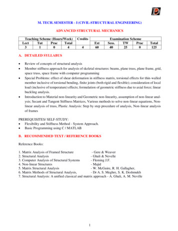
Please cite this article in press as: Ly et al., Psychedelics Promote Structural and Functional Neural Plasticity, Cell Reports (2018), y of psychedelics to promote neuritogenesis in more matureneurons by starting to treat Drosophila larvae during the late second instar. Again, psychedelics increased the branching of classI neurons, although the effect was less dramatic than thatobserved when treatment was started during the first instar (Figure 1M–1O). Although different developmental stages might bemore or less susceptible to the effects of psychedelics, it isalso possible that the smaller effect size observed after administering compounds starting at the later time point was simply theresult of treating the larvae for a shorter period of time. Regardless, it was quite surprising to observe compound-inducedchanges in neuronal structure after initiating treatment duringthe late second instar because class I neurons are stereotypedand typically possess relatively few higher-order branches(Grueber et al., 2002). Moreover, our results demonstrate thatpsychedelics can promote changes in neuronal structureacross vertebrate (rats) and invertebrate (Drosophila) species,suggesting that they act through an evolutionarily conservedmechanism.Psychedelics Promote Spinogenesis andSynaptogenesisIn addition to dendritic atrophy, loss of dendritic spines is a hallmark of depression and other neuropsychiatric disorders (Christoffel et al., 2011; Duman and Aghajanian, 2012), so we next assessed the effects of psychedelics on spinogenesis. We treatedmature rat cortical cultures for 24 hr with DOI, DMT, and LSD asrepresentative compounds from the amphetamine, tryptamine,and ergoline classes of psychedelics, respectively. All threecompounds increased the number of dendritic spines per unitlength, as measured by super-resolution structured illuminationmicroscopy (SIM) (Figures 2A, 2B, and S6), with LSD nearlydoubling the number of spines per 10 mm. Additionally, treatmentcaused a shift in spine morphology, favoring immature (thin andfilopodium) over more mature (mushroom) spine types (Figure 2C). Colocalization of pre- and postsynaptic markersfollowing treatment demonstrated that psychedelics promotedsynaptogenesis by increasing the density, but not the size of synapses (Figure 2D–2F). This increase in synapse density wasaccompanied by an increase in the density of VGLUT1 puncta,but not PSD-95 puncta, following compound administration(Figures 2G and 2H).Encouraged by our in vitro results, we next assessed the effects of a single intraperitoneal dose of DMT on spinogenesisin the PFC of adult rats using Golgi-Cox staining. We chose toadminister a 10 mg/kg dose of DMT for three reasons. First, allavailable data suggested that this dose would produce hallucinogenic effects in rats with minimal safety risks (Glennon et al.,1980, 1983; Glennon, 1999; Gatch et al., 2009; Smith et al.,1998; Appel et al., 1999; Winter et al., 2007; Carbonaro et al.,2015; Helsley et al., 1998; Strassman et al., 1994; Nair andJacob, 2016). Second, we have previously shown that a10 mg/kg dose of DMT produces positive effects in rat behavioral tests relevant to depression and PTSD (Cameron et al.,2018). Finally, we wanted to directly compare the effects ofDMT with ketamine, and seminal studies conducted by Li et al.(2010) had previously demonstrated that a 10 mg/kg dose of ketamine produced a robust increase in dendritic spine density inthe PFC of rats. We observed a significant increase in the densityof dendritic spines on cortical pyramidal neurons 24 hr afterdosing with DMT (Figures 2I and 2J). This effect was comparablewith that produced by ketamine at the same dose (Figure 2J).Importantly, this DMT-induced increase in dendritic spinedensity was accompanied by functional effects. Ex vivo slice recordings revealed that both the frequency and amplitude ofspontaneous excitatory postsynaptic currents (EPSCs) wereincreased following DMT treatment (Figures 2K–2M). Interestingly, 10 mg/kg and 1 mg/kg doses produced similar responsesdespite the fact that they are predicted to be hallucinogenic andsubhallucinogenic, respectively (Strassman et al., 1994; Nair andJacob, 2016).Because the half-life of DMT is exceedingly short ( 15 min),these results confirm that structural and functional changesinduced by DMT persist for hours after the compound hasbeen cleared from the body. Moreover, they demonstrate thatDMT produces functional effects on pyramidal neurons of thePFC that mirror those produced by ketamine (Li et al., 2010).Because the PFC is a key brain region involved in extinctionlearning (Quirk et al., 2006), and both ketamine and DMT havebeen shown to facilitate fear extinction (Cameron et al., 2018;Girgenti et al., 2017), our results suggest a link between the plasticity-promoting and behavioral effects of these drugs. Becausefear extinction can be enhanced by increasing levels of brainderived neurotrophic factor (BDNF) in the PFC (Peters et al.,2010), and ketamine’s behavioral effects have been shown tobe BDNF-dependent (Lepack et al., 2014), we next sought todetermine the role of BDNF signaling in the plasticity-promotingeffects of classical psychedelics.Psychedelics Promote Plasticity through a TrkBand mTOR-Dependent MechanismThe role of BDNF in both neuritogenesis and spinogenesis is wellknown (Cohen-Cory et al., 2010), and several reports suggestthat psychedelics are capable of increasing levels of neurotrophic factors (He et al., 2005; Martin et al., 2014; Nichols andSanders-Bush, 2002; Vaidya et al., 1997). Therefore, we treatedcortical neurons with BDNF, DOI, and a combination of thetwo to see whether they had any additive or synergisticeffects. Dose-response studies using recombinant BDNF(Figures 3A–3C) revealed that a 50 ng/mL treatment increasedneuritogenesis to a comparable extent as DOI (10 mM). Moreover, a combination of the two did not confer any added benefit,suggesting that they operate through a related mechanism(Figures 3D–3F). Next, we treated cortical neurons with DOI,DMT, and LSD for 24 hr before measuring BDNF gene and protein expression using droplet digital PCR (ddPCR) and ELISA,respectively. Although psychedelics did not increase the expression of BDNF transcript (Figure 3G), they did result in a 2-fold increase in BDNF protein levels, although this effect was not statistically significant (Figure 3H). When cortical cultures wereco-treated with ANA-12 (Cazorla et al., 2011), a selective antagonist of BDNF’s high-affinity receptor TrkB, the ability of psychedelics or BDNF to stimulate neuritogenesis and spinogenesiswas completely blocked (Figure 4).Activation of TrkB is known to promote signaling throughmTOR (Takei et al., 2004), which plays a key role in structural4 Cell Reports 23, 1–13, June 12, 2018CELREP 5086
Please cite this article in press as: Ly et al., Psychedelics Promote Structural and Functional Neural Plasticity, Cell Reports (2018), FHLKJGMFigure 2. Psychedelics Promote Spinogenesis, Synaptogenesis, and Functional Plasticity(A) Representative images of cortical neurons (DIV19) treated with compounds for 24 hr, demonstrating that psychedelics increase the number of dendritic spines(blue, MAP2; orange, F-actin).(B) Quantification of spine density (n 56–65 neurons).(C) Relative proportions of spine types following treatment of cortical cultures with psychedelics (n 16–21 neurons).(D) Representative images of cortical neurons (DIV19) treated for 24 hr, demonstrating that psychedelics increase synaptogenesis (green, VGLUT1; magenta,PSD-95; yellow, MAP2). White areas in the VGLUT1 PSD-95 images indicate colocalization of pre- and postsynaptic makers and are indicated by gray arrows.(E–H) Quantification of synapse density (E), synapse size (F), presynaptic density (VGLUT1) (G), and postsynaptic density (PSD-95) (H) following 24-hr treatment ofcortical neurons (DIV19) (n 39–42 neurons).(I) Representative images of Golgi-Cox-stained pyramidal neurons from the PFC of rats 24 hr after receiving a 10 mg/kg dose of DMT.(J) Quantification of spines from (G), demonstrating that DMT (10 mg/kg) increases spinogenesis in vivo to a comparable extent as ketamine (10 mg/kg) (n 11–17neurons).(K and L) Whole-cell voltage-clamp recordings of layer V pyramidal neurons from slices obtained 24 hr after DMT treatment (10 mg/kg and 1 mg/kg) demonstratethat DMT increases both spontaneous excitatory postsynaptic current (sEPSC) frequency (K) and amplitude (L) (n 11–38 neurons from 3 animals).(M) Representative traces for the 10 mg/kg experiments quantified in (K) and (L).*p 0.05, **p 0.01, ***p 0.001, ****p 0.0001, as compared to vehicle control. Data are represented as mean SEM. See also Figure S6.Cell Reports 23, 1–13, June 12, 2018 5CELREP 5086
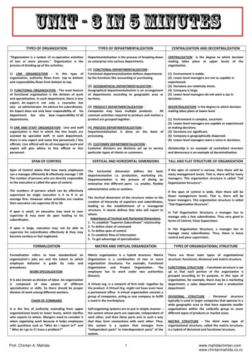
Please cite this article in press as: Ly et al., Psychedelics Promote Structural and Functional Neural Plasticity, Cell Reports (2018), GHFigure 3. Psychedelics and BDNF Promote Neuritogenesis via a Related Mechanism(A–C) Dose response of recombinant BDNF on neuritogenesis. AUC of the Sholl plots (A), Nmax of the Sholl plots (B), and total number of branches (C) of treatedcortical neurons (n 11–12 neurons per treatment, DIV6) indicate that the highest concentration of BDNF (50 ng/mL) is more effective at promoting neuritogenesisthan lower concentrations (5.0 and 0.5 ng/mL).(D) Sholl analysis (n 5–10 neurons) demonstrating that DOI (10 mM) increases neuritogenesis to a comparable extent as recombinant BDNF (50 ng/mL). Acombination of DOI (10 mM) and BDNF (50 ng/mL) did not have any additive or synergistic effects.(E) AUC of the Sholl plots in (D).(F) Nmax of the Sholl plots in (D).(G and H) Cultured cortical neurons (DIV18) were treated with compounds for 24 hr, and then BDNF gene (G) and protein (H) expression was assessed via ddPCR(n 4) and ELISA (n 3–4), respectively.*p 0.05, **p 0.01, ***p 0.001, ****p 0.0001, as compared to vehicle control. Data are represented as mean SEM.plasticity (Jaworski et al., 2005; Kumar et al., 2005), the production of proteins necessary for synaptogenesis (Hoeffer andKlann, 2010), and the effects of ketamine (Dwyer and Duman,2013; Li et al., 2010). Treatment with rapamycin, an mTOR inhibitor, completely blocked psychedelic-induced neuritogenesis(Figure 5), thus confirming that mTOR activation plays a rolein the plasticity-promoting effects of classical serotonergicpsychedelics.The 5-HT2A Receptor Mediates the Effects ofPsychedelics on Structural PlasticityFinally, we sought to determine whether the 5-HT2A receptorplayed any role in the plasticity-promoting effects of DOI, DMT,and LSD because this receptor is known to be primarily responsible for the hallucinogenic effects of classical psychedelics (Nichols, 2004, 2016). Furthermore, the psychoplastogenic potenciesof these and related compounds correlate well with their 5-HT2Areceptor affinities (Figure S3) (i.e., a higher 5-HT2A binding affinitygenerally predicts more potent psychoplastogenic effects). Control experiments demonstrated that 5-HT2A receptors were expressed on cultured rat cortical neurons at both day in vitro 6(DIV6) and DIV19 (Figure 6A). Next we found that co-treatmentwith ketanserin, a selective 5-HT2A antagonist, completely abrogated the ability of DMT, LSD, and DOI to promote both neuritogenesis and spinogenesis (Figures 6B–6F). Ketanserin was alsoable to block the effects of psilocin as well as the non-classicalpsychedelic noribogaine and enactogen MDMA (Figure 6G).These initial experiments were performed using doses ofpsychoplastogens that produced maximal effects on structural plasticity (circa 10 mM) in combination with a 10-foldexcess of ketanserin (100 mM). At these concentrations, wecould not rule out the possibility of other receptors contributing to the antagonistic effects of ketanserin. Therefore, wetreated cultured cortical neurons with a significantly lowerdose of LSD (10 nM) and attempted to block its ability to promote neurite outgrowth using increasing doses of ketanserin(Figure 6H). We found that ketanserin blocks the psychoplastogenic effects of LSD by 50% when treated at 10 nM. Thisis consistent with the fact that the binding affinities of ketanserin and LSD for the 5-HT2A receptor are roughly equivalent(low nanomolar). Increasing the concentration of ketanserin to100 nM, 10-fold higher than the concentration of LSD used inthis experiment, completely prevented LSD-induced neuritogenesis. At 100 nM, ketanserin is relatively selective for the5-HT2A receptor, although, at this concentration, we cannotrule out the possible involvement of 5-HT2C, adrenergic, orhistamine receptors.As a final note, the concentration responses of most psychoplastogens had Hill slopes that deviated from 1.0 (Figure S3), implying polypharmacology. Because psychedelicshave relatively high affinities for 5-HT2A receptors, it islikely that the effects of psychedelics are mediated primarilythrough 5-HT2A receptors at low concentrations and modulated by other targets at high concentrations. Interestingly,6 Cell Reports 23, 1–13, June 12, 2018CELREP 5086
Please cite this article in press as: Ly et al., Psychedelics Promote Structural and Functional Neural Plasticity, Cell Reports (2018), https://doi.org/10.1016/j.celrep.2018.05.022Figure 4. Psychedelic-Induced Changes inNeuronal Structure Are Mediated by TrkBABCDEFthe concentration response of DMT was the only one to exhibita Hill slope greater than 1.0, indicating some form ofcooperativity.DISCUSSIONClassical serotonergic psychedelics are known to causechanges in mood (Griffiths et al., 2006, 2008, 2011) and brainfunction (Carhart-Harris et al., 2017) that persist long after theacute effects of the drugs have subsided. Moreover, severalpsychedelics elevate glutamate levels in the cortex (Nichols,2004, 2016) and increase gene expression in vivo of the neurotrophin BDNF as well as immediate-early genes associated withplasticit
4Genome Center, University of California, Davis, Davis, CA 95616, USA . Neuropsychiatric diseases, including mood and anxiety disor-ders, are some of the leading causes of disability worldwide . of treatment before they experience any beneficial effects (Rush et al., 2006). Depression, post-traumatic stress disorder