Transcription
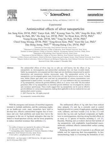
Nanomedicine: Nanotechnology, Biology, and Medicine 3 (2007) 95 – 101www.nanomedjournal.comExperimentalAntimicrobial effects of silver nanoparticlesJun Sung Kim, DVM, PhD,a Eunye Kuk, MS,b Kyeong Nam Yu, MS,a Jong-Ho Kim, MS,gSung Jin Park, BS,a Hu Jang Lee, DVM, PhD,c So Hyun Kim, DVM, PhD,dYoung Kyung Park, DVM, MS,d Yong Ho Park, DVM, PhD,dCheol-Yong Hwang, DVM, PhD,e Yong-Kwon Kim, PhD,f Yoon-Sik Lee, PhD,gDae Hong Jeong, PhD,b,4 Myung-Haing Cho, DVM, PhDaaLaboratory of Toxicology, Seoul National University, Seoul, KoreaDepartment of Chemistry Education, College of Education, Seoul National University, Seoul, KoreacInstitute of Animal Medicine, College of Veterinary Medicine, Gyeongsang National University, Chinju, KoreadDepartment of Microbiology, Seoul National University, Seoul, KoreaeDepartment of Internal Medicine, College of Veterinary Medicine, Seoul National University, Seoul, KoreafLaboratory of Micro Sensors and Actuators, School of Electronic Engineering and Computer Science, Seoul National University, Seoul, KoreagOrganic Synthesis Laboratory, School of Chemical and Biological Engineering, Seoul National University, Seoul, KoreaReceived 28 July 2006; accepted 15 December 2006bAbstractThe antimicrobial effects of silver (Ag) ion or salts are well known, but the effects of Agnanoparticles on microorganisms and antimicrobial mechanism have not been revealed clearly.Stable Ag nanoparticles were prepared and their shape and size distribution characterized by particlecharacterizer and transmission electron microscopic study. The antimicrobial activity of Agnanoparticles was investigated against yeast, Escherichia coli, and Staphylococcus aureus. In thesetests, Muller Hinton agar plates were used and Ag nanoparticles of various concentrations weresupplemented in liquid systems. As results, yeast and E. coli were inhibited at the low concentrationof Ag nanoparticles, whereas the growth-inhibitory effects on S. aureus were mild. The free-radicalgeneration effect of Ag nanoparticles on microbial growth inhibition was investigated by electronspin resonance spectroscopy. These results suggest that Ag nanoparticles can be used as effectivegrowth inhibitors in various microorganisms, making them applicable to diverse medical devices andantimicrobial control systems.D 2007 Elsevier Inc. All rights reserved.Key words:Ag nanoparticle; Antimicrobial effectsWith the emergence and increase of microbial organismsresistant to multiple antibiotics, and the continuing emphasis on health-care costs, many researchers have tried todevelop new, effective antimicrobial reagents free of resistance and cost. Such problems and needs have led to theresurgence in the use of Ag-based antiseptics that may belinked to broad-spectrum activity and far lower propensityto induce microbial resistance than antibiotics [1].No conflict of interest was reported by the authors of this paper.J.S. Kim and E. Kuk contributed equally to this work.4 Corresponding author. Department of Chemistry Education, Collegeof Education, Seoul National University, Seoul, Korea.E-mail address: jeongdh@snu.ac.kr (D.H. Jeong).1549-9634/ – see front matter D 2007 Elsevier Inc. All rights reserved.doi:10.1016/j.nano.2006.12.001The antibacterial effects of Ag salts have been noticedsince antiquity [2], and Ag is currently used to controlbacterial growth in a variety of applications, including dentalwork, catheters, and burn wounds [3,4]. In fact, it is wellknown that Ag ions and Ag-based compounds are highlytoxic to microorganisms, showing strong biocidal effects onas many as 12 species of bacteria including E. coli [5].Recently, Mecking and co-workers showed that hybrids ofAg nanoparticles with amphiphilic hyperbranched macromolecules exhibited effective antimicrobial surface coatingagents [6].Reducing the particle size of materials is an efficient andreliable tool for improving their biocompatibility. In fact,nanotechnology helps in overcoming the limitations of size
96J.S. Kim et al. / Nanomedicine: Nanotechnology, Biology, and Medicine 3 (2007) 95–101Fig 1. Absorption spectra of Ag nanoparticle solutions (A). The solid line is for Ag nanoparticle solution as prepared, the dashed one is for the ten-timeconcentrated one after diluted back to the original concentration, and the dotted one is for the solution left after the Ag nanoparticles are removed bysedimentation. The maximum potential peaks of Ag nanoparticles were measured at -0.33mV (B). The effective zeta-potential in aqueous solution weremeasured by particle characterizer bDelsa 440 SXQ (Coulter Ltd., Miami, FL), and the mean values were averaged from 3 times assay data.and can change the outlook of the world regarding science[7]. Furthermore, nanomaterials can be modified for betterefficiency to facilitate their applications in different fieldssuch as bioscience and medicine. In this study our researchteam, consisting of experts in several disciplines, hasinvestigated the antimicrobial effects of Ag nanoparticlesagainst representative microorganisms of public concern. Apossibility of free-radical involvement near the Ag nanoparticle surface in the antimicrobial activity of Ag nanoparticles was discussed based on electron spin resonance(ESR) measurements. Here, we report that Ag nanoparticles can be applied effectively in the control ofmicroorganisms and the prevention of deleterious infections. Our results support the hypothesis that Ag nanoparticles can be prepared in a simple and cost-effectivemanner and are suitable for formulation of new types ofbactericidal materials.Materials and methodsPreparation of Ag nanoparticlesAg nanoparticles were made according to the recipedescribed in the literature [8,9]. Briefly, a 100-mL aqueoussolution of 1.0 10–3 M silver nitrate was mixed with a 300-mLaqueous solution of 2.0 10-3 M sodium borohydride. Triplydistilled water was used for solutions, and both solutions werechilled to ice temperature before mixing. By mixing bothsolutions, Ag ions were reduced and clustered together to formmonodispersed nanoparticles as a transparent sol in aqueousmedium. The Ag solution was yellow because of the absorptionat 390 nm. The solution was stirred repeatedly whenever somedark color appeared for approximately an hour until it becamestabilized. At this point this solution of Ag nanoparticles was sostable that it did not change color for as long as several monthswithout any stabilizing agent. Because the particle concentrationof the solution is only 3.3 nM, it was concentrated 10 timesusing a rotary vacuum evaporator. Then, by diluting thissolution, each sample of different concentration was used toinvestigate the concentration dependence of the antifungal effectof Ag nanoparticles.Assay for antimicrobial activity of Ag nanoparticlesagainst microorganismsThe antimicrobial activity of Ag nanoparticles was evaluatedagainst yeast (isolated from bovine mastitis), E. coli O157:H8(ATCC 43886), and S. aureus (ATCC 19636) by modification ofthe agar disk diffusion method of the National Committee forClinical Laboratory Standards (NCCLS; now renamed as Clinicaland Laboratory Standards Institute, CLSI, 2000). Approximately107 colony-forming units of each microorganism were inoculatedon Muller Hinton agar (MHA) plates, and then 20 AL of Agnanoparticles were spread in a concentration of 0.2 to 33 nM.Itraconazol (for yeast, 33 nmol) and gentamicin (for E. coli andS. aureus, 33 nmol) were used as positive controls. The plates wereincubated for 24 hours at 378C.To evaluate the growth inhibition of Ag nanoparticles, wedesigned a new method. After a 24-hour incubation, each plate wasanalyzed by LAS3000 (FUJI, Tokyo, Japan); Briefly, we analyzedthe microorganism density at the center of the plate with Agnanoparticles (A) and at the outer edge of the plate without Agnanoparticles (B). The differences of microorganism densitybetween (A) and (B) were measured and divided by the numberof areas analyzed. The results on the three plates corresponding to aparticular sample were averaged and this value regarded as theminimal inhibitory concentration (MIC) of Ag nanoparticlesagainst each microorganism.Electron spin resonance spectroscopyThe Ag nanoparticles were aggregated by stirring the yellowcolloid solution with a Zn bar. Stirring with a Zn bar inducedaggregation of the Ag colloid particles by breaking the chargebalance between Ag nanoparticles without adding anything else.Other methods of obtaining aggregated Ag nanoparticles, such asadding salt or chemicals, were excluded so as to permit an accurateevaluation of the effect of Ag nanoparticles alone. Upon stirringthe solution of Ag nanoparticles with a Zn bar, the solution turneddark brown, after which the aggregated Ag nanoparticles slowlysettled down. The precipitated Ag nanoparticles were collected as apowder and packed into a glass capillary tube. Free-radical
J.S. Kim et al. / Nanomedicine: Nanotechnology, Biology, and Medicine 3 (2007) 95–10197Fig 2. (A) A TEM image of Ag nanoparticles dispersed on a TEM copper grid (a, scale bar: 30 nm). (B) A histogram showing size distribution ofAg nanoparticles.Table 1MIC results of Ag nanoparticlesyeast (ATCC19636)E.coli (ATCC43890)St. aureus (Bovine mastitis)MIC of Ag nanoparticlesN 6.6 nMN 3.3 nMN 33 nMin the same concentration of 33 nM. N-acetylcysteine (NAC) asantioxidant was added in the plates at 10 and 50 nM [10]. Theplates were incubated for 24 hours at 378C.ResultsCharacterization of the synthesized Ag nanoparticlesFig 3. Growth inhibition of Ag nanoparticles against yeast. Itraconasol,distilled water, and solution devoid of Ag stand were used for the positivecontrol, negative control, and vehicle control. The concentration of goldnanoparticles was 30 nM. Each point represents the mean F SD. **:Significantly different from positive control ( P b .01).generation from Ag nanoparticles were recorded in an ESRspectrometer JES-TE 200 (JEOL, Tokyo, Japan). The ESR settingand experimental conditions are described in the figure legends.Antioxidant effect of Ag nanoparticles and silver nitrate inantimicrobial activityTo confirm the effects of Ag nanoparticles, a comparative studyof Ag nanoparticles and silver nitrate on antimicrobial activityagainst E. coli O157:H8 was performed. Approximately 107colony-forming units of E. coli were inoculated on MHA plates,and then 20 AL of Ag nanoparticles and silver nitrate were spreadThe prepared aqueous solution of Ag nanoparticlesshowed an absorption band at 391 nm as shown inFigure 1, which is a typical absorption band of sphericalAg nanoparticles due to their surface plasmon [8]. Thestability of the concentrated solution was checked byobserving its absorption spectrum after rediluting 10 times.The absorption spectrum of the rediluted solution depictsalmost identical spectral features to the spectrum of theoriginal solution of Ag nanoparticles (Figure 1, A). Thisconfirms that the Ag nanoparticles are not further dimerizedor agglomerated with many particles together, in that a newabsorption band, appearing to the red side of the band at 390 nm because of a localized surface plasmon betweentwo or more Ag nanoparticles in contact with one anotherwhen dimerization or aggregation of Ag nanoparticlesoccurs, is not observed. The absorption spectrum of thesolution remaining after Ag nanoparticles had been completely sedimented and removed by stirring the solution witha Zn rod was obtained to confirm that there were no Agnanoparticles in the solution. Then this solution was used as
98J.S. Kim et al. / Nanomedicine: Nanotechnology, Biology, and Medicine 3 (2007) 95–101Fig 4. Growth inhibition of Ag nanoparticles in E. coli. Gentamycin,distilled water and solution devoid of Ag stand for a positive control,negative control and vehicle control. The concentration of gold nanoparticles is 30 nM. Each point represents the mean F SD. *: Significantlydifferent from positive control ( P b .05). **: Significantly different frompositive control ( P b .01).a vehicle control to confirm that the other salts such asnitrate, borate, and sodium ions included in the Ag nanoparticle solution during preparation of the nanoparticles didnot affect the antimicrobial activity. Surface zeta potential ofAg nanoparticles was measured to be slightly negative(Figure 1, B). In the colloid solution, there exist nitrate andborate ions adsorbed on the surface of Ag nanoparticles withthe result that the surface charge of the Ag nanoparticles willbe slightly negative. Shape and size distribution of thesynthesized Ag nanoparticles were characterized by transmission electron microscopic (TEM) study. A few drops ofAg nanoparticle solution were dropped onto a TEM grid, andthe residue was removed by a filter paper beneath the TEMgrid. The TEM image shown in Figure 2, A was obtained byhigh-resolution TEM (JEOL, JEM-2000E7). As can be seenby the shape and size distribution in Figure 2, B, the particlesare highly monodispersed with an average diameter of 13.5nm and a standard deviation of 2.6 nm.Antimicrobial activity of Ag nanoparticles againstmicroorganismsAntimicrobial tests were performed against yeast, E. coli,and S. aureus on MHA plates treated with differentconcentrations of Ag nanoparticles (from 0.2 to 33 nM).Yeast was isolated from bovine mastitis. Comparing withthe positive control, itraconazole, Ag nanoparticles of 33 nMshowed a similar growth inhibition effect against yeast, andsignificant growth inhibition was observed from 13.2 nM(Figure 3). These results revealed that the MIC of Agnanoparticles against yeast may be estimated between6.6 nM and 13.2 nM in this condition (Table 1). ForFig 5. Growth inhibition of Ag nanoparticles in S. aureus. Gentamycin,distilled water and solution devoid of Ag stand for a positive control,negative control and vehicle control. The concentration of gold nanoparticles is 30 nM. Each point represents the mean F SD. **: Significantlydifferent from positive control ( P b .01).E. coli, we used E. coli O157:H7, which is known as anotorious pathogen causing hemorrhagic enteritis. In ourresults, Ag nanoparticles were most effective against E. coli(Figure 4 and Table 1). The MIC of Ag nanoparticlesagainst E. coli may be estimated between 3.3 nM and6.6 nM, and the growth inhibition effect was observed in aconcentration-dependent manner. For S. aureus, however,Ag nanoparticles showed a mild growth-inhibitory effecteven in high concentration, and there was no statisticallysignificant inhibitory effect compared with the control(gentamicin) in this condition (Figure 5). MIC of Agnanoparticles against S. aureus was estimated to be morethan 33 nM (Table 1). Also, there is no antimicrobialactivity in solution devoid of Ag nanoparticles used as avehicle control, reflecting that antimicrobial activity wasdirectly related to the Ag nanoparticles. To determinewhether the growth-inhibitory effect of Ag nanoparticles isa specific event or not, we used gold (Au) nanoparticles( 30 nM) as another control of nanosized metals. Aunanoparticles showed no growth-inhibitory effect againstvarious microorganisms in our experimental conditions(Figures 3-5).ESR study of Ag nanoparticlesThe mechanism of the growth-inhibitory effects of Agnanoparticles on microorganisms has not been well understood. One possibility is that the growth inhibition may berelated to the formation of free radicals from the surface ofAg. Uncontrolled generation of free radicals can attackmembrane lipids and then lead to a breakdown of membranefunction [11]. To obtain insight into this possibility, wemeasured the ESR spectra of Ag nanoparticles. Ag samples
J.S. Kim et al. / Nanomedicine: Nanotechnology, Biology, and Medicine 3 (2007) 95–10199Fig 6. The ESR spectrum of Ag nanoparticles recorded at room temperature. m1 and m2 indicate the control peak of Mn and the peak (mT: 336.337) indicatesthe released free radical from Ag nanoparticles. The ESR spectral was obtained at room temperature. Instrumental setting of JEOL JES-TE 200 spectrometer:microwave power, 8.00 mW, MOD, 100 kHz, and time constant: 0.3 sec.radical generation of Ag nanoparticles may be responsiblefor the antimicrobial effects.Antioxidant effect of Ag nanoparticles and silver nitrate inantimicrobial activityTo determine the relationship between free-radical andantimicrobial activity, we used the antioxidant NAC to testwhether the antioxidant could influence antimicrobialactivity induced by Ag nanoparticles. In Figure 7, Agnanoparticles and silver nitrate showed similar growthinhibitory effect against E. coli. However, such inhibitoryeffect was abolished by the addition of NAC. NAC alonedid not affect the antimicrobial activity.DiscussionFig 7. Comparative study of Ag nanoparticles and silver nitrate in growthinhibition with/without N-acetylcystein (NAC). Solution devoid of Agstand for a control. The concentration of Ag nanoparticles and silver nitrateis 33 nM. Each point represents the mean F SD. **: Significantly differentfrom control ( P b .01).were prepared in powder form by stirring the Ag nanoparticles solution with a Zn bar, causing the Ag nanoparticles to aggregate. In Figure 6, peaks of m1 and m2indicate the control peaks of standard manganese, and thecentral peak (mT: 336.337) indicates the existence of freeradicals from Ag nanoparticles, thus supporting that free-It is well known that Ag ions and Ag-based compoundshave strong antimicrobial effects [12], and many investigators are interested in using other inorganic nanoparticles as antibacterial agents [4,12-14]. Theseinorganic nanoparticles have a distinct advantage overconventional chemical antimicrobial agents. The mostimportant problem caused by the chemical antimicrobialagents is multidrug resistance. Generally, the antimicrobialmechanism of chemical agents depends on the specificbinding with surface and metabolism of agents into themicroorganism. Various microorganisms have evolveddrug resistance over many generations. Thus far, theseantimicrobial agents based on chemicals have been
100J.S. Kim et al. / Nanomedicine: Nanotechnology, Biology, and Medicine 3 (2007) 95–101effective for therapy; however, they have been limited touse for medical devices and in prophylaxis in antimicrobialfacilities. Therefore, an alternative way to overcome thedrug resistance of various microorganisms is neededdesperately, especially in medical devices, etc. Ag ionsand Ag salts have been used for decades as antimicrobialagents in various fields because of their growth-inhibitorycapacity against microorganisms. Also, many otherresearchers have tried to measure the activity of metalions against microorganisms. Studies of Russel and Hugoon the antimicrobial properties of Ag and Cu [15], and ofMarsh on Zn were reported [16]. However, Ag ions orsalts has only limited usefulness as an antimicrobial agentfor several reasons, including the interfering effects of saltsand the antimicrobial mechanism of [the continuousrelease of enough concentration of Ag ion from the metalform.] In contrast, these kinds of limitations can beovercome by the use of Ag nanoparticles. However, touse Ag in various fields against microorganisms, it isessential to prepare the Ag with cost-effective methods andto know the mechanism of the antimicrobial effect.Besides, it is important to enhance the antimicrobial effect.In this study we report that Ag nanoparticles can beprepared cost effectively and that these Ag nanoparticlesare homogeneous and stable (Figures 1 and 2). Thenanosize allowed expansion of the contact surface of Agwith the microorganisms, and this nanoscale has applicability for medical devices by surface coating agents.In this study, to evaluate the antimicrobial effects againstvarious microorganisms, we used three representative microorganisms, yeast, E. coli and S. aureus. There were distinctdifferences among them. When Ag nanoparticles were testedin yeast and E. coli, they effectively inhibited bacterialgrowth. In our results, Ag nanoparticles showed antimicrobial activity against yeast and E. coli (Figures 3 and 4) thatwas similar to that found by Sondi and Salopek-Sondi [17].In contrast, the inhibitory effect of Ag nanoparticles wasmild in S. aureus as compared with other microorganisms;these results suggest that the antimicrobial effects of Agnanoparticles may be associated with characteristics ofcertain bacterial species. Gram-positive and gram-negativebacteria have differences in their membrane structure, themost distinctive of which is the thickness of the peptidoglycan layer. We think that the lower efficacy of the Agnanoparticles against S. aureus may derive from thedifference as a point of membrane structure. To confirm thishypothesis, further comparative study between variousgram-negative and gram-positive bacterial species is needed.The peptidoglycan layer is a specific membrane feature ofbacterial species and not mammalian cells. Therefore, if theantibacterial effect of Ag nanoparticles is associated with thepeptidoglycan layer, it will be easier and more specific to useAg nanoparticles as an antibacterial agent.To identify the difference of antimicrobial activitybetween Ag nanoparticles and other metallic nanoparticles,we carried out the growth inhibition test with 30 nM Aunanoparticle. We also wanted to estimate the potential sideeffects of other components which might be included in theAg nanoparticles solution. Thus, we prepared vehiclecontrol, in which the Ag nanoparticles were removed bysedimentation. Our results indicate no crucial antimicrobialactivity in either Au nanoparticles or vehicle control,suggesting that the antimicrobial activity of Ag nanoparticles is an Ag-specific event in our experimentalconditions (Figures 3-5).The mechanism of the inhibitory effects of Ag ions onmicroorganisms is partially known. Some studies havereported that the positive charge on the Ag ion is crucialfor its antimicrobial activity through the electrostaticattraction between negative charged cell membrane ofmicroorganism and positive charged nanoparticles[14,18,19]. In contrast, Sondi and Salopek-Sondi [17]reported that the antimicrobial activity of silver nanoparticles on Gram-negative bacteria was dependent on theconcentration of Ag nanoparticle, and was closely associated with the formation of dpitsT in the cell wall of bacteria.Then, Ag nanoparticles accumulated in the bacterialmembrane caused the permeability, resulting in cell death.However, because those studies included both positivelycharged Ag ions and negatively charged Ag nanoparticles, itis insufficient to explain the antimicrobial mechanism ofpositively charged Ag nanoparticles. Therefore, we expectthat there is another possible mechanism. Amro et al.suggested that metal depletion may cause the formation ofirregularly shaped pits in the outer membrane and changemembrane permeability, which is caused by progressiverelease of lipopolysaccharide molecules and membraneproteins [20]. Also, Sondi and Salopek-Sondi speculate thata similar mechanism may cause the degradation of themembrane structure of E. coli during treatment with Agnanoparticles [17]. Although their inference involved somesort of binding mechanism, still unclear is the mechanism ofthe interaction between Ag nanoparticles and component(s)of the outer membrane. Recently, Danilczuk and co-workers[21] reported Ag-generated free radicals through the ESRstudy of Ag nanoparticles. We suspect that the antimicrobialmechanism of Ag nanoparticles is related to the formation offree radicals and subsequent free radical–induced membranedamage. To confirm the production of free radical, weanalyzed the Ag nanoparticles by ESR. In Figure 6, weobserved an Ag-specific ESR spectrum. The obtained peakof Ag nanoparticles in an ESR assay corresponded with thatobtained by Danilczuk et al [21]. To determine therelationship between free-radical and antimicrobial activity,we used the antioxidant NAC to test whether the antioxidantcould influence Ag nanoparticles–induced antimicrobialactivity. Figure 7 shows that similar antimicrobial activitywas observed between Ag nanoparticles and silver nitrateagainst E. coli. However, the antimicrobial activity of Agnanoparticles and silver nitrate was influenced by NAC. Theresults of ESR and antioxidant study suggest that freeradicals may be derived from the surface of Ag nano-
J.S. Kim et al. / Nanomedicine: Nanotechnology, Biology, and Medicine 3 (2007) 95–101particles and be responsible for the antimicrobial activity inour experimental conditions.In conclusion, Ag nanoparticles prepared by the costeffective reduction method described here have greatpromise as antimicrobial agents. Applications of Ag nanoparticles based on these findings may lead to valuablediscoveries in various fields such as medical devices andantimicrobial systems.AcknowledgmentsThis work was supported by the NSI-NCRC program ofKOSEF. D.H.J. was supported by BK21 program. H.J.L. wassupported by the Korea Research Foundation Grant funded bythe Korean Government (MOEHRD) (R05-2004-10627-0).References[1] Jones SA, Bowler PG, Walker M, Parsons D. Controlling woundbioburden with a novel silver-containing Hydrofiber dressing. WoundRepair Regen 2004;12(3):288 - 94.[2] Silver S, Phung LT. Bacterial heavy metal resistance: new surprises.Annu Rev Microbiol 1996;50:753 - 89.[3] Catauro M, Raucci MG, De Gaetano FD, Marotta A. Antibacterial andbioactive silver-containing Na2O CaO 2SiO2 glass prepared bysol-gel method. J Mater Sci Mater Med 2004;15(7):831 - 7.[4] Crabtree JH, Burchette RJ, Siddiqi RA, Huen IT, Handott LL,Fishman A. The efficacy of silver-ion implanted catheters in reducing peritoneal dialysis-related infections. Perit Dial Int 2003;23(4):368 - 74.[5] Zhao G, Stevens Jr SE. Multiple parameters for the comprehensiveevaluation of the susceptibility of Escherichia coli to the silver ion.Biometals 1998;11:27 - 32.[6] Aymonier C, Schlotterbeck U, Antonietti L, Zacharias P, Thomann R,Tiller JC, et al. Hybrids of silver nanoparticles with amphiphilichyperbranched macromolecules exhibiting antimicrobial properties.Chem Commun (Camb) 2002;24:3018 - 9.[7] Mirkin CA, Taton TA. Semiconductors meet biology. Nature2000;405:626 - 7.[8] Creighton JA, Blatchford CG, Albrecht MG. Plasma resonanceenhancement of Raman scattering by pyridine adsorbed on silver 21]101gold sol particles of size comparable to the excitation wavelength.J Chem Soc Faraday Trans II 1979;75:790 - 8.Suh JS, DiLella DP, Moskovits M. Surface-enhanced Ramanspectroscopy of colloidal metal systems: a two-dimensional phaseequilibrium in p-aminobenzoic acid adsorbed on silver. J Phys Chem1983;87:1540 - 4.Lee MG, Lee KT, Chi SG, Park JH. Costunolide induces apoptosis byROS-mediated mitochondrial permeability transition and cytochromec release. Biol Pharm Bull 2001;24(3):303 - 6.Mendis E, Rajapakse N, Byun HG, Kim SK. Investigation of jumbosquid (Dosidicus gigas) skin gelatin peptides for their in vitroantioxidant effects. Life Sci 2005;77:2166 - 78.Furno F, Morley KS, Wong B, Sharp BL, Arnold PL, Howdle SM,et al. Silver nanoparticles and polymeric medical devices: a newapproach to prevention of infection. J Antimicrob Chemother 2004;54:1019 - 24.Abuskhuna S, Briody J, McCann M, Devereux M, Kavanagh K,Fontecha JB, et al. Synthesis, structure and anti-fungal activity ofdimeric Ag(I) complexes containing bis-imidasole ligands. Polyhedron 2004;23:1249 - 55.Hamouda T, Myc A, Donovan B, Shih A, Reuter JD, Baker Jr JR. Anovel surfactant nanoemulsion with a unique non-irritant topicalantimicrobial activity against bacteria, enveloped viruses and fungi.Microbiol Res 2000;156:1 - 7.Russel AD, Hugo WB. Antimicrobial activity and action of silver.Prog Med Chem 1994;31:351 - 70.Marsh PD. Microbiological aspects of the chemical control of plaqueand gingivitis. J Dent Res 1992;71:1431 - 8.Sondi I, Salopek-Sondi B. Silver nanoparticles as antimicrobial agent:a case study on E. coli as a model for Gram-negative bacteria.J Colloid Interface Sci 2004;275:177 - 82.Dibrov P, Dzioba J, Gosink KK, H7se CC. Chemiosmotic mechanismof antimicrobial activity of Ag( ) in Vibrio cholerae. AntimicrobAgents Chemother 2002;46:2668 - 70.Dragieva I, Stoeva S, Stoimenov P, Pavlikianov E, Klabunde K.Complex formation in solutions for chemical synthesis of nanoscaledparticles prepared by borohydride reduction process. NanostructMater 1999;12:267 - 70.Amro NA, Kotra LP, Wadu-Mesthrige K, Bulychev A, Mobashery S,Liu G. High-resolution atomic force microscopy studies of theEscherichia coli outer membrane: structural basis for permeability.Langmuir 2000;16:2789 - 96.Danilczuk M, Lund A, Saldo J, Yamada H, Michalik J. Conductionelectron spin resonance of small silver particles. Spectrochimaca ActaPart A 2006;63:189 - 91.
fLaboratory of Micro Sensors and Actuators, School of Electronic Engineering and Computer Science, Seoul National University, Seoul, Korea gOrganic Synthesis Laboratory, School of Chemical and Biological Engineering, Seoul National University, Seoul, Korea Received 28 July 2006; accepted 15 December 2006