Transcription
Hindawi Publishing CorporationEvidence-Based Complementary and Alternative MedicineVolume 2012, Article ID 828521, 10 pagesdoi:10.1155/2012/828521Research ArticleRegulation of Proinflammatory Mediators viaNF-κB and p38 MAPK-Dependent Mechanisms in RAW 264.7Macrophages by Polyphenol Components Isolatedfrom Korea Lonicera japonica THUNBKwang-Il Park,1 Sang-Rim Kang,2 Hyeon-Soo Park,1 Do Hoon Lee,1Arulkumar Nagappan,1 Jin A Kim,3 Sung Chul Shin,4 Eun Hee Kim,5Won Sup Lee,6, 7 Hyon-Jong Chung,1 Su Jin An,1 and Gon Sup Kim11 ResearchInstitute of Life Science and College of Veterinary Medicine, Gyeongsang National University, Gazwa,Jinju 660-701, Republic of Korea2 Department of Biological Engineering, School of Natural Science, Kyonggi University, Yeongtong, Suwon 443-760, Republic of Korea3 Korea National Animal Research Resource Center and Korea National Animal Bio-Resource Bank,Gyeongsang National University, Gazwa, Jinju 660-701, Republic of Korea4 Department of Chemistry and Research Institute of Life Science, Gyeongsang National University, Jinju 660-701, Republic of Korea5 Department of Nursing Science, International University of Korea, Jinju 660-759, Republic of Korea6 Department of Internal Medicine, Institute of Health Sciences, Gyeongsang National University School of Medicine,Jinju 660-702, Republic of Korea7 Gyeongnam Regional Cancer Center, Gyeongsang National University Hospital,Jinju 660-702, Republic of KoreaCorrespondence should be addressed to Gon Sup Kim, gonskim@gnu.ac.krReceived 21 October 2011; Revised 10 February 2012; Accepted 15 February 2012Academic Editor: Bashar SaadCopyright 2012 Kwang-Il Park et al. This is an open access article distributed under the Creative Commons Attribution License,which permits unrestricted use, distribution, and reproduction in any medium, provided the original work is properly cited.Lonicera japonica THUNB., which abundantly contains polyphenols, has been used as a traditional medicine for thousandsof years in East Asian countries because of the anti-inflammation properties. This study aimed to investigate the antiinflammatory mechanism of polyphenol components isolated from Korea L. japonica T. by nuclear factor-kappaB (NF-κB) andmitogen-activated protein kinases (MAPKs) pathway. Polyphenols significantly decreased lipopolysaccharide- (LPS-) inducedmRNA and protein expression of inducible nitric oxide synthase and cyclooxygenase-2, as well as mRNA expression of tumornecrosis factor-alpha, interleukin- (IL-) 1β, and IL-6. Moreover, polyphenols inhibited nuclear translocation of NF-κB p65,phosphorylation/degradation of the inhibitor of κB, and phosphorylation of p38 MAPK, whereas the extracellular signal-regulatedkinase and Janus N-terminal kinase were not affected. These results indicate that polyphenol components isolated from Korea L.japonica T. should have anti-inflammatory effect on LPS-stimulated RAW 264.7 cells through the decrease of proinflammatorymediators expression by suppressing NF-κB and p38 MAPK activity.1. IntroductionLonicera japonica THUNB. (Caprifoliaceae) has been usedin East Asian countries including Korea, China, and Japanas the treatment of inflammation and tumor for thousandsof years. Crude extracts of L. japonica have hepatoprotectiveand anti-inflammatory effects associated with suppressionof nuclear factor-kappaB (NF-κB) activation through reduction of I-kappaB degradation in lipopolysaccharide- (LPS-)challenged rats [1]. L. japonica T. extract exhibits neuroprotective effects associated with the suppression of hydrogenperoxide-induced apoptosis via phosphorylation of mitogenactivated protein kinases (MAPKs) and phosphoinositide 3kinase (PI3K)/Akt in neuroblastoma cells [2]. A number of
2compounds isolated from Lonicera species, including luteolin, quercetin, biflavonoids, and dicaffeoylquinic acid, havevarious pharmacological properties such as antimicrobial,antioxidative, antiviral, and anti-inflammatory effects [3, 4].Inflammation encompasses multiple processes by anorganism in response to injury that is related to harmfulstimuli such as infection by a pathogen, exposure toendotoxin (e.g., LPS), or chemical exposure. Inflammatoryresponses are pivotal in host defense against stimuli byactivating immune cells, in an effort to maintain homeostasis[5]. Macrophages play a critical role in the initiation ofinflammatory and immune responses by releasing proinflammatory mediators such as tumor necrosis factor-alpha(TNF-α), interleukin-6 (IL-6), cyclooxygenase-2 (COX-2),and inducible nitric oxide synthase (iNOS) [6]. Whenmacrophages are activated by stimuli, Toll-like receptor4 (TLR4) involvement with the myeloid differentiationfactor (MyD88) preludes the MyD88-dependent pathwaytriggering of the activation of transforming-growth-factorbeta- (TGF-β-) activated kinase 1 (TAK1). TAK1 activationcauses the phosphorylation of MAPKs and IκB kinase (IKK).Eventually, NF-κB is activated by these cell signals [7]. However, chronic inflammation leads to the upregulation of proinflammatory mediators in affected cells and overexpressionof the proinflammatory mediators such as iNOS and COX2, and various cytokines including TNF-α, IL-1β, and IL6 can cause many inflammation-related diseases, includingatherosclerosis, colitis, rheumatoid arthritis, inflammatorybowel disease, and cancer [8–10].NF-κB plays a crucial role in chronic inflammatorydiseases [11–13]. NF-κB is a heterodimer composed of p50and p65, which functions principally as a transcriptionalactivator. NF-κB is changed to the active form by cytokines,LPS, and oxidative stress, whereupon it regulates COX-2and iNOS through the initiation of transcription of targetgenes. However, the inherent mechanism underlying theeffect of L. japonica T. on LPS-induced inflammation remainsincompletely understood.In the present study, we investigated whether polyphenol components isolated from L. japonica T. have antiinflammatory activity via the production of COX-2, iNOS,and cytokines such as IL-1β, TNF-α, and IL-6 by inactivationof the NF-κB and MAPK pathways in LPS-treated RAW 264.7macrophages.2. Materials and Methods2.1. Extraction and Isolation of Polyphenol Components ofLonicera japonica T. L. japonica T. was obtained from theAnimal Bio-Resources Bank. The lyophilized plant material(100 g) was ground into powder and extracted in 500 mLof 70% methanol at 50 C for 12 h. After the extract wasfiltered in a Büchner funnel under reduced pressure andconcentrated to 100 mL under reduced pressure at condition of 40 C using a rotator evaporator, the concentratedmixtures were reconstituted in methanol (0.01 g/mL) andstored at 20 C until analysis. The concentrated extractswere washed with n-hexane (100 mL), extracted with ethylEvidence-Based Complementary and Alternative Medicineacetate (100 mL). After three extraction cycles, the organiclayer was dried over anhydrous MgSO4 . The solvent wasremoved under reduced pressure. The sticky residue wasloaded onto a silica gel column (3.7 20 cm) and elutedwith methanol/dichloromethane (1 : 5, 300 mL). The solventwas abolished to give different mixtures of polyphenolsaccording to the original plant components (0.64% of thedried leaves; 0.28% of stems; 0.35% of flowers). The mixturesof polyphenols were reconstituted in methanol (0.01 g/mL),filtered through 0.45 μm cellulose membranes, transferredinto siliconized vials, and stored at 20 C until HPLCanalysis.High-performance liquid chromatography (HPLC) wasconducted using a 1100 series LC system equipped with aG1322A degasser, G1312A pump, G1313A autosampler, anda G1316A oven (Agilent Technologies, Palo Alto, CA, USA).Chromatographic separation was performed on a ZorbaxStableBond Analytical SB-C18 column (4.6 250 mm, 5 μm;Agilent Technologies). The binary solvent system consistedof 1% aqueous formic acid (A) and methanol (B) with alinear gradient of 10–50% B over 50 min, decreased to 10%B over 10 min, followed by 10 min of isocratic elution at10% B. The flow rate was 0.5 mL/min, with the columntemperature maintained at 30 C and an injection volumeof 10 mL in each experiment. PAD spectra were measuredover the wavelength range of 240–600 nm in a 2 minstep. Chromatographic data were collected and manipulatedusing ChemStation, Rev.B.0302. Tandem mass spectrometry(MS/MS) experiments were conducted on a 3200 Q TRAPLC-MS/MS system (Applied Biosystems, Foster City, CA,USA) with a Turbo V source and a Turbo Ion Spray probeoperation at 450 C. The mass spectrometer was operatedin the negative ion mode. BioAnalyst, version 1.4.2, andAnalyst software, version 1.4.2 (AB Sciex, Zagreb, Croatia)were used for instrumental control and data acquisition,respectively. Nitrogen at a pressure of 60 psi was used asa nebulizing and drying gas. The capillary voltage was setat 5.5 kV with a source temperature of 400 C. The massspectra were obtained over m/z 100–1, 500 with a step sizeof 0.1 amu.2.2. Chemicals. Fetal bovine serum (FBS), antibiotics(streptomycin/penicillin), and Dulbecco’s modified Eagle’smedium (DMEM) were purchased from Gibco (GrandIsland, NY, USA) and Hyclone (Logan, UT, USA).Escherichia coli O111:B4 LPS and (3-(4,5-dimethyl-2yl)2,5-diphenyltetrazolium bromide) (MTT) were purchasedfrom Sigma-Aldrich (St. Louis, MO, USA). Antibodies toanti-COX-2 and iNOS were purchased from Santa CruzBiotechnology (Santa Cruz, CA, USA), and anti-β-actinwas purchased from Chemicon (Temecula, CA, USA).An antibody sampler kit of phospho-ERK1/2, p38, c-JunN-terminal kinase (JNK) MAPKs, phospho-IκB, IκB, andNF-κB p65 were obtained from Cell Signaling Technology(Danvers, MA, USA).2.3. Macrophage Cell Culture and Treatment. RAW 264.7murine macrophage cells (Korea Cell Line Bank, Seoul,
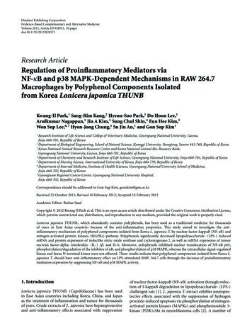
Republic of Korea) were cultured in DMEM (2 mM Lglutamine, 100 units/mL penicillin and 100 μg/μL, and 10%fetal bovine serum) in a 5% CO2 humidified incubatorat 37 C. RAW 264.7 cells were grown in six-well platesat a density of approximately 1 106 cells per well. Thepolyphenol compounds were dissolved in dimethylsulfoxide(DMSO) and filtered through 0.45 μm cellulose membranes.Cells were pretreated with the polyphenol componentsisolated from L. japonica T. at various concentrations (10, 50,100, and 200 μg/mL) and then stimulated with 1 μg/mL LPSfor 30 min, 6 h, and 24 h.2.4. Assay for Cell Viability. The cell viability inhibitoryeffect of polyphenols on RAW 264.7 cells was assessed bythe mitochondrial respiration-dependent reduction methodof MTT to formazan. Cells seeded in 12-well plates (1 104 cells/well) were pretreated with various concentrationsof polyphenols (10, 50, 100, 200, and 300 μg/mL) and thentreated with LPS (1 μg/mL) at 37 C in 5% CO2 for 24 h. Aftertreatment, 100 μL of MTT (5 mg/mL) dissolved in DMEMwas added to each well, followed by incubation for 3 h.The medium was aspirated, and the formazan crystals weredissolved in 500 μL of DMSO for 15 min. The optical densityof each well was measured at 540 nm. The assay was carriedout in triplicate.2.5. Western Blot Analysis. Raw 264.7 cells were seeded insix-well plates at a density of approximately 1 106 cellsper well and incubated with or without LPS in the presenceor absence of polyphenols. The cells were washed twicewith ice-cold PBS and resuspended in lysis buffer containing50 mM Tris-HCl (pH 8.0), 150 mM NaCl, 0.5% sodiumdeoxycholate, 0.1% sodium dodecyl sulfate (SDS), 1% NP40, protease inhibitor cocktail, 0.5 M EDTA, and phosphataseinhibitor. The cell lysate was obtained from the supernatantafter centrifugation at 13,000 rpm, 4 C, and 30 min. Theprotein concentration was determined using a Bradford assaykit (Bio-Rad, Hercules, CA, USA). Equal amounts of proteinswere blotted onto an Immobilon-P 0.45 mm polyvinylidene fluoride (PVDF) membrane (Millipore, Billerica, MA,USA) following separation by 10% SDS-polyacrylamide gelelectrophoresis (SDS-PAGE). The transferred proteins wereincubated overnight at 4 C with a dilution of primaryantibody (anti-iNOS, COX-2, phospho-I-κB, NF-κB p65,phospho-ERK1/2, phospho-JNK, or phospho-p38) and βactin and then washed five times with Tris-buffered salinecontaining 0.5% Tween-20 (TBS-T, pH 7.4) for 10 min.After washing, the blots were incubated with horseradishperoxidase-conjugated secondary antibody for 1 h and againwashed five times in TBS-T. The membranes were developedusing an ECK kit (GE Healthcare Life Sciences, Buckinghamshire, UK). The intensity of each band was quantitativelydetermined using Image J software (http://rsb.info.nih.gov).2.6. RNA Extraction and Reverse Transcription-PolymeraseChain Reaction (RT-PCR). RAW 264.7 cells were pretreatedwith polyphenols for 1 h and then stimulated with 1 μg/μLLPS for 6 h. Total RNA was isolated using TRIzol reagent3Abosorbance (mAU)Evidence-Based Complementary and Alternative 00600400200010510152 3 4205625 30 35Time (min)1015 1612 131811 14 17894045505560Figure 1: HPLC chromatogram and chemical structures ofpolyphenol components isolated from L. japonica T. HPLC chromatogram of L. japonica T.: (1) caffeoylquinic acid dimer, (2)caffeoylquinic acid, (3) caffeoylglycerol, (4) 5-p-coumaroylquinicacid, (5) feruloylquinic acid, (6) dicaffeoylquinic acid, (7) dicaffeoylquinic acid, (8) kaempferol 3-O-glucoside, (9) kaempferol-Orutinoside, (10) dicaffeoylquinic acid, (11) apigenin-7-O-glucoside,(12) apigenin rutinoside, (13) feruloyl caffeoylquinic acid, (14) trihydroxymethoxyflavone, (15) kaempferol, (16) isorhamnetin glucoside, (17) caffeic acid derivative, and (18) feruloyl caffeoylquinicacid.(GeneALL Biotechnology, Seoul, Republic of Korea) andreverse-transcribed into cDNA by commercially availablecDNA synthesis kits (iScript cDNA Synthesis Kit; Bio-Rad)according to the manufacturer protocol. cDNA (1 μg/μL) wasused to perform RT-PCR. The primer sequences used forquantification of iNOS, COX-2, TNF-α, IL-1β, IL-6, andglyceraldehyde 3-phosphate dehydrogenase (GAPDH) andthe PCR conditions were the same as previously described[14]. The PCR products were separated by 1.5% agarose gelelectrophoresis and visualized by ethidium bromide (EtBr)staining. The gels were then analyzed with ultraviolet transillumination. The quantity of each mRNA was calculated byusing Image J software and normalized to the amount of thehousekeeping GAPDH gene.2.7. Statistical Analysis. All experiments were reiterated atleast three times. The results of multiple observations areexpressed as the mean SD. Statistical significance wasdetermined by one-way analysis of variance (ANOVA) usingSPSS version 10.0 for Windows (SPSS, Chicago, IL, USA) formultiple comparisons. A value of P 0.05 was consideredstatistically significant.3. Results3.1. Characterization and Quantification of Polyphenol Components in Lonicera japonica THUNB. Polyphenol components were isolated from L. japonica T. using HPLC. Based onthe HPLC retention time characteristics and the ultravioletvisible spectra of standard compounds in a library, thecomponents isolated from L japonica T. were present as 18peaks and were identified (Figure 1). The 18 polyphenolsthat were comprised of 11 hydroxycinnamic acids and sevenflavonoids were recorded at 280 nm. The quantificationvalues of the 18 components are shown in Table 1.
Evidence-Based Complementary and Alternative MedicineNumber123 456789101112 131415 161718Quantity .631.27aData are the mean SD of triplicate determinations by HPLC-UV methodat the 280 nm.3.2. Effect of Polyphenols on Cell Viability. The cytotoxicityof polyphenols isolated from L. japonica T. on the viabilityof RAW 264.7 cells was measured in the concentrationrange of 10–300 μg/mL using the MTT assay. Cell viabilitywas not significantly changed by any of the polyphenolconcentrations compared with the negative control (datanot shown). Cotreatment of polyphenols and LPS (1 μg/mL)was not cytotoxic at any of the concentrations used. Weused 200 μg/mL for maximum concentration of polyphenolsisolated from L. japonica T., and this concentration was usedin subsequent experiments.2mRNA relative expression(COX-2/GAPDH)Table 1: Concentration of polyphenols of L. japonica THUNB.(mg/kg)a . 1.61.2 0.80.40LPS (1 μg/mL)Polyphenols (μg/mL) 1050100200COX-2GAPDH(a)Relative fold of COX-2/β-actin41.6 1.41.2 10.8 0.60.40.20LPS (1 μg/mL)Polyphenols (μg/mL) 1050100200COX-2β-actin(b)3.3. Inhibitory Effect of Polyphenols on COX-2 mRNA andProtein Expression in LPS-Stimulated RAW 264.7 Cells. LPSis associated with a marked increase in COX-2 expression inRAW 264.7 cells. This was also the case presently; expressionof COX-2 mRNA was markedly increased upon exposureto LPS for 6 h. Pretreatment with 200 μg/mL polyphenolsdecreased the mRNA expression of LPS-induced COX-2(Figure 2(a)). Also, studies were conducted to determinewhether the expression of COX-2 protein paralleled itsmRNA. RAW 264.7 cells treated with LPS displayed amarkedly induced expression of COX-2 protein, while itsprotein was not detectable in unstimulated cells. In responseto LPS treatment, the expression of COX-2 protein wassignificantly increased and polyphenols markedly suppressedCOX-2 protein induction at concentrations of 100 and200 μg/mL (Figure 2(b)).3.4. Inhibitory Effect of Polyphenols on iNOS mRNA andProtein Expression in LPS-Stimulated RAW 264.7 Cells. Asshown in Figure 3(a), LPS induced a significant increase inexpression of iNOS protein and mRNA. However, cotreatment of polyphenols and LPS inhibited the expression ofFigure 2: Effect of polyphenols on COX-2 mRNA and proteinexpression in RAW 264.7 macrophages. RAW 264.7 cells werepretreated with the indicated concentrations of polyphenols for 1 hand treated with LPS (1 μg/mL). (a) After 6 h, total RNA of the cellswas subjected to RT-PCR. (b) After 24 h, equal amounts of proteinwere subjected to 10% SDS-PAGE. Expression of COX-2 proteinswas determined by Western blot analysis. Data are the mean SDof triplicates. The asterisk ( ) indicates a significant difference fromthe control group (P 0.05) and indicates significant differencefrom the LPS-treated group (P 0.05).iNOS mRNA at 100 and 200 μg/mL of concentrations inLPS-stimulated RAW 264.7 cells. Furthermore, based onthis result, we assessed whether polyphenols affected theexpression of iNOS protein. RAW 264.7 cells induced highlevels of iNOS protein when treated with LPS for 24 h, whilethe expression of iNOS protein was hardly detectable inun-stimulated cells. Addition of polyphenols significantlyinhibited the LPS-induced iNOS protein production in adose-dependent manner (Figure 3(b)).
Evidence-Based Complementary and Alternative MedicinemRNA relative expression(iNOS/GAPDH)1.6 1.41.210.80.6 0.4 0.20LPS (1 μg/mL)Polyphenols (μg/mL) 1050100200iNOSGAPDHRelative fold of iNOS/β-actin(a) 0.80.70.6 0.5 0.4 0.30.20.10LPS (1 μg/mL)Polyphenols (μg/mL) 105010020053.6. Inhibition of Polyphenols on LPS-Induced Degradationand Phosphorylation of IκB-α and Nuclear Translocation ofthe NF-κB p65 Subunit in RAW 264.7 Cells. In order tounderstand the mechanisms underlying the inhibitory effectof polyphenols on LPS-induced inflammatory mediatorssuch as COX-2, iNOS, and various cytokines, we furtherassessed the protein level of NF-κB p65 in the cytosolicand nuclear fractions, as well as degradation and phosphorylation of IκB-α by Western blot analysis. The amountof NF-κB p65 in the nucleus was dramatically increasedupon exposure to LPS alone and markedly decreased inthe cytosolic fraction. However, in the nuclear fractions ofRAW 264.7 cells, the expression levels of NF-κB p65 proteinwere significantly attenuated by treatment with polyphenolsat various concentrations (10, 50, 100, and 200 μg/mL),suggesting that polyphenols inhibit the translocation of NFκB p65 protein from the cytosol to the nucleus (Figures5(a) and 5(b)). Since the degradation and phosphorylationof IκB-α are a principal step in NF-κB activation by LPS,we examined the effect of polyphenols on LPS-induceddegradation and phosphorylation of IκB-α protein. IκB-αwas degraded in RAW 264.7 cells by a 30 min treatmentwith LPS, and this degradation was noticeably preventedby 100 and 200 μg/mL of polyphenols (Figure 5(c)). Thephosphorylated forms of IκB-α were also barely detectable inthe un-stimulated RAW 264.7 cells (Figure 5(d)). However,polyphenols inhibited LPS-induced phosphorylation of IκBα in a dose-dependent manner, similar to results fromnuclear translocation of NF-κB p65, while LPS alone ledto a significant increase in the phosphorylation of IκB-α(Figure 5).iNOSβ-actin(b)Figure 3: Effect of polyphenols on iNOS mRNA and proteinexpressions in RAW 264.7 macrophages. RAW 264.7 cells werepretreated with the various concentrations of polyphenols for 1 hand treated with LPS (1 μg/mL). (a) After 6 h, total RNA of the cellswas subjected to RT-PCR. (b) After 24 h, equal amounts of proteinwere subjected to 10% SDS-PAGE. Expression of COX-2 proteinswas determined by Western blot analysis. Data are the mean SDof triplicates. The asterisk ( ) indicates a significant difference fromthe control group (P 0.05) and indicates significant differencefrom the LPS-treated group (P 0.05).3.5. Inhibitory Effect of Polyphenols on mRNA Expression ofProinflammatory Cytokines in LPS-Stimulated RAW 264.7Cells. To determine whether treatment with polyphenolsaffected the expression of pro-inflammatory cytokines,expressions of TNF-α, IL-1β, and IL-6 mRNAs were assessedby RT-PCR. When cells were treated with LPS (1 μg/mL), allthree mRNA levels significantly increased. However, mRNAexpressions of TNF-α, IL-1β, and IL-6 were significantlydecreased over that resulting from a single LPS stimulation by treatment of polyphenols at 200 μg/mL and 100–200 μg/mL, respectively (Figure 4).3.7. Effect of Polyphenols on LPS-Induced Phosphorylationof MAPKs in RAW 264.7 Cells. Inflammation is triggeredby intracellular signaling pathway events, which is relatedto the phosphorylation of MAPKs. To further understandthe underlying possible mechanisms involved in the antiinflammatory effect of polyphenols, we investigated whetherpolyphenols inhibited LPS-induced phosphorylation ofMAPKs including p38 MAPK, ERK 1/2, and JNK. As shownin Figure 6, LPS treatment caused a strong increase in thephosphorylation of p38 MAPK, ERK 1/2. and JNK. However,co-treatment with 100 and 200 μg/mL polyphenols reducedthe LPS-induced phosphorylation of p38 MAPK, whereas thephosphorylation of ERK and JNK was not affected.4. DiscussionTraditional Chinese medicine has used various herbal plantsfor centuries. A variety of compounds originating fromherbal plants have various effects such as anticancer, antiinflammation, and antiviral activities. Especially, L. japonicaT. has long been used as a medicinal herb in Korea becauseof its anti-inflammatory and analgesic properties [15, 16]. L.japonica T. contains a hundred bioactive compounds including polyphenols. We identified 18 polyphenols isolated fromKorea L. Japonica T. for investigating bioactive compoundby using HPLC analysis. Among them, the dicaffeoylquinic
6Evidence-Based Complementary and Alternative Medicine1.8 mRNA relative expression(IL-6/GAPDH)mRNA relative expression(TNF-α/GAPDH)1.51 0.50LPS (1 μg/mL)Polyphenols (μg/mL) 1050100200 1.51.20.9 0.6 0.30LPS (1 μg/mL)Polyphenols (μg/mL)TNF-αIL-6GAPDHGAPDH 1050100200(a)(b)mRNA relative expression(IL-1β/GAPDH)2.5 21.5 1 0.50LPS (1 μg/mL)Polyphenols (μg/mL) 1050100200IL-1βGAPDH(c)Figure 4: Effect of polyphenols on expression of pro-inflammatory cytokines. RAW 264.7 cells were treated with polyphenols extract atvarious concentrations (10, 50, 100, and 200 μg/mL) for 1 h before treatment with LPS (1 μg/mL) to induce inflammation. After 6 h ofincubation, mRNA levels of (a) TNF-α, (b) IL-6, and (c) IL-1β were measured by RT-PCR. Data are the mean SD of triplicates. Theasterisk ( ) indicates a significant difference from the control group (P 0.05) and indicates significant difference from the LPS-treatedgroup (P 0.05).acid is a major component and has anti-inflammatory effectby reducing the production of inflammatory mediatorsin lymphoma cells and suppressing COX-2 expression inmacrophages [17].Inflammation is a host primary response to infectionor injury; macrophages and mast cells induce the release ofinflammatory mediators such as COX-2, iNOS, and inflammatory cytokines. Normally, inflammation protects the hostfrom external challenge, but prolonged inflammation causesvarious diseases. Inflammatory stimuli affect the four majorintracellular signaling pathways: NF-κB and the three MAPKpathways [18].iNOS and COX-2 are important in the regulation ofinflammation. COX is the target protein for analgesicand anti-inflammatory therapies that have been used forhundreds of years. The COX enzyme consists of at least twoisoforms: COX-1 and COX-2. In mammals, the expression ofCOX-1 is constitutive in normal tissues such as production ofprostaglandin precursors for thromboxane in platelets andthe regulation of blood flow. In contrast to COX-1, COX2 protein is only slightly expressed in most resting tissues.However, upon inflammatory stimulation, the expressionlevels of COX-2 protein and mRNA are increased [19].Blocking NF-κB and MAPKs activation by luteolin isolatedfrom the flowers of L. japonica inhibits inflammatory mediators such as cytokines and COX-2 release from mast cells[20]. This study investigated that polyphenols suppressed theexpressions of COX-2 protein and mRNA in LPS-induced
Evidence-Based Complementary and Alternative Medicine71.21 0.80.6 0.40.20 LPS (1 μg/mL)Polyphenols (μg/mL) 10 50 100 200Relative fold ofp65/β-actin (NE)Relative fold ofp65/β-actin (CE)1.2 1 0.80.6 0.40.20LPS (1 μg/mL)Polyphenols (μg/mL)P65 (CE)P65 (NE)β-actinβ-actin 1050100200 (a)(b)1.4 0.81.2 Relative fold ofp-IκB/β-actinRelative fold ofIκB/β-actin10.6 0.40.2 10.8 0.60.40.20LPS (1 μg/mL)Polyphenols (μg/mL) 10 50 100 2000LPS (1 μg/mL)Polyphenols (μg/mL)IκBP-IκBβ-actinβ-actin(c) 1050100200(d)Figure 5: Effect of polyphenols on activation of NF-κB in cytosol and nuclear translocation of NF-κB p65. The cells were pretreated with theindicated concentrations of polyphenols for 1 h and treated with LPS (1 μg/mL). After 30 min incubation, the expression of (a) cytosolic and(b) nucleic NF-κB as well as (c) IκB and (d) phosphorylated IκB was examined by Western blot. Data are the mean SD of triplicates. Theasterisk ( ) indicates a significant difference from the control group (P 0.05) and indicates significant difference from the LPS-treatedgroup (P 0.05).RAW 264.7 cells (Figure 2). The results showed that theprotein and mRNA expressions of COX-2 were decreased bypolyphenols in LPS-induced RAW264.7 cells.NOSs are a family of enzymes that catalyze the production of NO from L-arginine. Three isoforms of NOShave been found: neuronal NOS (nNOS), endothelialNOS (eNOS), and inducible NOS (iNOS). NOS existsin neuronal tissues (nNOS) and vascular endothelial cells(eNOS), whereas inducible NOS (iNOS) is detected ina variety of cell types including macrophages, microglialcells, astrocytes and keratinocytes. With infectious andproinflammatory stimuli, overexpression of iNOS proteinand mRNA occurs. Poncirin, a flavanone glycoside isolated from Poncirus trifoliata, suppresses the expressionsof iNOS, COX-2, and cytokines by blocking the NF-κBpathway in LPS-induced RAW 264.7 macrophages [21].Also, inactivation of NF-κB by kaempferol downregulatesiNOS and TNF-α expression in aged rat gingival tissues[22]. Our results showed that the levels of iNOS proteinand mRNA were dose-dependently manner-suppressed bypolyphenols in LPS-induced RAW 264.7 cells (Figure 3).Additionally, to investigate the anti-inflammatory effect bypolyphenols, the mRNA levels of proinflammatory cytokinesincluding TNF-α, IL-1β, and IL-6 were analyzed. This datarepresented that LPS-stimulated proinflammatory cytokineproduction was significantly attenuated by polyphenols ina dose-dependent manner (Figure 4). These results indicatethat polyphenols suppressed the expression of iNOS andproinflammatory cytokines which were important role ininflammatory response.
8Evidence-Based Complementary and Alternative Medicine Relative fold of phosphop38/β-actin1.61.2 0.80.40LPS (1 μg/mL)Polyphenols (μg/mL)LPS (1 μg/mL)Polyphenols (μg/mL) 1050100200 1050100200P-ERKP-JNKβ-actinRelative fold of phosphoERK/β-actin1.2P-p38 0.80.40LPS (1 μg/mL)Polyphenols (μg/mL) 1050100200Relative fold ofJNK/β-actin1.2 0.80.40LPS (1 μg/mL)Polyphenols (μg/mL) 1050100200Figure 6: Effect of polyphenols on phosphorylation of MAPKs in RAW 264.7 macrophages. RAW 264.7 cells were pretreated with theindicated concentrations of polyphenols for 1 h and treated with LPS (1 μg/mL) for 15 min. The cell lysates were analyzed by Western blotfor the detections of phosphorylation of ERK1/2, JNK, and p38 MAPK. Data are the mean SD of triplicates. The asterisk ( ) indicates asignificant difference from the control group (P 0.05) and indicates significant difference from the LPS-treated group (P 0.05).NF-κB plays a central role as an inducible transcriptionfactor and is critical for regulation of gene expression inresponse to inflammation and immunity. NF-κB proteinsexist normally in the cytoplasm of inactivated cells, butthese must be translocated into the nucleus to functionin stimulated cells. The NF-κB pathway is induced byLPS and pro-inflammatory cytokines such as TNF-α andIL-1β [23]. This process leads to the activation of RelA(p65)/p50 complexes. These complexes are controlled by theinhibitor of κB (IκB) proteins, which bind NF-κB, therebyblocking nuclear translocation. When cells are exposed toextracellular stimuli, IκB proteins are phosphorylated b
4Department of Chemistry and Research Institute of Life Science, Gyeongsang National University, Jinju 660-701, Republic of Korea 5Department of Nursing Science, International University of Korea, Jinju 660-759, Republic of Korea 6Department of Internal Medicine, Institute of Health Sciences, Gyeongsang National University School of Medicine,
