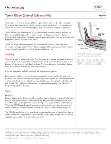
Transcription
Tennis Elbow and the Cervical SpineC. Chan Gunn, MDThe exact cause of tennis elbow, a common condition, is still obscure. While the condition may well be entirelydue to a local disorder at the elbow, the results of a study of 50 patients whose condition was resistant to 4 weeksof treatment directed to the elbow suggest that the underlying condition may have been (at least in thesepatients) a reflex localization of pain from radiculopathy at the cervical spine. Clinical, radiologic andelectromyographic findings supported this suggestion. The pain was demonstrated to be muscular tenderness,which was maximal and specific at motor points. Treatment directed to the cervical spine appeared to give reliefin the majority of patients. The more resistant the condition, the more severe were the radiologic andelectromyographic findings in the cervical spine.Tennis elbow, a common affliction, the exact cause of which is unknown, has been considered to be a selflimiting condition, seldom persisting for longer than 12 months, yet symptoms may continue longer despite alltypes of conservative treatment or even surgery.This paper reports a new approach: in a series of 50 patients treatment was directed to the cervical spine after atleast 4 weeks of treatment of the elbow had failed, and it was successful in most.Patients and symptomsThe 50 patients with tennis elbow, 37 men and 13 women, were referred by attending physicians to therehabilitation clinic of the Workers' Compensation Board of British Columbia for management. In many thecondition had not responded to the usual conservative office measures, such as injections of steroids and localanesthetics, manipulation, ultrasound, friction massage and immobilization. Their age distribution was asfollows: 21 to 30 years, 8 patients; 31 to 40 years, 10; 41 to 50 years, 13; 51 to 60 years, 15; and over 60 years,4.All were right-side-dominant, but three had only left-side complaints. Eleven had bilateral lateral epicondylarsymptoms, 12 had concurrent medical epicondylar symptoms and 7 had bilateral medical and lateral epicondylarsymptoms. The time lapse between onset of symptoms and referral to the clinic was 8 weeks or less in 24, 8 to12 weeks in 11 and more than 12 weeks in 15.The clinical types of tennis elbow, classified by onset and injury, were the following:1. Acute type, precipitated by indirect trauma (Cyriax's typeI) - - for example, probable avulsion due to acutepull of forearm extensor muscles at their origin (four patients).2. Subacute type, following indirect trauma (Cyriax's type II), from repeated and forcible extensionmovements at the wrist (six patients).3. Insidious onset (Cyriax's type III), with no specific single incident (11 patients).4. Acute onset following blunt trauma (Cyriax's type IV) (six patients).5. Associated with cervical "strain", with history of phyerextension or flexion injury to cervical spine or neck"strain" (13 patients).6. Associated with jolt or traction to shoulder (three patients).7. Not classified elsewhere - - for example, as part of multiple injuries, when exact mechanism is notdetermined (seven patients).
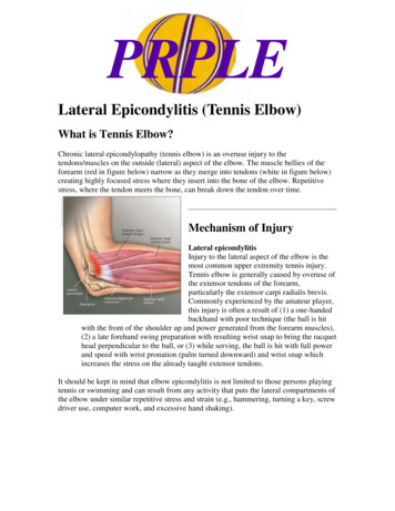
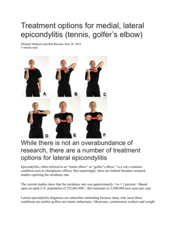
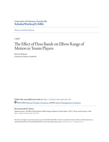
topExaminationIn addition to eliciting the classic signs, the examiner palpated carefully the entire elbow region with a bluntpoint or the tip of a finger. Points of maximal tenderness found (usually four) were accurately determined andmarked. These points seemed to correspond to the sites of muscle motor points in the region and were confirmedas such by electric stimulation. A motor point is defined as the site where a muscle twitch may be evoked inresponse to minimal electric stimulation. This point, a fixed anatomic site, lies close to where the motor nerveenters the muscle. Many of the motor points of the wrist extensor lie around the lateral epicondyle of thehumerus, where there is also a rich supply of sensory nerve fibre endings (Fig I). Other upper-limb motor pointswhere tenderness might be found were likewise examined (Figs. 2, 3 and 4). Since the tender muscles havecommon root derivations of their motor nerves (Table I), the cervical spine was also examined.Electromyographic examination was performed in 42 patients, and in all patients with a history ofacroparesthesia or in whom carpal tunnel syndrome or ulnar nerve tardiness was suspected, motor nerveconduction velocity tests were done to exclude such conditions.Physical findingsThe wrist extensor motor points generally found to be tender were those of the brachioradialis, extensor carpiradialis, supinator, extensor digitorum and extensor carpi ulnaris; these points are closely situated in an area ofabout 5 cm in diameter. Tenderness in other areas was of similar quality and often equal intensity; frequentlyboth sides were involved. The frequency of tenderness at the various motor points is shown in Table II.Eighteen patients showed slight limitation of lateral rotation or lateral tilting of the cervical spine to the affectedside. On that side, in all patients, the apophyseal joints of involved levels were tender to digital pressure(commonly C5 and C6) when carefully examined showed resistance to passive motion.topTable I - Spinal cord root derivations of motor nerves supplying arm and shoulder muscles.MuscleMotor nerve root derivation
Infraspinatus, s majorTricepsExtensor carpi radialisFlexor carpi radialisPronator quadratiusFlexor carpi ulnarisDorsal interosseiC5,6C5,6C5,6C5,6C5-8, T1C6-8, T1C6-8C6-8C8, T1C7,8, T1C8, T1Table II - - Frequency of tenderness at various motor points.Muscle of tender motor pointFrequency* of id (any of three points)Pectoralis majorBiceps branchialisExtensor carpi radialisExtensor carpi ulnarisExtensor digitorumBranchirradialisTriceps (any of three points)Adductor pollicis brevisFlexor carpi ulnaris3639122813155757501261316*Out of a possible 100 -- that is, 50 X patients X 2 sides.topMuscle atrophy, especially of the trapezius, supraspinatus, triceps and deltoid, was noted in 15 patients, andpartial loss of sensation to pinprick over the associated dermatome was detected in 1 patient.In 10 patients an autonomic (pilomotor and sudomotor) reflex was elicited when the patient was exposed to coldair: the skin over the dermatome involved - generally C5,6 - showed "goose pimples" or an erector pili effect(cutis anserina). This reflex (sometimes accompanied by excessive perspiration in the axillae) could also beinduced by digital frictional pressure over many of the tender motor points. Deep reflexes were always normal.
topRadiologic findingsRadiographs of the elbow invariably showed no significant findings, but radiographs of the cervical spine in 34patients (average age, 47 years) showed changes commensurate with age.Electromyographic findingsAll 42 patients showed some abnormal electromyographic findings of early neuropathy or radiculopathy inaffected myotomes. Discharge of action potentials due to mechanical excitation was often increased, and withvoluntary activity the mean duration of the action potentials appeared prolonged, but the amplitude was normalor reduced. Typically, polyphasic potentials appeared in abnormal numbers. The interference pattern wasreduced and in severe cases lost altogether. In some patients, action potentials of individual units could beidentified even during maximum contraction.TreatmentWhen first assessed, the condition of 23 of the 50 patients had not improved with at least 4 weeks of standardtreatment measures. Two had had bilateral surgical procedures but pain had persisted. In view of the apparentrelation of elbow symptoms to disorders of the cervical spine, our approach to relieving the symptoms in thisgroup (group A) was directed immediately to the neck.The other 27 patients (group B), who had not previously received local treatment of the elbow, were first givenultrasound, friction massage, ice and other therapy, and when symptoms persisted after 4 weeks, treatment wasdirected instead to the neck.Treatment of the cervical spine included one or more of the following:1. Mobilization (Maitland's grades I to IV).2. Cervical traction.3. Isometric cervical exercises.4. Heat or ultrasound, or both, applied to apophyseal joints if excessive tenderness was spresent.top
Response to cervical treatmentOf the patients in group A the average duration of cervical treatment in the 22 who responded was 4.7 weeks; 1patient still had symptoms on discharge after 9 weeks. Of the patients in group B the average total duration oftreatment (elbow and neck) at the clinic in the 25 who responded was 11.1 weeks, and the average duration ofneck treatment was 5.8 weeks; 2 patients still had symptoms on discharge after 18 to 20 weeks. The time lapsebetween onset of symptoms and beginning of treatment did not appear to influence duration or outcome oftreatment.Relation to electromyographic findings: Of the 42 patients who had an electromyographic examination theaverage duration of treatment in the 39 who responded was 5.3 weeks (4.7 weeks in the 20 with mildelectromyographic abnormalities and 7.2 weeks in the 19 with moderate to severe abnormalities); 3 patients withmoderate to severe abnormalities continued to have symptoms.Relation to radiologic findings: The average duration of treatment was as follows: in the 16 patients with normalradiologic findings, 4.8 weeks; in the 19 patients with minor radiologic abnormalities (restricted motion, earlydegenerative changes), 4.7 weeks; and in the 12 patients with moderated to severe radiologic abnormalities(severe osteoarthrosis, narrowing of disc space, foramina encroachment), 7.31 weeks. The tree patients whocontinued to have symptoms had moderate to severe radiologic abnormalities.Results on discharge from the clinicGood: These 29 patients were able to resume their previous occupation.Satisfactory: These 14 patients returned to light duties or changed to a suitable, nonaggravatingoccupation.Fair: Four patients were discharged with residual discomfort. Some relief was obtained with a cervicaltraction apparatus used at home. All returned to a suitable, nonaggravating occupation.Poor: Three patients, with severe radiologic and electromyographic abnormalities, continued to complainof symptoms on discharge.Follow-upOf the 47 patients who responded to treatment 44 were assessed at 3 and 6 months (time of writing) afterdischarge. They had had no further symptoms and had not sought further medical attention. Of the three who hadsymptoms on discharge, two were asymptomatic within 3 months and one, with severe cervical spondylosis,continued to have symptoms, even at a 1-year follow-up.topDiscussionIt is obviously not possible to draw definite conclusions from this small series because the condition of tenniselbow is often self-limiting, yet the findings challenge some current concepts.For instance, women in this series were proportionately affected more than twice as often as men. Although theproportion of women affected was only 26% (13 patients), this is more than twice the usual proportion ofwomen attending the clinic for other injuries - - 12% (598 of 4990 patients in 1974).Bilateral and medial epicondylar symptoms are said to be unusual, yet 22% (11 patients) in this series hadbilateral lateral epicondylar symptoms and 24 % (12 patients) had concurrent medial epicondylar symptoms;14% (7 patients) had bilateral lateral and medial epicondylar symptoms.In this study a force overload to the extensor muscles, direct or indirect, was found to be not the onlyprecipitating factor; in 26% (13 patients) cervical "strain" was associated, and 84% (42 patients) showed some
electromyographic evidence of cervical radiculopathy as well as physical signs in muscles of the myotomesinvolved.While the pain may have presented at the bony epicondyle, maximum tenderness was more commonly found inthe muscles at the several motor points that are close together and situated over bony prominences, where theyare subjected to tension or pressure.Other physical signs found, related to the cervical spine, were selective atrophy of muscles, especially the tricepsand supraspinatus (15 patients), altered dermatomal sensation (1 patient) and presence of an autonomic reflex(10 patients).These findings led us to conclude that, at least in this group of selected patients, the condition of tennis elbowwas related to disorders of the cervical spine; therefore, when treatment to the elbow failed, neck treatment wastired - - with good results. It is probable that in many patients some degree of cervical degeneration preceded theelbow condition.In this series, treatment of the cervical spine was followed by good or satisfactory relief of elbow symptoms in86% (43 patients) in an average of 5.25 weeks. In four patients the continual use of a cervical traction apparatusat home provided relief. Tow of the three patients who had symptoms at the time of discharge subsequentlyimproved within 12 weeks. Recovery time may be related to the degree of trauma sustained. Denny-Brown andBrenner have shown that mild percussive trauma to a nerve leads to swelling and local edema of the nerve,together with dissolution of the myelin, and recovery takes at least 4 to 5 weeks; however, if the trauma issufficiently severe to lead to Wallerian degeneration, recovery takes at least 12 weeks. In the one patient in thisseries with persistent pain, treatment probably failed to relieve the causative factor (severe cervical spondylosis).The time lapse between onset of symptoms and beginning of treatment bore no relation to the outcome oftreatment.We are grateful to the commissioners of the Board and Dr. A.S. Little, director of medical services. Workers'Compensation Board of British Columbia, for their support and advice; and to Mr. G. Page, supervisor of thephysiotherapy department. Miss C. Patterson. Mrs. J.E. Gunnyon and the other members of the staff for theircooperation and involvement in the project.topReferencesCYRIAX JH: Pathology and treatment of tennis elbow. J Bone Joint Surg [Am] 18; 921, 1936COONRAD RW, Hooper WR: Tennis elbow; its course, nature history, conservative and surgical management. J Bone JointSurg[Am] 55: 1177, 1973SAVASTANO AA. Kaminek S. Knowles K. et al: Treatment of resistant tennis elbow by a combined surgical procedure. Int Surg57: 470, 1972BOYD HB, 'McLeod AC: Tennis elbow. J Bone Joint Surg [Am] 55; 1183, 1973CHUSID JG; Correlative Neuroanatomy and Functional Neurology, 15the ed, Los Altos, CA, Lange, 1973, p 237COERS C: Note sur une technique de prelevement des biopsies neuromuscularies. Acta Neuol Psychiatr Beig 53; 759, 1953GOLDIE I: Epicondylitis lateralis humeri (epicondylalgia or tennis elbow); a pathogenetical study. Acta Chir Scand [Suppl] 339:1, 1964CYRIAX JH: Textbook of Orthopaedic Medicine, vol 1, 5th ed. London, Bailliere Tindall. 1969, p 167BONICA JJ: Management of myofascial pain syndromes in general practice. JAMA 1164: 732, 1957BUCHTHAL F. PINELLI P: Analysis of muscle action potentials in muscular atrophy of neuogenic origin. Neurology (Minneap)3: 591, 1953MAITLAND GD: Vertebral Manipulation, 3rd ed. London, Butterworth, 1973, pp 76-95DENNY-BROWN D. BRENNER C: The effect of percussion on nerve. J. Neurol Neurogurg Psychiatry 7: 76, 1944topclose window iSTOP - Institute for the Study and Treatment of Pain - 2002
least 4 weeks of treatment of the elbow had failed, and it was successful in most. Patients and symptoms The 50 patients with tennis elbow, 37 men and 13 women, were referred by attending physicians to the rehabilitation clinic of the Workers' Compensation Board of British Columbia for management. In many the