Transcription
Franz et al. Journal of Biological 8-z(2020) 14:26RESEARCHOpen AccessA biomedical Engineering Laboratorymodule for exploring involuntary musclereflexes using ElectromyographyKarly S. Franz1,2, Kramay Patel1,3,4 and Dawn M. Kilkenny1,5*AbstractBackground: Undergraduate biomedical engineering (BME) students interested in pursuing a career in researchand development of medical or physiological monitoring devices require a strong foundation in biosignal analysisas well as physiological theory. Applied learning approaches are reported to be effective for reinforcingphysiological coursework; therefore, we propose a new laboratory protocol for BME undergraduate physiologycourses that integrates both neural engineering and physiological concepts to explore involuntary skeletal musclereflexes. The protocol consists of two sections: the first focuses on recruiting soleus motor units throughtranscutaneous electrical nerve stimulation (TENS), while the second focuses on exploring the natural stretch reflexwith and without the Jendrassik maneuver. In this case study, third-year biomedical engineering students collectedelectromyographic (EMG) activity of skeletal muscle contractions in response to peripheral nerve stimulation using aBioRadio Wireless Physiology Monitor system and analyzed the corresponding signal parameters (latency andamplitude) using the MATLAB platform.Results/protocol validation: Electrical tibial nerve stimulation successfully recruited M-waves in all 8 studentparticipants and F-waves in three student participants. The students used this data to learn about orthodromic andantidromic motor fiber activation as well as estimate the neural response latency and amplitude. With the stretchreflex, students were able to collect distinct signals corresponding to the tendon strike and motor response. Fromthis, they were able to estimate the sensorimotor conduction velocity. Additionally, a significant increase in thestretch reflex EMG amplitude response was observed when using the Jendrassik maneuver during the knee-jerkresponse. A student exit survey on the laboratory experience reported that the class found the module engagingand helpful for reinforcing physiological course concepts.(Continued on next page)* Correspondence: dawn.kilkenny@utoronto.ca1Institute of Biomedical Engineering, University of Toronto, 164 College StRoom 407, Toronto, ON M5S 3G9, Canada5Institute for Studies in Transdisciplinary Engineering Education & Practice,University of Toronto, 35 St. George Street, Toronto, ON M5S 1A4, CanadaFull list of author information is available at the end of the article The Author(s). 2020 Open Access This article is licensed under a Creative Commons Attribution 4.0 International License,which permits use, sharing, adaptation, distribution and reproduction in any medium or format, as long as you giveappropriate credit to the original author(s) and the source, provide a link to the Creative Commons licence, and indicate ifchanges were made. The images or other third party material in this article are included in the article's Creative Commonslicence, unless indicated otherwise in a credit line to the material. If material is not included in the article's Creative Commonslicence and your intended use is not permitted by statutory regulation or exceeds the permitted use, you will need to obtainpermission directly from the copyright holder. To view a copy of this licence, visit http://creativecommons.org/licenses/by/4.0/.The Creative Commons Public Domain Dedication waiver ) applies to thedata made available in this article, unless otherwise stated in a credit line to the data.
Franz et al. Journal of Biological Engineering(2020) 14:26Page 2 of 11(Continued from previous page)Conclusion: This newly developed protocol not only allows BME students to explore physiological responses usingnatural and electrically-induced involuntary reflexes, but demonstrates that budget-friendly commercially availabledevices are capable of eliciting and measuring involuntary reflexes in an engaging manner. Despite somelimitations caused by the equipment and students’ lack of signal processing experience, this new laboratoryprotocol provides a robust framework for integrating engineering and physiology in an applied approach for BMEstudents to learn about involuntary reflexes, neurophysiology, and neural engineering.Keywords: Electromyography, Biomedical engineering, Undergraduate physiology, Laboratory protocol, TENS,Electrical stimulation, Stretch reflexIntroductionDespite diversity across different undergraduate biomedical engineering (BME) curricula, most programs includephysiology as a core part of the student learningexperience [1] upon recognition of its importance inpromoting interdisciplinary engagement [2]. Beyond understanding fundamental anatomical and physiologicalconcepts, it is important for BME students interested inpursuing graduate education or a professional career designing and developing medical devices to learn effectiveacquisition of biosignals [3], because these responses canbe monitored and measured to provide real-time feedback regarding system function.Growth of applications in commercial, industrial, medical, and defense sectors has led to an increased demandin graduates well-versed in biological signal physiology,acquisition, interpretation, and application [4]. Therefore, it is critical to provide opportunities for our students to develop skills in biosignal data acquisition andanalysis, with strong ability to translate outcomes tophysiological function. Applied laboratory exercises forscientific education are widely documented to enhancelearning because they allow students to explore theorywithin a practical, concrete context [5] and allow translation of concepts and development of professional skills[6]. Unfortunately, the dominant mode of curriculumdelivery across BME programs remains in lecture format[1], likely due to time and budgetary constraints in relevant programs. Unfortunately, in the context of biosignalstudy, lack of practical experience restricts the opportunity for BME students to merge data outcomes withphysiological concepts and translate them into tangibleapplications.Because active learning approaches have been found tobe particularly effective for teaching physiological concepts [7] meanwhile encouraging students to think at ahigher level and engage in enhanced discussion with instructors [7], we are motivated to develop and implement economical solutions and include active learningapproaches in our curriculum that focuses on physiological organ system integration. The sensorimotor system and its relationship with skeletal muscle reflexes isoften included in introductory physiology course curricula because it provides an excellent model of physiological integration. During our course module onneurophysiology, students explored relevant conceptsthrough the lens of engineering by using different approaches to evoke and analyze involuntary reflexes inskeletal muscles of the lower extremity. In addition toaccomplishing this task using affordable, commerciallyavailable equipment, students correlated their own electrical biosignals to mechanical activity, observed physiological variability in the acquired signals across the classpopulation, applied engineering skills by processing thesignals for enhanced analysis, and translated data outcomes to lecture concepts through continued discussion.Sensorimotor reflexes can be involuntary and areevoked naturally, through the stretch reflex, or artificiallyusing an electrical stimulation. Additionally, applicationof electrical stimuli to induce physiological responses isa prominent area of BME research that is of stronginterest to our students. Electrical stimulation can beused to restore lost function via functional electricalstimulation (FES), for rehabilitative purposes, or formuscle physiotherapy using neuromuscular electricalstimulation (NMES) [8–10]. While electrical stimulationapplications are vast and can be implemented using amultitude of approaches, the fundamental concept is todeliver electrical pulses to the body to trigger compoundaction potentials in nerve fibers [8, 9]. Transcutaneouselectrical nerve stimulation (TENS) is a cost-effective approach that is used in several applications to noninvasively interface with the peripheral nervous system[11]. In this way, TENS can be used to evoke involuntarymotor responses through electrical stimulation. Depending on the stimulation amplitude and an individual’snerve activation threshold, motor neurons can be recruited by stimulating sensory neurons that initiate a response that is similar to the natural stretch reflex knownas the Hoffman Reflex [12]. At higher stimulation amplitudes, direct motor unit activation manifests as short latency M-waves while supramaximal stimulation causesantidromic motor neuron activation resulting in largelatency F-waves in the EMG signal.
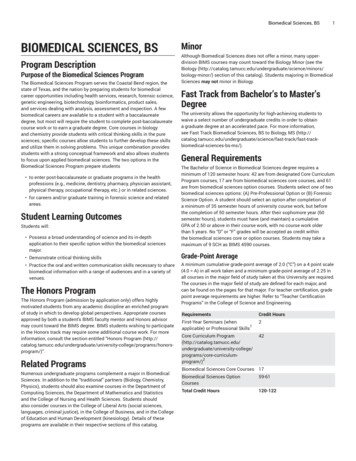
Franz et al. Journal of Biological Engineering(2020) 14:26Involuntary reflexes also occur naturally within thebody. These natural stretch reflexes are spinal mechanisms induced by sensory stimuli that lead to skeletalmuscle contractions. Like the H-reflex, stretch reflexes are a spinal mechanism that involve the entiresensorimotor loop and are commonly used in clinicalsettings to assess upper and lower neuron function[13]. This naturally occurring reflex can be reinforcedand amplified using the Jendrassik Maneuver which iswhen an individual voluntarily clenches their teethand pulls on their interlocked fists before the tendontap [13].This newly developed laboratory experience exploresthe use of EMG to detect involuntary motor reflexesevoked by TENS, and also the knee-jerk reflex when occurring naturally or amplified using the Jendrassik Maneuver. The laboratory exercise was developed with threemain objectives: 1) to support student learning ofphysiological course concepts; 2) to teach students howto use a wireless physiological monitoring system (theBioRadio; Great Lakes Neurotechnologies, Cleveland,OH) in conjunction with MATLAB (MathWorks, Natick, MA) to acquire and analyze relevant biosignals; and3) to create a satisfying experience that promotes selfperception of the individual’s engineering skills. The described protocol demonstrates the opportunity for students to directly explore relevant physiological and basicelectrical interfacing concepts related to the lower extremities using multiple analytical platforms, and demonstrates that physiological responses previouslydescribed can be elicited and measured using budgetfriendly commercially available devices. Survey responsefeedback indicated that most of the students felt this labprotocol was an engaging approach to immerse in learning neurophysiology while applying their engineeringknowledge. This newly developed protocol integratesneurophysiology and engineering concepts to provide aricher, more impactful experience for BME students tolearn about involuntary physiological reflexes by exploring different approaches to evoke and analyze involuntary skeletal muscle reflexes.MethodsParticipantsTwenty-four third-year undergraduate students in aBME program at the University of Toronto participated in piloting the described laboratory protocol foran introductory physiology course. Students weredivided into 8 groups (three students per group) andmeasured EMG signals from one volunteer withineach group. This protocol was approved by the Research Ethics Board at the University of Toronto(REB #37563).Page 3 of 11Biosignal acquisitionPart 1: muscle recruitment using TENSSoleus EMG activity was measured using the BioRadio(Great Lakes Neuro Technologies, Cleveland) and collected using the compatible BioCapture Research Systemsoftware (version 5.5.640). The skin covering the soleusmuscle was first cleaned using an alcohol wipe to preparefor electrode application (Covidien Kendall mini foamelectrode, diameter 3 cm). Two electrodes, the sensingand reference, were subsequently placed on the cleanedarea and connected to a single channel on the BioRadio(Fig. 1). The signal was grounded by placing the third electrode on the medial malleolus. Prior to placing the TENSelectrodes on the legs, students verified that an EMG signal was detected by voluntarily contracting the soleusmuscle multiple times and observing a corresponding increase in signal amplitude on the live BioCapturerecording.Once the soleus EMG activity was observed, studentsset up the TENS device (Classic TENS Unit, Body Clock,London) to stimulate the tibial nerve. Two square TENSelectrodes (50 50 mm) were placed vertically on thepopliteal fossa with the cathode connected proximallyand the anode distally along the leg (Fig. 1). Continuousbiphasic stimulation at 2 Hz with a 250 μs pulse widthand an intensity between 1 and 80 mA was used to activate sensory and motor fibers of the tibial nerve. Studentswere instructed to gradually increase the stimulation intensity and take note of the amplitude at three differentthresholds: sensory, motor, and maximum tolerable. Thesensory threshold was defined as the minimum stimulation amplitude that caused electrical stimulus sensationwith no motor activity. The motor threshold was definedas the minimum intensity needed to elicit an observablemuscle twitch and the maximum tolerable was the higheststimulation amplitude that the student could apply without pain. Instructors emphasized that the activationthresholds would be different for each student and thatcorrect neural recruitment should be painless.After verifying that both the TENS and the BioRadiowere properly set up, students began data collection. Students were instructed to record 5–7 stimulation pulses ateach defined threshold (sensory, motor, and maximal)with a 5–10 s break between each stimulation level. Recorded EMG signals were digitally sampled (2 kHz), amplified (gain 1000), and filtered (high pass Butterworth, 4thorder, 30 Hz cutoff) during acquisition. Student EMG response data to tibial nerve stimulation and the differentstimulation thresholds were collated and averaged to report the proposed method effectiveness.Part 2: natural muscle recruitmentEMG activity associated with the involuntary and naturally occurring knee-jerk response was collected. Again,
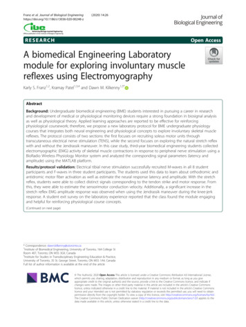
Franz et al. Journal of Biological Engineering(2020) 14:26Page 4 of 11Fig. 1 Placement of Simulating and Recording Electrodes. Two TENS stimulating electrodes are placed vertically along the superficial tibial nervein the popliteal space. The soleus EMG activity was measured with sensing and reference electrodes aligning with the muscle fibers andconnected to the positive and negative channel 1 (CH1) terminals of the BioRadio. The EMG signal was grounded with a third electrode placedon the medial malleolusan alcohol wipe was used to prepare the skin for electrode application above the rectus femoris, patella, andpatellar tendon (Fig. 2). Two wet snap electrodes wereplaced longitudinally along the rectus femoris to measure the EMG response to the patellar reflex. Specifically,students were instructed to place the first electrode approximately four finger-widths proximal to the patellaand the second placed two finger-widths above the first.Another pair of electrodes was placed on the patellartendon and patella, respectively, to capture the hammerstrike as a signal artifact. All sensing and reference electrodes were connected to their respective two channelson the BioRadio and both signals were grounded with alead connected to the lateral epicondyle.Once electrodes were properly placed, the participantsplaced their knee joint at a 90-degree angle and relaxedtheir leg muscles. Prior to data collection, students practiced eliciting a knee-jerk response on the target subject.They also verified that a signal spike associate with thehammer strike and the respective rectus femoris contraction were observed in BioCapture. Upon signal verification, students then collected the EMG response totwo different movements: the natural stretch reflex andthe stretch reflex with the Jendrassik maneuver. Studentsperformed each movement between 5 and 7 times withan approximately 5-s break between plexor taps and 10s break between movements. As with the previous section, student EMG response to the different movementsand the hammer stimulus data were collated and averaged as an indicator of the method success.EMG signal processing and analysisAll acquired signals were exported as comma separatedvalue files and imported into MATLAB2015b (TheMathWorks Inc., Natick, Massachusetts) for analysis.Part 1: TENS-EMGAll EMG responses were epoched into 2 s windows centered around the TENS pulse artifact. Next, epocheddata at the different stimulation thresholds (i.e., sensorythreshold, motor threshold, and maximum tolerablethreshold) were averaged for each student and the baseline deflection points were identified. The latency wasthen calculated as the peak-to-peak difference betweenthe stimulus artifact and the subsequent wave responsethat corresponded to the muscle twitch. The twitch response amplitude was also measured and defined as theabsolute maximum peak from the baseline (0 mV).
Franz et al. Journal of Biological Engineering(2020) 14:26Page 5 of 11Fig. 2 Placement of Recording Electrodes to Measure the Stretch Reflex Response. Two electrodes were placed longitudinally to measurecontractions of the rectus femoris using the BioRadio (CH1). The other electrodes were connected to channel 2 (CH2) of the BioRadio and wereplace on the patella and patellar tendon. These electrodes were placed for detection of the tendon-tap that elicited the muscle contractionPart 2: stretch reflexEMG signals were high pass filtered to remove motionartifacts (Butterworth, 6th order, cut off frequency 20Hz), rectified, and subsequently low pass filtered (Butterworth, 4th order, cut off frequency 100 Hz) in order toremove high frequency noise. EMG data was epochedinto 2 s windows centered around the temporal peak ofthe plexor artifact. The epoched EMG and hammer signals were each averaged for the natural and Jendrassikreflex trials for each student. Response latency was measured as the difference between the stimulus onset andpositive peak corresponding to the rectus femoris contraction. Amplitude was also defined as the maximumvoltage of the rectified signal from the baseline.Statistical analysis was conducted using JMP Software(version 14.1, SAS Institute Inc., Cary, NC). The Wilcoxon Signed-Rank test was used to compare amplitudeand latency data in response to different stimulationlevels or stretch reflex movements. All values are reported and plotted as mean SEM and a p-value 0.05was considered significant.Survey collection and analysisA survey instrument was created to assess student perceptions of this newly developed practical experience.Participation was advertised to students during lecture 1week before the lab experience, and the survey was administered to participants by a teaching assistant. Alldata was anonymized and collated for assessment by aresearch volunteer not associated with data collection orthe course.Survey questions related to the specific objectives ofthe lab experience were used for the analysis and questions were grouped accordingly (Table 1; See ‘Additionalfile 1’ for Survey Instrument). The objectives were defined as: 1) how much the lab supported students inlearning the course material; 2) to teach the studentshow to use the BioRadio and MATLAB for biosignalacquisition and analysis; and 3) to create a satisfying experience that promotes self-perception of the individual’sengineering skills.Table 1 outlines the primary laboratory objectives andthe survey questions used to qualitatively assess the students’ opinion on the experience. The survey questionswere grouped under the relevant objectives outlinedabove.Students responded to each question by selecting ‘verypoor’, ‘poor’, ‘okay’, ‘good’, or ‘excellent’. A research volunteer not associated with the data collection coded theordinal responses provided by each student on a 1–5
Franz et al. Journal of Biological Engineering(2020) 14:26Page 6 of 11Table 1 Survey Questions Relating to Each Lab ObjectiveObjectivesSurvey QuestionsObj. 1: Supported LearningPhysiological Concepts How would you rate the effectiveness with which course concepts were explained in Lab 2, the EMG labexercise? How would you rate the extent to which this laboratory experience improved your understanding ofimportant course concepts related to skeletal muscle lectures? How would you rate your confidence in the related subjects after completing this laboratory experience? How would you rate the contribution of this laboratory experience to the value of your learning in theBME350H1 course (specifically, learning related to the muscular system)?Obj. 2: Using the BioRadio andMATLAB for Biosignals How would you rate your confidence in your BioRadio EMG data acquisition skills after completing thislaboratory experience? How would you rate your confidence in application of your coding skills after completing this laboratoryexperience? How would you rate your comprehension of EMG signal processing after completing this laboratoryexperience?Obj. 3: Self-perception andSatisfaction How would you predict your comfort level in navigating this laboratory experience independently (if youdid not have a partner)? How would you rate the ease at which you were able to navigate through this exercise? How would you rate the interactivity and your level of engagement with the laboratory experience? How would you rate your critical thinking and evaluation skills after completing this laboratory experience?scale respectively. The student responses to the questionswere grouped into the three categories as shown inTable 1. All responses for each category were used togenerate a distribution for each objective. Next, the objectives were compared to determine whether there wasa significant difference in the response distributions.Pair-wise comparisons between objectives (Obj1XObj2,Obj1XObj3, Obj2XObj3) was accomplished using aPearson Chi-squared test in JMP. A p-value less than0.05 was considered significant. To determine the average response for each objective, the mean SEM all thestudent responses in each category was calculated. Todetermine a general response to the lab for each student,the mean SEM response to survey questions 1–11 foreach student was calculated. The response to Question12 (‘In general, how would you rate this type of laboratory experience as an effective way to learn?’) of the survey was used to provide the general opinion of the labamong students (expressed as a percentage of the number of students who selected the same ordinal responseover the total number of students).ResultsTENS EMG responseThe EMG response to electrical stimulation using theTENS device was collected in eight student participants.Students stimulated the tibial nerve several times whileattempting to elicit different muscle responses at thesensory (total n 103), motor (n 142), and maximumtolerable (n 148) intensities (Fig. 3). At the sensorythreshold, no H-reflex was observed in any participants.At the motor and maximum tolerable stimulationthresholds, EMG responses were observed in all students. At both threshold levels, an M-wave was elicitedin all students approximately 13.2 0.53 ms after the applied stimulation. There was a significant difference inthe amplitude of the M-wave response (Wilcoxonsigned-rank; Z 3.31; p 0.01) at both intensities, withthe maximum tolerable amplitude response (2.74 1.40mV) being over three times that at the motor threshold(0.87 0.52 mV).While a M-wave response was observed in all students,only three were able to evoke an F-wave approximately39.3 0.43 ms after stimulation (mean absolute amplitude 1.56 0.33 mV; Fig. 3).Stretch reflex EMG responseStretch reflex data from 6/8 students was used for thisanalysis. One dataset was removed from the analysis because the Jendrassik EMG signal was contaminated withthe hammer artifact, making the two signals indistinguishable from one another. Another dataset was removed due to data corruption during acquisition. Theremaining 6 student datasets were used for subsequentmethods analysis (Fig. 4a).As expected, the Jendrassik amplitude (0.50 0.17 mV)response was significantly (Z 2.00; p 0.05) largerthan in the natural reflex (0.16 0.06 mV) as shown inFig. 4b. The latency did not differ significantly betweenthe responses (Z 0.4; p 0.69). However, the naturalreflex was marginally more responsive (56.7 3.8 ms)compared to the Jendrassik maneuver (60.7 3.3 ms).Student survey feedbackA total of 24 students (100%) completed the exit surveyon their lab experience with questions related to howwell the lab supported course theoretical comprehension(specifically, neurophysiological concepts), improvedtheir ability to acquire and analyze biosignals using theBioRadio system and MATLAB, as well as fostered application of their engineering skills. Most students rated‘good’ or ‘excellent’ for all questions related to the three
Franz et al. Journal of Biological Engineering(2020) 14:26Page 7 of 11Fig. 3 Electromyographic Response to Electrical Simulation of the Tibial Nerve. Plot representing the average Soleus muscle response at thesensory, motor, and maximum tolerable stimulation intensities. At the sensory threshold, no H-reflex was observed. The motor threshold evokedan M-wave in all 8 students and three participants also elicited an F-wave response at the maximum tolerable thresholds. With increasingstimulation intensity, there was a visibly larger amplitude response. Shaded regions represent SEMcategories (Fig. 5a). For a general opinion on the lab asan effective way to learn, 23 of 24 students indicated thatthis new lab protocol was ‘good’ (45.8%) or ‘excellent’(50.0%).Pearson chi-square analysis revealed a significantdifference between the ‘Self-perception/Satisfaction’ and‘Course Learning’ categories compared to the ‘UsingBioradio/MATLAB’ category, (χ2 9.50; p 0.02 and χ2 8.84; p 0.03 respectively). The average response for‘Course Learning’ (mean coded response 4.21 0.07) and‘Self-perception/Satisfaction’ (mean coded response4.21 0.07) was ‘good’ in comparison to ‘Using theBioRadio/MATLAB’ (mean coded response 3.90 0.08)which leaned more towards ‘okay’. The differences inthe distributions are attributed to more students stating‘good’ or ‘excellent’ in response to Obj. 1 compared toObj. 2 (total count 86 vs. 53). Whereas when comparingthe responses to Obj. 2 versus Obj. 3, over half the students provided an ‘okay’ rating compared to ‘excellent’,respectively (total count 18 vs. 37). The majority of students indicated that the lab was ‘good’ (55.6%) or ‘okay’(25.0%) in teaching use of the required lab equipment.Fig. 4 Electromyographic Response to the Patellar Hammer Strike. a Hammer (red) and EMG response (blue) signals were both measured duringdata collection. Due to electrode proximity and equipment limitations, it is possible that the EMG signal bled into the measured stimulus.However, a distinct difference is observed between the hammer strike onset and peak EMG response. b Aligning the EMG peak reflex responsereveals that the Jendrassik maneuver caused a visibly larger response than that of the natural stretch reflex. Shaded regions represent SEM
Franz et al. Journal of Biological Engineering(2020) 14:26Page 8 of 11Fig. 5 Class Response to Laboratory Experience Based on the Student Survey Instrument. a A contingency plot aggregates survey questions intothree categories related to the primary objectives provided by this lab. Specifically, questions assessed how the lab helped students learn andapply course content, assessed effectiveness in teaching students how to use specific equipment (BioRadio/MATLAB) for biosignal acquisition andprocessing as well as defined student engagement; b The average student response ( SEM) for each objective shows the class was ‘okay’ or‘good’ for all three objectives; c The mean SEM response of each student (represented as p#) for the questionnaire (regardless of category). Theshaded blue region indicates the global mean StdDev for the class. This data shows most of the students reported ‘good’ for theirquestionnaire response; however, three students (p3, p7, and p13) felt the experience to be “okay”When looking at each individual student’s average response regardless of category, there were three studentswho rated significantly lower (average 3.54 0.11) compared to the rest of the class (average 4.24 0.04; χ2 41.99; p 0.01).DiscussionIn this study, we describe a new BME laboratory experience that integrates neural engineering and neurophysiological concepts to explore involuntary muscle reflexesusing affordable, commercially available devices. Students exploited an affordable TENS device, the commonpatellar stretch reflex, and BioRadio Wireless Physiological Monitoring systems to acquire EMG signals. Thisprotocol brings other unique elements to the sensorimotor learning experience for our students compared tomany teaching methods described in the literature. First,students are required to capture biosignals from musclesin the lower extremities involved in the common patellarstretch reflex, a target not commonly used in teachingprotocols likely due to the preferred ease of access tomusculature in the face or upper extremities [14, 15]. Bytargeting the lower extremities in this protocol, we couldexplore involuntary reflexes that are induced by electrical stimulation (due to the superficial nature of thetibial nerve) and compare this response to the naturallyinduced stretch reflex. It is a unique feature of thispr
evoked naturally, through the stretch reflex, or artificially using an electrical stimulation. Additionally, application of electrical stimuli to induce physiological responses is a prominent area of BME research that is of strong interest to our students. Electrical stimulation can be used to restore lost function via functional electrical