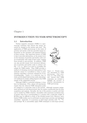
Transcription
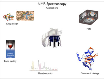
UNESCO/IUPAC Course 2005/2006Jiri BrusNMR Spectroscopy of Polymers Brus J1. partAt the very beginning the phenomenon of nuclear spin resonance wasstudied predominantly by physicists and the application was relativelylimited. However, the discovery and understanding of chemical shift,which is the difference between resonance frequency of variousstructure units, made it possible to employ this method in chemistry asan analytical tool to describe structure of small organic molecules. Withthe development of the advanced experimental procedures, andhardware of spectrometers, NMR spectroscopy become unique methodfor structural biology to describe structure and dynamics of complexbiomacromolecules. Nowadays many applications can be find inindustry (pharmaceutical industry), metabonomic research, materialengineering etc. But one of the most important current application ismagnetic resonance imaging – very powerful diagnostic method.
Approximately 13 billion years ago large amount of energy wasconverted to the matter during the process known as The Big Bang.And from these times say from the Hadron or Electroweak epoch allfundamental particles, elementary particles and composite particlesincluding protons and majority of heavier atomic nuclei posses mass,charge and spin. The internal nuclear spin was for the first timepredicted by Wolfgang Pauli in 1924 as the fourth quantum number.Although the spin, the property of many particles has not entiremacroscopic analogy in many cases it can suggest rotation. Rotationand spinning is very common motion around us. But in contrast tomacroscopic objects which can rotate around a rotation axis andgradually and fluently change their angular momentum, the energystates of spins are quantized, and all states and transitions, eigenvaluesand eigenvectors are thoroughly described only by quantum mechanics.
In principle majority of all nuclei can be considered as NMR activebecause they posses nuclear spin, however, we can easily use onlythose with nuclear spin one-half. This follows form the fact that thesenuclei can exist only at two energy states (levels), and because weobserve transitions between these levels, the resulting NMR spectrumis relatively narrow and easy to understand. The nuclei with highernumber-spin can be found at larger number of energy levels andconsequently we observe a wide range of transitions and the resultingNMR spectrum is complicated and broad.In quantum mechanics, nuclear spin can be expressed as angularmomentum which is related to nuclear magnetic moment. In the staticmagnetic field orientation of nuclear magnetic moment is not stable butit is continuously changing. This leads to the well-known precession ofspins. The spins rotate around the direction of magnetic field on a cone.Frequency of this rotation is the well-known resonant frequency. Thismeans that the frequency of the excitation filed must be exactly thesame to successfully perform NMR experiment.Spins placed into the magnet rapidly orientate into the two possibledirections (parallel and anti-parallel), and originally degenerated energylevel splits. In NMR experiment we observe transitions between theselevels. Transition is induced by a pulse of rf field with frequency close toLarmor frequency. The difference in population at the energy levelsdetermines the sensitivity of the experimental method. As you may seethe sensitivity is very low and this is the crucial problem of NMRspectroscopy.
For instance, the difference given by Boltzmann distribution in acollection of 1 million spins at the magnetic field about 2T is only 16. Ingeneral this difference increases with decreasing temperature andincreasing magnetic filed.Theoretically it is possible to cool-down the samples, however inpractice this procedure is limited by freezing points of solvents or lowsolubility, possible phase transitions etc. That is why the second wayhow to increase the sensitivity of NMR experiments is much better andthis is general way which is usually used. On the other hand this way isvery expensive.
Probably it will not be surprising that NMR spectroscopy can be alsoconsidered as a certain consequence of the research development inradar technology during the Second War. And that is why one of thefirst signals of water was observed early after the war at 1949 by FelixBloch. Approximately ten years after he and E.M. Purclell were awardedby Nobel Prize.One of the first commercial NMR spectrometer equipped byelectromagnet.
A bit better version.NMR spectrometer with a cryomagnet.
A current routine, everything is automated.Research laboratories are equipped by several huge spectrometerswith very high intensity of magnetic fields.
At the beginning of any NMR experiment there is nothing. There is nomacroscopic magnetization, there is no precession as well. However,the spins placed into the strong magnetic field became oriented andstart to rotate around the static magnetic field. The sum of all thevectors of nuclear magnetic moment produces longitudinalmagnetization. However, this magnetization cannot be detected. That iswhy the system must be perturbed. The spin systems is irradiated by avery short radio-frequency pulse. This leads to the phase coherence inthe rotation of spins around the magnetic field. The spins start to rotateat the same time from the same orientation. Consequently the htthismagnetization contains required information about the structure ofmolecules.The transverse magnetization rotates around the magnetic field with thefrequency which is close to theoretical Larmor precession frequency. Inthe detection coil a voltage is induced and subsequently recorded as socalled Free Induction Decay. This FID contains all structuralinformation, however, we are not able to understand it, we cannot readit. The clue to this problem gave Joseph Fourier in late 18 century. Hismathematic procedure, Fourier transformation, converts the timefunctions to the functions of frequency. Almost two hundred years laterthis transformation was used by Richard Ernst to convert FID in to theform of a classical frequency spectrum. In the resulting spectra it is thenpossible to resolve individual structure units in molecules. Thesespectra can be understood as unique pictures of molecular structure.The main job of NMR researchers is a conversion of these spectra to a3D model of the molecules.
Why can we resolve individual atoms in molecules?This possibility follows from the presence of electron clouds aroundeach nucleus. Electrons in magnetic field produce very weak magneticfields which can increase or decrease the intensity of static magneticfield produced by the magnet. That is why the nuclei are placed in socalled effective magnetic field and due to this fact frequency of theprecession of transverse magnetization is slightly affected. We canobserve differences in precession frequencies for different atoms. Forinstance, increasing electronegativity of a neighboring atom increasesvalue of the chemical shift. Simply speaking position of signals in NMRspectra strongly depends on the electron density around theinvestigated atoms.However, one crucial problem follows from the fact that the differencesbetween resonant frequencies of various nuclei are very small. In manycases it is necessary to resolve two signals differing in frequency byonly 0.1Hz. This is very complicated.
Nuclear spins produce very weak signals and magnetization fluxthrough the detection coil is almost comparable with the noise producedby electronic components.Nevertheless solution state NMR have become routine technique tocharacterize organic molecules. Thanks to fast isotropic moleculartumbling only isotropic values of chemical shift are detected and allmolecules in the solution are equivalent. Consequently the resultingNMR spectra are highly resolved with line-width usually below 0.1 Hz.Ideally every structure unit in the molecule is resolved andcharacterized by its own chemical shift.
Typical 1H NMR spectrum contains four basic parameters. i) Thenumber of signals - this should corresponds to the number of basicstructure units. ii) Then the signal intensity - this should reflects thenumber of equivalent hydrogen atoms in one structural group. iii) Theposition of signal on NMR scale - this is the previously mentionedchemical shift. Here we can see typical values of chemical shifts ofbasic units. iv) And finally this is the multiplicicty of signals. This is thenumber of signals in the multiplet which reflects the number of hydrogenatoms in the neighboring function groups.The situation should be the same also in polymer systems, howeverthere are some differences. Clear and well defined multiplicity rapidlydisappears and at the same time signals became broader and broaderas a result of increasing molecular weight and viscosity of solution.Narrow signals then reflect low-molecular-weight impurities. However,shorter oligomers still exhibit high-resolution pattern. Standard onedimensional experiment performed on copolymer polypeptidepolyethylene oxide produces traditional 1H NMR spectrum. One can seeresidual signal of water and signals reflecting traces of residual ether.Polyethylene oxide block is reflected by the main signal and several lowintensive signals correspond to the nonequivalent terminal units. Theseterminal units can be found also for polypeptide block. The spectrumthen provide information about the basic structure, composition andpurity of the polymer systems.
For peptides and proteins the position of signals of α-protons alsoreflects secondary structure, the conformation of the backbone. Inprinciple it is relatively easy to resolve α-helix or β-sheet. There is alarge database containing values of chemical shifts and there are tablesof these values for random coil unfolded structures. Deviation fromthese random coil range toward higher values indicate formation of βsheet conformation while deviations toward lower values ratherindicates formation of α-helix. This procedure is known as the ChemicalShifts Indexation (CSI).Unambiguous signal assignment can be achieved by two-dimensionalspectroscopy.Simply speaking, to obtain 1D spectrum one response of the sample toat least single pulse during one detection period must be detected.The acquisition of 2D spectra requires detection of the response of asample to at least two pulses during two detection periods. In practice aseries of 1D spectra with gradually prolonging t1 period is recorded. Asmagnetization evolves after the first pulse, the intensities of signalsdetected during the second period are modulated by the length of thisperiod. In fact the intensities of these signals oscillate. Consequentlythe second FT of this oscillation produces a two-dimensional spectrum.First of all this spectrum significantly enhances spectral resolution.Information originally stored in one dimension are expanded on theplane.
What can be seen in the 2D spectrum? And what is the interpretation ofthe signals?Diagonal signals directly correspond to the signals which are detectedin 1D spectrum. However, if information about the polarization state of ahydrogen atom in one structure unit is transferred to the neighboringhydrogen atom than the off-diagonal signal appears. Such informationor polarization is transferred via bonding electrons and the longestdistance is 3 or 5 chemical bonds. For instance starting from the signalwhich probably corresponds to Val methyl protons, the off-diagonalsignal correlates with the structure unit which is connected via threechemical bonds. So by this way we found out the signal of CH betahydrogen and subsequently the other correlation signal indicates alphaproton. This procedure must be repeated for all signals. And by this waycomplete assignment of all signals in the 1H NMR spectra can beperformed.In general we proceed by the method trial-and-error. At first we must atleast assign one signal for instance the CH proton with double bond.We find the correlation signal reflecting the neighboring proton number10 than proton number 9 and so on and so on. It seems to fit well withour assumption. In general we try to start from well defined signal ofstructure unit with extraordinary chemical shift. Owing to we can traceproton-proton interaction network with steps which are long only threechemical bonds. That is why we can describe primary structure ortopology of molecules. However, some times the interaction network isbroken and in many cases the resolution in this region is very poor.That is why we need additional increase in the spectral resolution.
To increase spectral resolution very often NMR spectra of other nucleiespecially of carbon 13 are measured. As you can see the signals arewell separated and every carbon atom is reflected by a single signal. Itis very good, in general it is true that with increasing number ofelectrons around a nucleus the dispersion of chemical shift increases.On the other hand this experiment is less sensitive because the isotopicabundance is only 1%During 70’s and 80’s last century, NMR researchers learned tomanipulate with the spin systems so skillfully that they could obtaindetailed information about interatomic distances, local geometry andsegmental dynamics of wide range of materials and natural products.Owing to this manipulation which is sometimes called as spingymnastics, the advanced techniques of NMR spectroscopy allow us todetermine global structure of proteins, nucleic acids and theircomplexes in solution.One of the most outstanding scientists which significantly developedthese techniques is Kurt Wüthrich which was awarded by Nobel Prize in2002. Consequently NMR spectroscopy crossed a borderline of physicsand chemistry and became inseparable part of structural biology.
At present the main research interest is focused to biologically activemacromolecules with limited solubility. At first this is concerned to themembrane peptides and proteins which control wide range of biologicalprocesses in living cells. For instance stabilization of membranepotentials, regulation of cell volume or transport of ions etc. The secondtype of the interesting systems provides amyloid proteins and Prionrelated proteins.It is very difficult to prepare suitable single crystals of theses membraneproteins or convert these membrane proteins to a solution. In this casejust only solid-state NMR spectroscopy can provide required structuralinformation.
magnetic resonance imaging - very powerful diagnost ic method. Although the spin, the property of many particles has not entire macroscopic analogy in many cases it can suggest rotation. Rotation and spinning is very common motion around us. But in contrast to macroscopic objects which can rotate around a rotation axis and .