Transcription
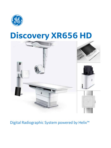
Discovery XR656 HDDigital Radiographic System powered by Helix
Table of contentsIntroduction . 1Detectors & Image Processing . 2Workflow and User Interface. 5Advanced Clinical Applications. 8Acquisition Workstation . 10Networking . 12Overhead Tube Suspension . 13Elevating Table . 14Digital Tilting Wallstand . 16X-Ray Generator . 17X-ray Source. 18Service Options . 19Accessories . 20Install and Room Considerations . 21Compliance to Standards . 22Warranty . 22
IntroductionOutstanding image quality & diagnostic confidence at low dose. Effortless precision. Powered by Helix advanced image processing.Are you looking for the best image quality at low dose from the very first X-ray? Are patient comfort, safety and userexperience top priorities in your clinical environment?Discovery XR656 HD is GE’s advanced digital radiographic system powered by Helixdesigned to help you achieve clinical excellence in X-ray, with ease and efficiency.TMadvanced image processing isOutstanding image quality at low doseDon’t miss a thing. Get the diagnostic clarity your need from the first X-ray image with: FlashPad HD high resolution, CsI, cassette size wireless digital detectors that capture extraordinaryanatomical detail at low dose with high DQE of 75% at 0 lp/mm and 100 microns, 5.0 lp/mm resolution Helix advanced image processing that delivers extraordinary anatomical detail and consistent performancein X-ray, despite variations in patient size, exposure technique, collimation, and metal implantsTMEnhance patient comfort, user experience and workflow efficiency: Auto Image Paste advanced application (optional), enhanced with Auto Spine, for seamless long boneand scoliosis exams offering outstanding precision and speed at the table and wall stand. AutoRAD workflow automation suite for fast and easy X-ray exams and effortless patient positioning,featuring Auto Positioning, Auto Tracking, Auto Protocol Assist, Auto Field-of-View, Auto QA, Quick Charge,Quick Share, and Quick Connect capabilities. Bariatric elevating table with 400 kg static and 320kg dynamic weight limit featuring a spacious table topthat lowers to below 50 cm Extensive Patient Safety features including collision and pinch sensors Redesigned console & user interface for intuitive and fast operation.Improve operational efficiency with powerful analytics and advanced service technologies that never sleepDiscovery XR656 HD is compatible with: GE’s -ray Quality Application featuring Repeat Reject Analytics. X-ray Quality App provides web-baseddashboards to manage quality assurance, uncover the root cause of rejected X-ray exams, plan targetedtraining and help reduce unnecessary radiation dose iCenter asset management software platform designed to help you optimize utilization of your X-rayequipment and balance workload using the full power of healthcare data analytics InSite remote service support. Remote diagnostics and troubleshooting for fast resolutions, often withouta field engineer visit.Discovery XR656 HD Available ConfigurationsThe Discovery XR656 HD is designed to handle standard 2D exams and advanced radiographic applications using GE’swireless flat panel digital detector. Various system configurations are available to align to your department’sradiographic requirements.All systems include: at least a single FlashPad HD detector, Helix advanced image processing, table and/or wall stand,systems cabinet, ceiling mounted tube support, acquisition review workstation, short-term storage, and quick in-roomviewing of images.Discovery XR656 HD datasheet1
Main Four ConfigurationsWallstand only system (standard or extended arm)One GE FlashPad HD wireless or tether detectorTable and wallstand (standard or extended arm)One GE FlashPad HD wireless or tether detectorTable and wallstand (standard or extended arm)Two GE FlashPad HD wireless or tether detectorsOTS only system (without Table and Wallstand)One GE FlashPad HD wireless or tether detectorA full range of accessories is also available, such as: mobile stretchers, weight bearing exam tools, patient tableaccessories and grids.Detectors & Image ProcessingDigital image acquisition supports fast and efficient exam procedures, eliminating time spent handling film andcassettes, as well as reliability issues inherent in cassette tray systems, thus helping to reduce overall exam times andimprove patient satisfaction.At the core of Discovery XR656 HD is GE’s high resolution FlashPad HD wireless flat panel detectors and Helix advanced image processing that provide outstanding image quality at low dose for general radiography applications.2Discovery XR656 HD datasheet
FlashPad HD: GE’s High-Resolution Flat-Panel Wireless Digital DetectorFlashPad HD detectors are ready when you are thanks to multiple features that enhance workflow:Quick ShareHassle-free management of multiple wireless detectors. Once registered, QuickShare allows the detector to workacross multiple compatible GE systems with no additional configuration required. Pairing enables registered detectorsto connect wirelessly to the host system within seconds.Quick ChargeIn-bin charging, the FlashPad HD detectors charge when they are in the table or wall stand housing. This gives youpeace of mind knowing that the detectors will be ready when you need them.Quick ConnectAdaptive wireless technology enables automatic channel switching to improve image transfer and avoid wirelessinterference with other equipment on the hospital networkAuto QAUsing the integrated system Quality Assurance Procedure (QAP), image quality checks can be easily performed by thecustomer. The QAP includes a phantom, optimized for Digital Image Quality testing and is included with the system.System changes are highlighted and can be corrected before they become a problem.FlashPad Wireless DetectorDetector technologyHD 3543 (14”x17”)Single panel (non-tiled) amorphoussilicon with a Cesium Iodide (CSI)scintillatorHD 2530 (10”x12”)Single panel (non-tiled) amorphoussilicon with a Cesium Iodide (CSI)scintillatorDimensionsISO 4090 cassette sizeISO 4090 cassette size384 x 460 x 15.5 mm282 x 332 x 15.5 mmPixel Matrix3524 x 42882508 x 3004Image Depth16 Bit16 BitPixel size100 µm100 µmLimiting Resolution5.0 lp/mm5.0 lp/mmTypical Dynamic Range6 uR –9 mR @ RQA56 uR – 9 mR @ RQA5Discovery XR656 HD datasheet3
Typical DQE (RQA5)75% (@ 0lp/mm)75% (@ 0lp/mm)60% (@ 1lp/mm)60% (@ 1lp/mm)40% (@ 3p/mm)40% (@ 3p/mm)70% (@ 1lp/mm)70% (@ 1lp/mm)40% (@ 2lp/mm)40% (@ 2lp/mm)15% (@ 4p/mm)15% (@ 4p/mm)Wireless IEEE 802.11n, 5GhzWireless IEEE 802.11n, 5GhzEthernet 1000MbpsTether 100MbpsAvailable options includeWireless, Docked, TetheredWireless, TetheredMax. Load capacity150 kg distributed weight,150 kg distributed weight,100 kg concentrated weight (45mmdiameter area)100 kg concentrated weight (45mmdiameter area)Weight (with Battery)3.2 kg (7 lbs)1.8 kg (4 lbs)Ingress ProtectionIPX4 (Protection against splashingwater)IPX4 (Protection against splashingwater)Battery Indicator andChargerDetector Battery FullCharging timeBattery Operation TimeYesYes2.5 hours2.5 hours4 hours (@ 60 exposures/hour)2 hours (@ 60 exposures/hour)Clip-on Grid (Optional)70 lines/cm,8:1 ratio, focus 130 cm(universal), SID range 90 cm–190 cm70 lines/cm,8:1 ratio, focus 130 cm(universal), SID range 90 cm–190 cm70 lines/cm, 6:1 ratio, focus 130 cm(universal) SID range 85 cm – 190 cm70 lines/cm, 6:1 ratio, focus 130 cm(universal) SID range 85 cm – 190 cmTypical MTF (RQA5)Communication interfaceHelix Advanced Image ProcessingHelix advanced image processing algorithms harness the full high-resolution power of FlashPad HD detectors todeliver exceptional image quality despite challenging exams conditions.Helix algorithms are designed to deliver outstanding resolution, excellent edge presentation, consistency, and noisehandling. The algorithms incorporate the following capabilities:Image processingAuto ShutteringAutomatically detects collimator edges and adjusts to the selected field of view: Raw RadiationRejectionGrid Line Reduction4ACED (Automatic Collimator Edge Detection) –providing masking of the image.ICED (Intelligent Collimator Edge Detection) An intelligent algorithm that reliessolely on image information to locate collimation edges present in an x-ray image.Identify raw radiation pixels and improve post processing image display.Suppresses grid lines on the image without impacting anatomical details.Advanced NoiseReductionSuppresses the mottle noise in denser areas of the anatomy while preserving detail inthe rest of the image. The algorithm takes account tissue penetration and dosereaching the receptor.Multi-resolutionprocessingImproved edge presentation, exquisite bone detail, differentiation of soft tissues andvisualization of metal.Discovery XR656 HD datasheet
Tissue EqualizationEnhance contrast in thick and thin regions of the anatomy is used to correct overpenetrated and under-penetrated areas within the image.Smart WindowingDelivers the correct display brightness and contrast without needing window level andwidth adjustments, Provides consistent brightness and contrast across variations inexposure technique.Multiplecustomizable looks4 Factory (GE pre-set) image processing selections (looks) optimized for eachanatomical view with the ability to define multiple5 Custom looks for each anatomical view/patient size combination.Workflow and User InterfaceWorkflow FeaturesWorklistCustomized Worklist column customizeWorklist auto-refreshEmergency patient feature – allows user to open exam and acquire images without aworklist entryPatient edit/auto-folderingBar code reader (Optimal) for patient data entry – can be used for patient selectionfrom the work listProtocols selectionSet of default adult and pediatric protocols allows quick selection of the appropriatetechniques for common procedures/exams with the ability to define unlimited numberof custom protocols.Imagemanagement“Patient Directory” provides fast access to the image and exam database for casereviews and file managementCopy exam – allows user to copy patient images into a second patient entry patiententryRead/Write (write once, multiple access) CD/DVD-ROM to be used as an imageexchange medium. Images are written to an archive CD/ DVD along with a DICOMviewer.Image Display andpreviewWindow width and levelGray scale invertInterpolated zoom with roamImage flips (horizontal, vertical) with automatic indicatorImage Rotate – 90 incrementsFree rotation – 360 Image orientation managementL/R markersFree text annotationManual shutteringAnnotation &MeasurementFull range of measurements toolsVOI LUT burn on sendSystem information annotations with configurable font size and display on/off:Discovery XR656 HD datasheet5
–Hospital/Institution Name–Date, Time (hh:mm:ss) of Acquisition–Measurements (when activated)–Contrast, Brightness Values (WW/WL annotations)–Processing Look–Anatomical View–Exposure Technique including kVp, mA, mAs, and time–Estimated Exposure Dose (Dose area product (DAP)) read out in dGy-cm2 units–Operator Entered Annotations–Patient ID, Patient Name–Patient Age and DOB (date of birth)–Edge annotationsImage reprocessingImage reprocessing based on anatomy, patient size and viewOff center imaging with automatic cropping and manual shuttering capabilitiesImage transferCompatible with DICOM 3.0 and IHE, the image can be tagged and automatically sent toPACS for storage and to printer for print. This capability is further increasing examthroughput and decreasing examination times for patients.PrintOrthopedic Magnification/PrintMulti-format printing – 1x1, 2x1, 1x2 and 2x2Print Preview functionAuto portrait and landscape printCustomizable Quick Tools Customizable menu with quick access tools Adapt your Quick Tools bar to include your most frequent operations for enhanced workflowAutomatic Reprocess Button for Manual ShutterA Quick Tool function to manually shutter and reprocess image in one click when needed. Quick Reprocess Function6 Easily preview all image processing looks on one screen Select, compare & apply desired look with an intuitive workflowDiscovery XR656 HD datasheet
Quick EnhanceA one-touch processing function that can reprocess images for a different custom look with no additional doseto the patient and no additional clicks and steps for the user.QuickEnhance is customizable by anatomy for multiple uses including instrument check, implant visualization, lineplacement.Discovery XR656 HD datasheet7
Detector Exposure Indicator Detector Exposure Indicator (DEI) is a tool for tracking patient over/under-exposure by estimating radiation exposurebehind the patient and is a relative measure of exposure to the detector Exposure index (EI) is proportional to detector exposure assuming that the x-ray technique used is the same as that ofthe calibration technique. Deviation index (DI) estimates the deviation of actual detector exposure from target detector exposure.Repeat/Reject Analysis (optional) An automated quality assurance tool that allows for repeat or reject images to be captured and categorized bytechnologistReports can be exported in DVD, CD or USB format for ease of use Discovery XR656 HD is also compatible with GE’s Xray Quality Application featuring Repeat Reject Analytics. X-ray Quality Application is a web-based solution that canconnect to multiple compatible radiography systems and help you identify root causes of rejects, enhance training,drive efficiency and help reduce dose.Auto Positioning Protocol LinkAuto-Positioning protocol link enables pre-determined receptor and corresponding auto-positioning protocol per view.Auto Field of ViewAuto Field of View enables the user to pre-define the collimation size on an individual view basis.Advanced Clinical ApplicationsHelix advanced image processing and the FlashPad HD detector with high DQE, low noise and high detectoracquisition speed allows the Discovery XR656 HD to offer the following advanced clinical application including AutoImage Paste:8Discovery XR656 HD datasheet
Auto ImagePaste on tableAuto Image Pasteat Wall StandAutoSpineAuto Image Paste has been enhanced with AutoSpine, an intelligent algorithm that follows the contour of the spine forvertical equalization enabling a natural balance of brightness & contrast along the patient body in lateral spine exams.Discovery XR656 HD datasheet9
Wallstand Auto Image Paste (optional) – Spine and Long Bone Imaging Fully automated acquisition and processing of a series of images with user defined start and stop locations on theanatomical regions of interest Image pasting and processing time for a 3-image exam is 22 seconds from first exposure. Average acquisition time for a 3-image exam (90cm coverage) is 10 seconds Allows 2 to 5 images to be pasted together with a maximum range of 150 cm Includes imaging of the spine for scoliosis evaluation and imaging of the legs for orthopedic evaluations Supports anatomies/view combinations of Spine Antero-posterior, Spine Postero-anterior, Spine Lateral, Leg Antero-posterior, Leg Postero-anterior Includes a patient stand with screen help to keep the patient comfortable during acquisitionTable Auto Image Paste (optional) – Spine and Long Bone Imaging Fully automated acquisition and processing of a series of images with user defined start and stop locations on theanatomical regions of interest Image pasting and processing time for a 3-image exam is 22 seconds from first exposure Average acquisition time for a 3-image exam (90cm coverage) is 10 seconds Allows 2 to 4 images to be pasted together with a maximum range of 110 cm Includes imaging of the spine for scoliosis evaluation and imaging of the legs for orthopedic evaluations Supports anatomies/view combinations of Spine Anteroposterior, Spine Postero-anterior, Spine Lateral, LegAnteroposteriorAcquisition WorkstationThe Acquisition Workstation is the primary interface to the network and provides image post-processing capabilities. The SystemController Module provides single point control, directing and coordinating overall system operation, while monitoring all systemmodules automatically through software.10Discovery XR656 HD datasheet
Workstation SpecificationsMonitorNon-touch24 in (61 cm) LCD Color Monitors (1920 x 1200 pixels) 250 cd/m2 calibrated brightnessCPUIntel Xeon processor, 3.5 GHz, 4 coresHard Disk Storage1 TB Enterprise-Class SATAImage Storage 17000 imagesProgrammable auto delete functionRAM16 GBImage processing timesFast preview images: 1 second (docked) or 2 seconds (wireless or Tether)Final Conditioned Image including Auto-Shuttering: 6 seconds (docked) or 8 seconds (wireless or tether)Expose to expose cycle time 5 seconds @ 70% HUTime to boot the systemafter normal shutdownSystem reset time 180 secondsImage Pasting Acquisitiontime 22 seconds (3 images) 230 secondsDiscovery XR656 HD datasheet11
NetworkingIHE Compliance for Scheduled Workflow Integration Profile. Images may be transmitted manually or automaticallythrough the DICOM interface to printers, archival devices, servers, or review workstations. System Access andAuthorization Control to support HIPAA CompliancePlease refer to the DICOM Conformance Statement for complete definition of supported DICOM connectivity services.DICOM 3.0 ServicesDICOM Modality Worklist(SCU)Interface with HIS/RIS with programmable auto refreshDICOM MPPS (SCU)Feedback the status of exams to the HIS/RISDICOM Storage (SCU)Manual and auto send image (DX or CR IOD) to multiple PACSDICOM Storage commitment(SCU)Send commitment state.DICOM Query/Retrieve (SCU)Query/Retrieve images from PACSDICOM Query/Retrieve (SCP)Provide Query/Retrieve service instance to other systemDICOM Media ExchangeCD/DVD DICOM image export and import.DICOM Grayscale PrintManual and Auto print with print layout options at the consoleVerification servicesC-Echo as SCU and SCPDICOM Dose StructureReport (Optional)Send Dose values for each study to an archiving system.IHE Integration ProfilesScheduled workflowAcquisition Modality: Patient Based Worklist Query/Broad Worklist QueryPatient InformationReconciliationAcquisition Modality:REM (Optional)Radiation Exposure Monitoring for Dose structure report Minimum Printer Requirements:– 10 and 12-bit printers– Printed images are not intended for diagnostic use unless produced with a printer capable of at least 1,000gradations of gray scale (or at least 10 bits)– Several popular printers have been validated for connectivity and image quality. Recommendations are availablefrom your sales representative.– Non-DICOM laser cameras will require an upgrade to DICOM connectivity GE Healthnet Services can provide physical network connectivity solutions – Layer 1 and 2 Ethernet (IEEE 802.3)interoperability – and include network components and physical installation TVA (Tip Virtual Assist) is a tool that realizes remote desktop sharing; it can support remote application training.(Option.)Auto Protocol Assist (optional)System will automatically transition directly to the Acquire screen when the protocol code downloaded from the HIS/RIS (automatically performed with worklist refresh) matches the exam code contained in the protocol database. Thistool eliminates the user steps required to select patient exam types and initiate an exam.12Discovery XR656 HD datasheet
Overhead Tube SuspensionThe Overhead Tube Suspension (OTS) system with motorized movement delivers excellent levels of operationalsupport designed for efficient operation and precise positioning.Overhead TubeSuspensionMotion controlManual and Auto positioning.5 axis servo motion. -longitudinal, lateral, vertical, tube angulation and columnrotation.Override modeAllowing the user to assume complete control for complex or emergencypositioning. Two-position key switch remains in NORMAL for automaticfunctioning. OVERRIDE for manual control.Auto-DetentsAssisting the user with locating and securing detentsAuto TrackingAutomated vertical tracking to align with Wallstand detector.Automated vertical tracking to maintain Table SID.Longitudinal travelrange2.3 m (for a 3.4 m rail)Lateral travel range1m (for 2m bridge)4.7m (for a 5.8 m rail)2m (for 3m bridge)3.3m (for 4m bridge)Vertical travel range180cm. Dual cable safety system and high precision telescoping column.Discovery XR656 HD datasheet13
Tube angulation range 135/-180 degrees. Continuous rotation with locking at any position. Fix detentsat 0, 90, and -90 when selected.Column rotation range /-135 degrees. Continuous rotation with locking at any position.Longitudinal SpeedUp to max. 30 cm/sLateral speedUp to max. 25 cm/sVertical speedUp to max. 25 cm/sTube angulation speedUp to max. 25 degree/sColumn rotation speedUp to max. 25 degree/sAuto Field of View (FOV)selections7 Default FOV-43 cm x 43 cm (17 in x 17 in)-35 cm x 43 cm (14 in x 17 in) / 43 cm x 35 cm (17 in x 14 in)-24 cm x 30 cm (9 in x 12 in) / 30 cm x 24 cm (12 in x 9 in)-18 cm x 24 cm (7 in x 9 in) / 24 cm x 18 cm (9 in x 7 in)OTS User InterfaceLCD Touch Screen, Auto Horizontal and Vertical UI display for Table andWallstand application. Providing the following functions to the user:-Lock, Detent Control-Technique Adjust (kVp, mAs)-Receptor Selection (table, wallstand, table top or cassette)-Collimator FOV Selection-Exam Inhibit Display-Position Display (SID, Tube Angle, Column Rotation, Detector Orientation)-Display of Patient Name and Date of Birth for In-Room VerificationRemote ControlInfrared Remote controller (Optional). Auto positioning and FOV Selection Auto-Positioning Package (included in base)– Auto-Positioning enables the users to select a predefined system position from the system console andautomatically move the equipment by simply holding the “Auto Positioning” buttons. This feature is designed to helpreduce user fatigue and increase the productivity of the operator.– Auto-Positioning will incorporate angulation of the tube, longitudinal, lateral, rotational and vertical positioning ofOTS, table detector longitudinal positioning, wallstand detector vertical and tilting positioning– Auto-Positioning is controlled at the acquisition workstation or with the IR remote control, allowing the user toremain in the room while moving the system– Auto-Positioning comes with more than 9 default positions. The user can create additional defined positions asneeded.– Pre-set positions at the table, wallstand and park position at various SIDs and vertical and horizontal orientations– Simultaneous movement of the OTS might be limited in some situations due to safety considerations during auto-positioningElevating TableA bariatric X-ray table enables you to serve the X-ray imaging needs of patients of all sizes and mobility levels.14Discovery XR656 HD datasheet
Elevating TableMotion controlMotor driven elevatingManual 8-way floating tabletopServo motion -Detector longitudinal.Motion safetyDouble-Press Safety Foot PedalTwo safety switches to disable motion during patient transferTwo emergency stop buttonsTable anti-collision safety deviceAuto TrackingYes. Automated Detector longitudinal tracking to align with Tube (includingtube longitudinal and angulation)Tabletop materialCarbon Fiber compositeTabletop Inherent filtration 0.7 mm Al equivalent @ 100 kVpTabletop size93 mm x 240 cm (37 in x 94 in)Tabletop longitudinaltravel range68 cmTabletop transversal travelrange28 cmDetector longitudinaltravel range80 cmMax. Patient CoverageLongitudinal 188 cm (74 in)Lateral 68 cm (26.8 in)Elevating range50 cm – 85 cm (19.7 in –33.5 in)Elevation from minimum tomaximum height 18 secondsMax. patient weight400 kg (882 lbs) static non-elevating320 kg (705 lbs.) dynamic elevating load centerFoot pedalLiquid proof, Degrees of protection is IP36High reliability wit parallel switch redundant designDiscovery XR656 HD datasheet15
Front foot pedal as defaultRear foot pedal as optionalFootprint100 cm x 71 cm (39.4 in x 28 in) including foots pedalsDetector housingSupport portrait and landscape exposure for FlashPad HD 35x43GridRemovable Grid with handle provided-70 lines/cm,12:1 ratio, focus 100 cm (40 in), SID range 90 cm–120 cm-70 lines/cm, 13:1 ratio, focus 120 cm (48 in) SID range 102 cm – 146 cm-70 lines/cm, 12:1 ratio, focus 110 cm (43 in) SID range of 95 cm–130 cm(Optional)AEC support3 Cell Ion chamber.Table AccessoriesTabletop digital detector holderPatient Hand GripsCompression BandDigital Tilting WallstandDigital tilting Wallstand is designed for use with the wireless digital detector, ion chamber and removablenonreciprocating gridDigital Tilting WallstandMotion controlMotorized detector housing vertical and tilting movement.Manual detector housing vertical and tilting movement compatible.Motion safetyElectromagnetic braking secures vertical motion.Anti-collision safety device for titling16Auto TrackingAuto-tracking detector from the OTSFoot switchVertical motion control.Remote ControlInfrared Remote controller (Optional). Vertical and tilting motion control.Discovery XR656 HD datasheet
Information Display PanelIndicates the Exposure hold, Grid Type, Tilting degree, and System readystatusMin. height from floor tocenter of detector28.5 cmVertical travel range150 cmMax. Patient CoverageVertical 192 cm (75.6 in)Titling range-20 to 90 -20 , 0 , 90 detent positionDetector housingSupport portrait and landscape exposure for FlashPad HD 35x43GridRemovable Grid with handle provided-70 lines/cm,13:1 ratio, focus 100 cm (40 in) SID range 90 cm–118 cm-70 lines/cm, 13:1 ratio, focus 120 cm (48 in) SID range 102 cm – 146 cm-70 lines/cm, 13:1 ratio, focus 180 cm (72 in) SID range 145 cm – 245 cm-70 lines/cm, 10:1 ratio, focus 130 cm (universal) SID range 90 cm–190 cm(Optional)-70 lines/cm, 10:1 ratio, focus 130 cm (universal) SID range 90 cm–190 cm.Horizontal orientation for stretcher, cross table and angulated exams(Optional)AEC support3 Cell Ion chamber for standard Wallstand4 Cell Ion chamber for extended arm WallstandThe standard right, left and center ion chambers are available when thewallstand is in the vertical position. The addition of the 4th ion chamberprovides for supine chest and abdominal imaging when a stretcher is used(any time the wallstand housing is horizontal).Extended Arm Option34 cm (13.4 in) longer than standard wallstand to accommodate stretcherworkAccessoriesIntegrated hand grips and lateral support bar for patient comfort andstabilizationX-Ray GeneratorThe digital radiographic imaging system is available with a high frequency generator.X-ray GeneratorNominal Output50 kW, 65kW or 80 kWTube voltage range40 to 150 kVTube current range10 to 630 mA for 50 kW system,10 to 800 mA for 65 kW system10 to 1000 mA
Discovery XR656 HD datasheet 5 Tissue Equalization Enhance contrast in thick and thin regions of the anatomy is used to correct over- penetrated and under-penetrated areas within the image. Smart Windowing Delivers the correct display brightness and contrast without needing window level and width adjustments, Provides consistent brightness and contrast across variations in