Transcription
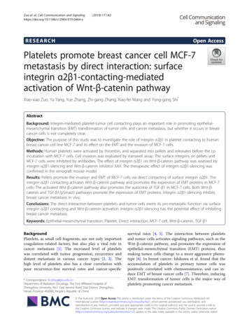
Zuo et al. Cell Communication and (2019) 17:142RESEARCHOpen AccessPlatelets promote breast cancer cell MCF-7metastasis by direct interaction: surfaceintegrin α2β1-contacting-mediatedactivation of Wnt-β-catenin pathwayXiao-xiao Zuo, Ya Yang, Yue Zhang, Zhi-gang Zhang, Xiao-fei Wang and Yong-gang Shi*AbstractBackground: Integrin-mediated platelet-tumor cell contacting plays an important role in promoting epithelialmesenchymal transition (EMT) transformation of tumor cells and cancer metastasis, but whether it occurs in breastcancer cells is not completely clear.Objective: The purpose of this study was to investigate the role of integrin α2β1 in platelet contacting to humanbreast cancer cell line MCF-7 and its effect on the EMT and the invasion of MCF-7 cells.Methods: Human platelets were activated by thrombin, and separated into pellets and releasates before the coincubation with MCF-7 cells. Cell invasion was evaluated by transwell assay. The surface integrins on pellets andMCF-7 cells were inhibited by antibodies. The effect of integrin α2β1 on Wnt-β-catenin pathway was assessed byintegrin α2β1-silencing and Wnt-β-catenin inhibitor XAV. The therapeutic effect of integrin α2β1-silencing wasconfirmed in the xenograft mouse model.Results: Pellets promote the invasion and EMT of MCF-7 cells via direct contacting of surface integrin α2β1. Theintegrin α2β1 contacting activates Wnt-β-catenin pathway and promotes the expression of EMT proteins in MCF-7cells. The activated Wnt-β-catenin pathway also promotes the autocrine of TGF-β1 in MCF-7 cells. Both Wnt-βcatenin and TGF-β1/pSmad3 pathways promote the expression of EMT proteins. Integrin α2β1-silencing inhibitsbreast cancer metastasis in vivo.Conclusions: The direct interaction between platelets and tumor cells exerts its pro-metastatic function via surfaceintegrin α2β1 contacting and Wnt-β-catenin activation. Integrin α2β1-silencing has the potential effect of inhibitingbreast cancer metastasis.Keywords: Epithelial-mesenchymal transition, Platelet, Direct interaction, MCF-7 cell, Wnt-β-catenin, TGF-β1BackgroundPlatelets, as small cell fragments, are not only importantcoagulation-related factors, but also play a vital role incancer metastasis [1]. The increased level of plateletswas correlated with tumor progression, recurrence anddistant metastasis in various cancer types [2, 3]. Thehigh level of platelets also has a close correlation withpoor recurrence-free survival rates and cancer-specific* Correspondence: fccshiyg@zzu.edu.cnDepartment of Radiation Oncology, The First Affiliated Hospital ofZhengzhou University, No.1 East Jianshe Road, Erqi District, Zhengzhou,Henan Province 450000, People’s Republic of Chinasurvival rates [4, 5]. The interaction between plateletsand tumor cells activates signaling pathways, such as theWnt-β-catenin pathway, and promotes the expression ofepithelial-mesenchymal transition (EMT) proteins, thusmaking tumor cells change to a more aggressive phenotype [6]. In breast cancer, Ishikawa et al. found that theaccumulation of platelets in primary tumor cells waspositively correlated with chemoresistance, and can induce EMT of breast cancer cells [7]. Therefore, inducingEMT transformation of tumor cells is the major way ofplatelets promoting cancer metastasis. The Author(s). 2019 Open Access This article is distributed under the terms of the Creative Commons Attribution 4.0International License (http://creativecommons.org/licenses/by/4.0/), which permits unrestricted use, distribution, andreproduction in any medium, provided you give appropriate credit to the original author(s) and the source, provide a link tothe Creative Commons license, and indicate if changes were made. The Creative Commons Public Domain Dedication o/1.0/) applies to the data made available in this article, unless otherwise stated.
Zuo et al. Cell Communication and Signaling(2019) 17:142Previous study mainly investigated the indirect interactions between platelets and tumor cells——secretingreleasates (mainly α-granules) of platelets to promoteEMT of tumor cells [8]. It has been reported thatplatelet-derived α-granules containing transforminggrowth factor-β1 (TGF-β1) increased the growth of primary tumors in murine models of ovarian cancer [9, 10].However, in recent years, the effect of direct interactionof platelet and tumor cells on promoting EMT has beenattracting attentions in the field of cancer treatment.Pang et al. demonstrated that the direct interaction between tumor cells and platelet fractions, named aspellets (with the activation marker of P-selectin, butwithout releasates [11]), promotes the EMT and extracellular matrix (ECM) degradation, inducing a more aggressive phenotype in tumor cells [12]. Labelle et al.demonstrated that direct platelet-tumor cell contactingand platelet-derived TGF-β1 synergistically activate theTGF-β1/Smad pathway in cancer cells, resulting in theirEMT transformation and enhanced metastasis [13].Whereas, the mechanism underlying the direct interaction between platelets and breast cancer cells is largelyunknown.Integrins mediate cell-to-cell contacting, and are keymediators in delivering signals between cells. They werediscovered by Hynes RO and his colleagues [14, 15].Also, integrins are suggested to be involved in the tumorgrowth and progression [16], and were regarded as atherapeutic target for tumors. As reported, the combinedblockade of both tumor integrin αvβ3 and platelet integrin αIIbβ3 inhibited platelet-cell contacting and suppressed angiogenesis and tumor growth of melanoma[17]. α2β1, a kind of integrin, has been shown to exist inthe surface of breast cancer cells, and is believed to mediate the direct interaction between tumor cells andplatelets [18]. However, how integrin α2β1 mediates theEMT by platelet-breast cancer cell contacting remainsunknown.Herein, this current study defined the role of integrinα2β1 in mediating the direct interaction between platelets and breast cancer cells, and explored its effect onthe promotion of EMT by controlling the correlated signaling molecules, thus implying the important role ofdirect contacting of platelets and breast cancer cells inpromoting breast cancer metastasis.Materials and methodsPreparation of platelets and platelet fractionsPlatelets and platelet fractions used in this study werecollected from human blood as previously described[13]. Briefly, healthy human blood was centrifuged at250G for 10 min. The plasma and buffy coat were gentlytransferred to a fresh tube, and the centrifugation wasrepeated at 2000G for 10 min. The platelet-rich plasmaPage 2 of 15in the bottom was then washed with the platelet washingsolution for 3 times. To prepare platelet fractions,platelets were stimulated by thrombin 0.5 U/ml for 15min at 37 C. The pellets (platelet membranes) were separated from the releasates (supernatant containing activecomponents) by centrifugation at 2800G for 7 min. Todetermine the extent of contamination of the thrombinactivate pellets with soluble platelet proteins, plateletfactor 4 (PF4) was measured using an ELISA assay(Abcam, Cambridge, UK) and compared with the PF4levels in the releasates and whole platelets solubilised bythe addition of 0.1% Triton-X 100 [12].P-selectin exposure analysisA minimum of 10,000 gated events was analyzed on aFACScalibur flow cytometer (BD Biosciences, San Jose,CA, US). The anti-CD41 conjugated with fluorescein(FITC, BD Pharmingen, San Jose, CA) and the antiCD62P conjugated with phycoerythrin (PE, BD Pharmingen) were used as antibodies. Then they were added tothe activated platelets with a final dilution of 1:20 (v/v).FITC and PE-conjugated isotype-matched antibodieswere used to control nonspecific labeling of antibodies.The samples were fixed with 0.4% paraformaldehyde andanalyzed by flow cytometry after antibody incubation for15 min at 37 C.Transwell invasion assayHuman breast cancer cell lines, MCF-7, MDA-MB-231,and SK-BR-3 (all from American Type Culture Collection, ATCC, Manassas, VA, US), were maintained at37 C, 5% CO2, grown in Dulbecco’s Modified Eagle’smedium (DMEM; ThermoFisher, Pittsburgh, PA, US)containing 1% penicillin-streptomycin (Gibco, NY, USA)and 10% fetal bovine serum (FBS; ThermoFisher) at pH7.2 overnight. The invasion assay was performed in 24well BD Biocoat Matrigel Invasion Chambers (8 μmpore size; BD Biosciences) [13]. Total 5 104 MCF-7cells were plated in transwell inserts. Then the DMEMbuffer (the buffer group), thrombin-activated platelets(the platelet group), releasates (the releasate group), pellets (the pellet group), and the supernatant of the coincubation of pellets with MCF-7 cells for 48 h [thepellet MCF-7 conditioned medium (CM) group] wereadded. Both upper and lower chambers containedDMEM. After 48 h, cells remaining in the upper part ofthe transwell were removed with a cotton swab. Migrated cells were then stained with Crystal Violet 0.5%and the total number of cells was counted with a ZeissAxiovert 200 microscope (Zaventem, Belgium). The invasion assay was also performed in MDA-MB-231 cellsand SK-BR-3 cells after adding platelets.
Zuo et al. Cell Communication and Signaling(2019) 17:142Page 3 of 15Direct interaction between tumor cells and platelets orplatelet fractionsTable 1 Primers and promoter sequences used in qRT-PCR andChIPThe medium of MCF-7 cells was changed for freshDMEM immediately prior to the treatment. Then theDMEM buffer (the buffer group), thrombin-activatedplatelets (the platelet group), releasates (the releasategroup), pellets (the pellet group), and the supernatant ofthe co-incubation of pellets with MCF-7 cells for 48 h(the pellet MCF-7 CM group) were added, and incubated for 40 h.qRT-PCR Primers (from 5′ to 3′)SnailR: CGGTGGGGTTGAGGATCTSlugE-cadherinTotal protein was extracted from the tumor cell line ortumor tissues using RIPA lysis buffer. The concentration oftotal protein was quantified using a BCA kit (ThermoFisher). The protein samples (20 μg/sample) were separatedby SDS-PAGE and then transferred to poly-vinylidenedifluoride (PVDF) membranes (Merck Millipore, Danvers,MA, US). After blocking with 5% fat-free milk in Trisbuffered saline containing 0.1% Tween 20 (TBST) for 1 h,the proteins were immunoblotted with primary antibodiesat 4 C overnight. After washing with TBST three times, theproteins were incubated with horseradish peroxidaseconjugated secondary antibodies for 1 h at roomtemperature. After washing with TBST three times, proteinsignals were determined using a substrate chemiluminescence detection system (ThermoFisher) with the Image LabSoftware (Bio-Rad, Hercules, CA, US). The primary antibodies used were anti-β-catenin (1:5000, Rabbit monoclonal antibody, Abcam, ab32572), anti-integrin α2 (1:10000,Rabbit monoclonal antibody, Abcam, ab133557), anti-βactin (1:500, Mouse monoclonal antibody, Abcam, ab8226),anti-Lamin B1 (0.1 μg/ml, Rabbit polyclonal antibody,Abcam, ab16048), and anti-pSmad3 (1:2000, Rabbit monoclonal antibody, Abcam, ab52903).Analysis of tumor cell contacting to platelets or pelletsThe platelet adhesion assay was performed to evaluatethe contacting between platelets/pellets and tumor cellsas previously described [18]. Briefly, 5 104 cells/mlbreast cancer cells were seeded in a 96-well plate.Washed thrombin-activated platelets or pellets labeledF: CGACCCAACCCAAGAATCTAR: AGGCTGTGCCTTCCTACAGAFibronectinWestern blottingF: AGATGCATATTCGGACCCACR: CCTCATGTTTGTGCAGGAGAQuantitative real-time PCR (qRT-PCR)Total RNAs were extracted from the tumor cell line ortumor tissues using isolation kit according to the manufacturer’s protocol. The cDNA was generated fromRNAs using a cDNA Reverse Transcription Kit (AppliedBiosystems, ThermoFisher). The expression of RNAswas analyzed by qRT-PCR using Power SYBR GreenPCR Master Mix (Applied Biosystems, ThermoFisher)with GAPDH as endogenous controls. Relative expression levels of all genes were calculated as 2–ΔΔCt. Theprimers used were shown in Table 1.F: CTTCCAGCAGCCCTACGACF: CCATCGCAAACCGCTGCCATR: AACACTTCTCAGCTATGGGCTTCOL1A1F: TAACTTCTGGACTATTTGCGGACTTTTTGGMMP9F: CCTGGAGACCTGAGAACCAATCtgfb1F: CGTGGAGCTGTACCAGAAATAGAPDHF: TGTTCGACAGTCAGCCGCR: GGGCGAGGGAGGAGAGAAR: GATTTCGACTCTCCACGCATCR: TCCGGTGACATCAAAAGATAAR: GGTGTCTGAGCGATGTGGCChIP Primers (from 5′ to 3′)Snail promoterF: CACTTCCTCTGGGAAGTCACCR: CCTTCCCTTATCCAGTGTTTACGGAGSlug promoterF: CTGCACCACATCTGGAAGCCAGR: CCAATCACAGCTGAGAGGTTCAGtgfb1 promoterF: GCAACTTCGACCGCTACGGR: CTGCGACCCCATACATTTACTGGAPDH promoterF: AGCTCAGGCCTCAAGACCTTR: AAGAAGATGCGGCTGACTGTwith fluorescein (FITC, BD Pharmingen) were added tothe cells for 30 min at 37 C. The non-adherent plateletsor pellets were discarded. Then the flow cytometry wasused to perform the platelet/pellet adhesion assay.Antibody inhibition of pellet or tumor cell membraneThe antibody inhibition of pellet and tumor cell membrane was performed as previously described [12]. Inbrief, the 5 108 CFDA-SE-conjugated thrombinactivated pellet were pre-incubated with 50μg/ml anti-Pselectin, anti-AK7 (integrin α2β1), or anti-CRC64 (integrin αIIbβ3) on ice for 30 min. Then they were added to3 105 MCF-7 cells in 100 μl of DMEM in a 96-wellplate and incubated at 37 C for 30 min. To prevent further binding before flow cytometry, 100 μl /well ice cold0.1% BSA/PBS was added and the flow cytometry wasperformed immediately. The antibody inhibition ofMCF-7 cell membrane was also performed as describedabove, and the antibodies used were anti-AK7 and anti-
Zuo et al. Cell Communication and Signaling(2019) 17:142CRC64. The anti-AK7 was used to inhibit the cell surface integrin α2β1 of MDA-MB-231 cells and SK-BR-3cells.Page 4 of 150.1% Triton X-100 and once in PBS alone. Then thebeads were suspended in SDS-PAGE sample buffer,heated to 95 C for 5 min, and analyzed by westernblotting.Integrin α2β1-silencing MCF-7 cell lineThe shRNA specific for the integrin α2β1 subunit(Sigma, St. Louis, MO, US) was cloned to the lentiviralplasmid vector pLKO.1-puro, and the pLKO.1-purolentiviral vector without shRNA (empty vector) was usedas the control [19]. Lentiviruses were produced inHEK293T cells by co-transfection of plasmid vector containing shRNA or control vector. Then the MCF-7 cellswere infected with shRNA lentivirus or empty lentivirusin the presence of polybrene (8 mg/ml) and selected withpuromycin (1–2 mg/ml) for 4–6 days.TGF-β1 ELISATGF-β1 levels were detected in MCF-7 cells co-culturedwith DMEM (the buffer group), platelets, releasates, orpellets for 40 h with the Quantikine TGF-β1 immunoassay kit (R&D Systems, Minneapolis, MN, US).Chromatin Immunoprecipitation assay (ChIP)The promoter of Snail and Slug binding by pSmad3 and/or β-catenin was determined by ChIP [20]. Briefly, afterthe co-culture with activated platelets with or withoutXAV (Wnt-β-catenin inhibitor) or SB (TGF-β1/Smad3inhibitor), the MCF-7 cells were fixed with 1% formaldehyde, sonicated on ice to shear the DNA into the fragments from 200 bp to 500 bp. The lysate was subjectedto immunoprecipitations with anti-pSmad3, anti-βcatenin, or non-specific rabbit IgG. The immunoprecipitated DNA was subjected to PCR to amplify a fragmentof Snail, Slug, or tgfb1 promoter. The PCR productswere run electrophoretically on a 1% agarose gel and visualized by ethidium bromide staining. The ChIPprimers used were shown in Table 1.Immunoprecipitation (IP)For pSmad3/β-catenin IP, three subconfluent 35-mmdishes of platelet-co-cultured MCF-7 cells were eachextracted with 1 ml each of PBS containing 0.1% TritonX-100, 1:1000 CLAP, 2.5 mM sodium pyrophosphate, 1mM β-glycerophosphate, and 1 mM sodium vanadate.The dishes were rocked at 4 C for 15 min, and the celllysates were centrifuged for 10 min at 4 C. Then theprecipitate was reextracted in a volume of 3 ml radioimmunoprecipitation assay buffer for 15 min at 4 C. Thesupernatants were pooled and divided into 500-μl aliquots after centrifugation for 10 min. Each aliquot wasthen incubated with appropriate combinations of immune and preimmune sera and protein A–Sepharosebeads for 14 h at 4 C. The beads were collected by briefcentrifugation and washed three times in PBS containingPromoter activityThe promoter activity was determined by the luciferasereporter assay. In brief, the promoter sequences of tgfb1,Snail, and Slug were cloned into the pGL3 vector. TheMCF-7 cells with different treatments were transientlytransfected with the vectors carrying tgfb1, Snail, or Slugpromoter sequences using Lipofectamine 2000 (Invitrogen, Pittsburgh, PA, US). After 48 h of transfection, luciferase activity was detected using a luciferase reporterassay system (Promega, Madison, WI) according to themanufacturer’s protocol and was normalized to therenilla luciferase activity.Pulmonary metastasis assaysMCF-7 cells were co-incubated with platelets (the MCF7 platelet group) or without platelets (the MCF-7group) for 40 h. MCF-7 cells which were stably interfering integrin α2β1 (Si-MCF-7) were obtained using thelentivirus vectors. The Si-MCF-7 were co-incubated withplatelets (the Si-MCF-7 platelet group) or withoutplatelets (the Si-MCF-7 group) for 40 h. On the otherhand, following the treatment of anti-AK7 for 30 min,the platelets were then co-incubated with MCF-7 cellsfor 40 h (the MCF-7 platelet/AK7 group). The coincubation between anti-AK7-untreated platelets andMCF-7 cells was the control (the MCF-7 plateletgroup). The tumor cell monolayer was washed threetimes to remove platelets and dead cells. Then thetumor cells (1.5 106 cells/100 μl PBS) were intravenously injected into the lateral tail vein of immunodeficient nude mice (6-week old, female). There were 5 micein each group.MDA-MB-231 cells were co-incubated with platelets for40 h (the MDA-MB-231 Platelet group). Following thesimultaneous inhibition of surface integrin α2β1, MDAMB-231/AK7 cells were co-incubated with platelet/AK7for 40 h (the MDA-MB-231/AK7 platelet/AK7 group).MDA-MB-231 cells cultured in the normal medium wasthe control (the MDA-MB-231 group). The tumor cellmonolayer was washed three times to remove plateletsand dead cells. Then the tumor cells (1 106 cells/100 μlPBS) were intravenously injected into the lateral tail veinof immunodeficient nude mice (6-week old, female).There were 5 mice in each group. The Balb/c nude miceused in this study were bought from Shanghai Lab AnimalResearch Center (Shanghai, China).After 5 weeks, mice were sacrificed and the lungs wereseparated. For hematoxylin-eosin (HE) staining, thepulmonary nodules were fixed in 10% formaldehyde of
Zuo et al. Cell Communication and Signaling(2019) 17:142NaCl/Pi buffer with a pH of 7.4. After being dehydratedin alcohol, they were embedded in paraffin. Paraffinblocks were sliced in 4 μm pieces and stained with HE.Statistical analysisStatistical analysis was performed using SPSS version18.0 (SPSS Inc., Chicago, IL, US) with a Student’s t-testor analysis of variance. The data were expressed as themean standard deviation (SD). P 0.05 was consideredstatistically significant. All experiments were performedin triplicate.ResultsActivated platelets promote MCF-7 EMT via directcontactingPlatelet and platelet fractions, including releasates (activecomponents of activated platelets) and pellets (plateletmembranes without active components), were preparedfrom human blood and used to perform the transwellinvasion assay, aiming at evaluating their impact on theinvasion ability of breast cancer cells. The resultsshowed that both platelets and pellets promoted theinvasion of MCF-7 cells, but releasates did not significantly affect the invasion of MCF-7 cells (Fig. 1a). TheP-selectin, an indicator for the assessment of platelet activation [21, 22] was analyzed by flow cytometry, andconfirmed the activation of platelets and pellets (Fig. 1b).The concentration of PF4 (the marker of α-granules)and TXB2 (the products of functional platelets), werealso detected. The result revealed that the PF4 andTXB2 content of pellets was significantly lower than thatof platelets and releasates, indicating the low level of αgranules and low platelet function in the pellet group(Fig. 1c). The mRNA levels of both EMT markers andECM proteins indicated that pellets promoted MCF-7EMT via direct interaction (Fig. 1d). Notably, to observethe secretion change of pellet after contacting MCF-7cells, the MCF-7 cells were incubated again with thesupernatant of the co-incubation of pellets and MCF-7(the pellet MCF-7 CM group). The result indicated thatthere was no significant difference between the releasategroup and the pellet MCF-7 CM group, suggesting thatthe secretion of pellets was not much changed after contacting MCF-7 cells.Platelets contact breast cancer cells via surface integrinα2β1MCF-7 cells were incubated with fluorescein-labeledplatelets or pellets for 30 min, and the fluoresceinpositive MCF-7 cells were analyzed by the flow cytometry to investigate the contacting ability between plateletsand tumor cells. The result confirmed the enhancementof contacting ability between MCF-7 cells and platelets/pellets (Fig. 2a). Meanwhile, the antibodies, includingPage 5 of 15anti-P-selectin, anti-AK7 (integrin α2β1), and antiCRC64 (integrin αIIbβ3), were used to inhibit the surface proteins on pellets to explore the direct contactingsite. The inhibition of the three pellet surface proteinsmarkedly increased the mRNA level of E-cadherin anddecreased the mRNA level of Snail. Interestingly, amongthe three proteins, the inhibition of integrin α2β1 mostsignificantly changed the expression of EMT markers(Fig. 2b). Meanwhile, the pellet-MCF-7 contacting wasdramatically suppressed by the inhibition of the threepellet surface proteins, especially by the inhibition of integrin α2β1 (Fig. 2c). On the MCF-7 cells, the integrinα2β1 and integrin αIIbβ3 had more proportions amongthe three surface integrins (Fig. 2d), therefore the antiAK7 and anti-CRC64 were used to inhibit the surfaceproteins on MCF-7 cells. In the MCF-7 cells, the inhibition of the two surface integrins markedly increased themRNA level of E-cadherin and decreased the mRNAlevel of Snail (Fig. 2e). In addition, the inhibition of integrin α2β1 most significantly changed the expression ofEMT markers and inhibited MCF-7-pellet contacting(Fig. 2f). Another two breast cancer cell lines (MDAMB-231 and SK-BR-3) were also used to co-incubatewith platelets with or without inhibiting surface integrinα2β1. The results showed that the contacting ability wasreduced after simultaneously inhibiting integrin α2β1 inplatelets and tumor cells in comparison with uninhibitedplatelets tumor cells (Additional file 1: Figure S1A).These data suggested that platelets contact breast cancercells via surface integrin α2β1.Integrin α2β1 contacting between platelets and breastcancer cells activates Wnt-β-catenin pathwayWe detected the expression of β-catenin in the MCF-7cytoplasm and nucleus after pellet-MCF-7 contacting,and found that the expression of β-catenin in the MCF7 nucleus was markedly increased after the plateletMCF-7 and pellet-MCF-7 contacting (Fig. 3a). Meanwhile, the blockade of Wnt-β-catenin pathway by XAVdramatically decreased the number of invasive cells afterthe platelet-MCF-7 and pellet-MCF-7 contacting(Fig. 3b). Likewise, by integrin α2β1-silencing (Fig. 3c &d), the number of invasive MCF-7 cells, as well as theexpression of β-catenin in MCF-7 cytoplasm and nucleus appeared a remarkable defect after the plateletMCF-7 and pellet-MCF-7 contacting (Fig. 3e & f). InMDA-MB-231 cells and SK-BR-3 cells, the invasive cellnumbers were significantly increased after the coincubation of platelets and tumor cells, while such response was negated after the simultaneous inhibition ofsurface integrin α2β1 in platelets and tumor cells (Additional file 1: Figure S1B). Meanwhile, the simultaneousinhibition reduced the enhancement of β-catenin andSnail which were increased by the co-incubation of
Zuo et al. Cell Communication and Signaling(2019) 17:142Page 6 of 15Fig. 1 Direct contact between platelet and MCF-7 triggers the EMT of cells. Platelets were activated by thrombin and the platelet fractions,including pellets and releasates, were respectively separated. a The number of invasive MCF-7 cells was increased by platelet and pellet withoutbeing affected by releasates or conditioned medium (CM) obtained from the co-incubation of pellets and MCF-7 cells. Invasion experiment usingthe DMEM buffer was used as control. b The level of P-selectin exposure of the platelets before and after thrombin stimulation was analyzed byflow cytometry. c The concentration of PF4 (the marker of α-granules) and TXB2 (the products of functional platelets) were detected by ELISAassay. d The relative mRNA levels of EMT markers and ECM-associated molecules were detected by qRT-PCR. Scalebar 100 μm.*p 0.05, **p 0.01platelets and tumor cells (Additional file 1: Figure S1C).These data indicated that human breast cancer invasionwas mediated by the platelet contacting in surface integrin α2β1, and subsequently activating Wnt-β-cateninpathway.Activated Wnt-β-catenin pathway promotes tgfb1transcription and TGF-β1 autocrine in MCF-7 cellsThe supernatant TGF-β1 level, the tgfb1 mRNA leveland the tgfb1 promoter activity after the pellet-MCF-7contacting were detected to elucidate the effect of Wntβ-catenin pathway activation. As shown in Fig. 4a, thesupernatant TGF-β1 level was markedly increased by thecontacting. The tgfb1 mRNA level appeared an enhancement after the platelet-MCF-7 and pellet-MCF-7 contacting (Fig. 4b). Although the TGF-β1 level after thepellet-MCF-7 contacting seemed lower than that afterthe platelet-MCF-7 and the releasate-MCF-7 contacting,there was no significant difference in the expression ofpSmad3, which is a downstream molecule of activated
Zuo et al. Cell Communication and Signaling(2019) 17:142Page 7 of 15Fig. 2 Surface integrin α2β1 mediated the contacting between platelet and MCF-7 cells. MCF-7 cells contacted with fluorescein-labeled plateletsor pellets by the co-incubation for 30 min. a The percentage of fluorescein-positive MCF-7 cells was analyzed by the flow cytometry. Fluoresceinlabeled pellets were co-incubated with MCF-7 cells after their surface proteins being inhibited by anti-P-selectin, anti-AK7, and anti-CRC64, theantibodies against P-selectin, integrin α2β1 and integrin αIIbβ3, respectively. b The relative mRNA levels of EMT markers in MCF-7 cells weredetermined by qRT-PCR and c the percentage of fluorescein-positive MCF-7 cells was analyzed by the flow cytometry. d The percentage ofsurface integrins on the MCF-7 cells. MCF-7 cells were co-incubated with fluorescein-labeled pellets after their surface proteins being inhibited byanti-AK7 and anti-CRC64. e The relative mRNA levels of EMT markers in MCF-7 cells were determined by qRT-PCR, and f the percentage offluorescein-positive MCF-7 was analyzed by the flow cytometry. *p 0.05, **p 0.01, ##p 0.01TGF-β1 (Fig. 4c). During the co-incubation between platelets and MCF-7 cells and the co-incubation between pelletsand MCF-7 cells, the pSmad3 expression at 0, 12, 24, and40 h was detected. With time increasing, the pSmad3 expression was gradually increased in both co-incubations,and the speed in the platelet/MCF-7 co-incubation seemsfaster than the pellet/MCF-7 co-incubation, whereas thepSmad3 expression at 40 h was not obviously different inthe two groups (Fig. 4d). These data indicated that thepellet-induced TGF-β1 secretion could activate Smad3 signaling pathway. After integrin α2β1-silencing or Wnt-βcatenin blockade, both tgfb1 mRNA level and TGF-β1 levelwere markedly reduced (Fig. 4e & f). Meanwhile, after theplatelet-MCF-7 and pellet-MCF-7 contacting, the tgfb1
Zuo et al. Cell Communication and SignalingFig. 3 (See legend on next page.)(2019) 17:142Page 8 of 15
Zuo et al. Cell Communication and Signaling(2019) 17:142Page 9 of 15(See figure on previous page.)Fig. 3 The effect of Wnt-β-catenin signaling and integrin α2β1-mediated contacting on the invasion of MCF-7 cells. a The expression of β-cateninprotein in the cytoplasm and nucleus of the MCF-7 after co-incubation with platelets, releasates, and pellets was detected by western blotting. bThe effect of platelets, releasates, and pellets on the MCF-7 cell invasion with or without the treatment of XAV, an inhibitor for Wnt-β-catenin.MCF-7 cells were transfected with Sh-integrin α2β1. Then the percentage of α2β1-positive MCF-7 cells (c), the expression of integrin α2β1 inMCF-7 cells (d), the number of invasive MCF-7 cells (e), and the expression of β-catenin in MCF-7 cytoplasm and nucleus (f) were detected. Scalebar 100 μm. *p 0.05, **p 0.01, ##p 0.01promoter activity was significantly inhibited by Wnt-βcatenin blockade (Fig. 4g & h).Both Wnt-β-catenin and TGF-β1/pSmad3 pathwayspromote MCF-7 EMTTo explore the causal relationship between signalingpathways and EMT, ChIP and IP were performed inMCF-7 cells after the platelet-MCF-7 contacting. TheChIP result demonstrated that β-catenin and pSmad3could interact with the promoter of Snail and Slug(Fig. 5aI). Blocking the Wnt-β-catenin pathway alone totally inhibited β-catenin and pSmad3 binding with thepromoter of Snail and Slug (Fig. 5aII), while blockingthe TGF-β1/pSmad3 pathway partly inhibited the interaction (Fig. 5aIII). As shown in Fig. 5b, IP confirmed thebinding between β-catenin and pSmad3, indicating thatTGF-β1/pSmad3 promoted Snail and Slug transcriptionvia β-catenin and pSmad3 binding. The promoter activity of Snail and Slug was partly inhibited by TGF-β1/pSmad3 blockade, while it was greater inhibited by Wnt-β-catenin blockade (Fig. 5c). In comparison with thetranswell invasion assay, the direct interaction betweenMCF-7 cells and platelets was more potent to MCF-7EMT. Besides, Wnt-β-catenin pathway played a moreimportant role
FACScalibur flow cytometer (BD Biosciences, San Jose, CA, US). The anti-CD41 conjugated with fluorescein (FITC, BD Pharmingen, San Jose, CA) and the anti-CD62P conjugated with phycoerythrin (PE, BD Pharmin-gen) were used as antibodies. Then they were added to the activated platelets with a final dilution of 1:20 (v/v).