Transcription
Vol. 1 No. 1 January - June 2014JOURNAL OF SIRIRAJ RADIOLOGYPatient Dose Measurement in Digital Mammographyat King Chulalongkorn Memorial HospitalKanlayanee Theerakul, B.Sc.(Radiation Technology)IntroductionBreast cancer remains a leading cause of cancerdeath among women in many parts of the world. InThailand, the estimated incidence rate of breast canceris 17.2 per 100,000 and the number of 12,000 new casesper year expected in 20081. Early detection of breastcancer is the key to successful long-term control of thedisease and good prognosis. Mammography is the mosteffective method to produce a high photographic sensitivity image, based on x-ray attenuated through the image receptor and absorbed as a latent image on therecording devices. Most standard mammography includestwo views per breast, the craniocaudal (CC) andmediolateral oblique (MLO) views. Mammography requiresthe highest quality of imaging techniques and fine detailover a wide spectrum of object contrasts in order tosuccessful identify cancerous growths in their earlieststages of development.*Department of Radiology King ChulalongkornMemorial HospitalKanlayanee TheerakulScreen-film imaging receptors have been the standard detector used in conventional mammography. Disadvantages with screen-film mammography are the radiation that the sensitivity for detecting breast cancer isdiminished in radiographically dense breasts, which limits its usefulness in high-risk younger women. New developments in detector technology and computers arealtering the landscape of mammography imaging. Digitalmammography offers the promise of revolutionizing thepractice of mammography through its superior dose andcontrast performance. For clinical use it is highly effective imaging method for detecting,diagnosing and managing a variety of breast diseases, especially cancer. It isan application where an emphasis on patient dose management and risk reduction is required2. The importantof digital imaging devices provide a dose index to givean indication of the exposure received by the detector.Nevertheless, the breast tissue has a relatively high sensitivity to some adverse effects of radiation and significant risk of radiation induced carcinogenesis associatedwith the radiation absorbed dose to the breast.85
ªï ’Ë 1 ‡ à¡ ’Ë 1 ¡ “§¡ - ¡‘ ÿπ“ π 2557The estimation of absorbed dose to the breast isan important part of the quality control of mammographicexamination. Knowledge of breast dose is essential forthe design and performance assessment of mammographic imaging systems. Minimizing radiation risk isimportant in general as manifested by the as low asreasonably achievable (ALARA) principle. Radiation riskis a factor in the benefit-risk ratio of mammography3. Toquantify the risk from radiation in mammography, theaverage glandular dose (AGD) is used. AGD is currentlyaccepted as an estimation of the patient dose in mammography. The Food and Drug Administration (FDA),American College of Radiology (ACR) and Mammography Quality Standards Act (MQSA) have established limited of 3.0 mGy for AGD in order to minimize the to theglandular tissue4.Literature ReviewYoung KC, Burch A, Oduko JM.5 reported çRadiationdoses received in the UK Breast Screening Programmein 2001 and 2002é. This paper reviews a large representative sample of dose measurements collected duringscreening in 2001 and 2002 for 53218 films. The average glandular dose was 2.23 mGy per oblique film and1.96 mGy per craniocaudal film. The MGD to the standard breast was found to vary from 0.76 mGy to 2.29mGy, with 97% of units below the recommended upperlimits of 2 mGy, illustrating the benefit of strict qualitycontrol. A reduction in dose of 3% was observed between the age bands 50-54 years and 60-64 years. Inmost case these higher doses were explained by designof one particular make of X-ray set and its mode ofoperation. Average doses for oblique views of averagesized breasts were fairly well correlated with the dose to86«“ “ —ß ’«‘ “»‘ ‘ “ the standard breasts.Kruger RL, Schueler BA.6 Report çA survey of clinical factors and patient dose in mammographyé. This report reviews a survey was conducted to estimate themean glandular dose (MGD) for women undergoing mammography and to report the distribution of doses, compressed breast thickness,glandular tissue content andmammographic technique factors used. From 24471mammograms, clinical data were collected. Exposure factors for each mammogram were collected automaticallyonto a floppy disk on each unit. All mammography unitswere calibrated individually using breast tissue equivalent attenuation slaps of varying glandular content, sothe breast glandular contented could be estimated onthe basis of exposure factors and compressed breastthickness. The MGD was estimated for each mammogram based on the normalized glandular dose and calculated entrance exposure in air. The survey found a median MGD of 2.6 mGy. The median breast glandular tissue content was 28% and the median compressed breastthickness was 5.1 cm. Also, patient attenuation data wereconverted to equivalent BR12 and acrylic thickness tohelp determine appropriate phantom thickness requiredfor mammography units automatic exposure control performance assessment.PurposeTo determine the entrance skin exposure (ESE) andthe average glandular dose (AGD) per exposure with gridfor each projection of mammography service at KingChulalongkorn Memorial Hospital.MaterialThe mammography equipment is a Selenia Dimensions, manufactured in 2009 by HologicLorad verified bydepartment of medical science, Thailand in 2011 and thePatient Dose Measurement in Digital Mammography at King Chulalongkorn Memorial Hospital
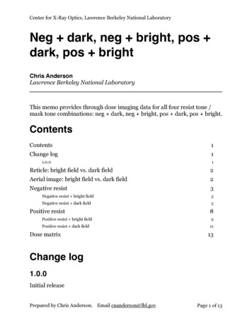
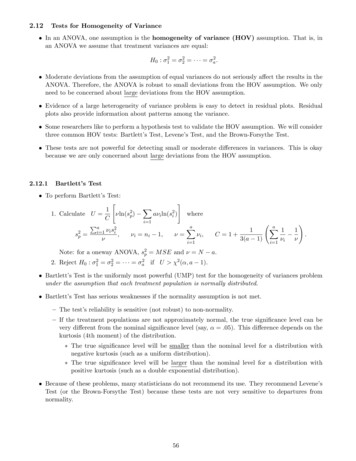
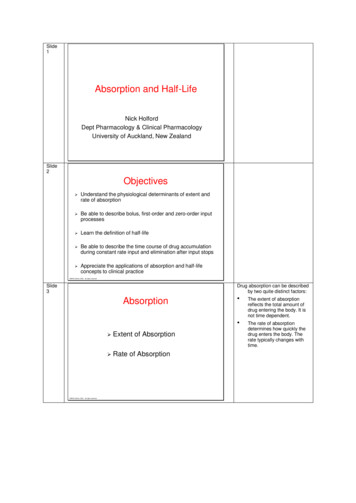
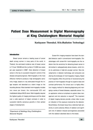
JOURNAL OF SIRIRAJ RADIOLOGYstudy involved 200 patients.MethodRetrospective study of patient dosimetry from adigital mammography system involving routine mediolateral oblique (MLO) and cranio caudal (CC) views acquired on the right and left breasts of female patients asin figure 1A, 1B, 1C and 1D.ResultThe average range, minimum and maximum valuesof the following parameters such as the tube voltageVol. 1 No. 1 January - June 2014(kVp), the tube current-time (mAs), compressed breastthickness (CBT) were collected. The entrance skin exposure (ESE) and average glandular dose (AGD) were determined from 200 mammography female patients. Theresults are displayed in Table 1. The average glandulardose (AGD) was 1.78 mGy for (RCC), 1.77 mGy for (LCC),1.86 mGy for (RMLO) and 1.98 mGy for (LMLO) respectively. The average entrance skin exposure (ESE) was6.79 mGy for (RCC), 6.83 mGy for (LCC), 7.15 mGy for(RMLO) and 7.83 mGy for (LMLO) respectively.ABCDFigure 1 A Right MedioLateralOblique (RMLO) B Left MedioLateral Oblique (LMLO) C Right CranioCuadal (RCC) D LeftCranioCuadal (LCC)Kanlayanee Theerakul87
ªï ’Ë 1 ‡ à¡ ’Ë 1 ¡ “§¡ - ¡‘ ÿπ“ π 2557«“ “ —ß ’«‘ “»‘ ‘ “ Table 1 Data collected from 200 female patients at the digital mammography systems, KingChulalongkorn Memorial )55.202186kVp29.422532(RCC)mAs As AGD(mGy)145.201.86630.794055.07Discussion and ConclusionThe average glandular dose can be estimated using a Perspex phantom exposed at the settings normallyused in clinical practice for a breast of the size and composition similar to that of the standard breast. The methodinvolves the determination of incident air kerma for thecorrect exposure and the half value layer of the x-raybeam. The tube output per mAs should first be measured and then multiplied by the conversion factor ofPerspex phantom for the standard breast. This methodwas used at the commissioning and reference testing ofthe mammography, ESE and AGD are displayed on thecomputer screen after exposure for this study. The mammogram was performed using the grid. The determination of patient dose in Digital mammography at KingChulalongkorn Memorial Hospital revealed that the meanAGD per image is 1.78 mGy for RCC view, 1.77 mGyforLCC view,1.86 mGy for RMLO view and 1.98 mGy forLMLO view. The ESE and AGD from MLO views weregreater than those from the CC views for the right andleft sides because the MLO CBT is slightly thicker 1799CBT(mm)56.7622102kVp29.472535(LCC)mAs 29.842149(LMLO)mAs AGD(mGy)150.791.98630.773976.14ESE7.831.8727.68the CC CBT. Overall, 91.41% of CC view and 89.14% ofMLO view were lower than the reference level of 3.0mGy. The result on ESE was comparable to the ESAK(entrance skin air kerma) value published in IAEA TECDOC1447 of 6.4 2.6 mGy after the QC procedure7. TheAGD is less than the limiting values of 3.0 mGy perexposure with grid 1.5 mGy without grid.The AGD per image is significantly different between the CBT as classified by glandular content groups.It increases with increasing CBT, while decreases withincreasing age. The results represent the digital mammography examination at one department is capable ofachieving acceptable dose levels for patient safety. ThemAs is significant affect on the AGD. The AGD in thedigital mammography system is related to auto filter modeof automatic exposure control system, which adjust theradiation dose, as defined by the mAs product, based onthe patient thickness. These are useful for the patientsthat the risk of cancer induction can be considered. It isan important for radiologists and technologist to increasethe awareness in the study for the patient especially thePatient Dose Measurement in Digital Mammography at King Chulalongkorn Memorial Hospital
Vol. 1 No. 1 January - June 2014JOURNAL OF SIRIRAJ RADIOLOGYadult one.In conclusion the optimization for the patient couldbe obtained by implementation the quality control program of the x-ray system and by determining the patientdose for both ESE and AGD with other parameters suchas kVp, mAs, CBT, type of target, filter and the compressed force. The patient dose should be comparedwith the reference level and the dose limit. The imagequality should be maintained with dose optimization forthe patients of every case.3.4.5.6.7.References1. Sriplung H, et al. Cancer incidence in Thailand 1995-1997. AsianPacific J Cancer Prev 2005;6:276-81.2. Andrew PS. Fundamentals digital mammography: Physics, tech-Kanlayanee Theerakul8.nology and practical consideratios [online]. Hologic Inc. (Distributor), 2005. Available from : htt://www.hologic.com/oem/pdf/R-BI-016 FundDig%20Mammo.pdf[2007,Apr17]Law J. Risk and benefit associated with radiation dose in breastscreening programmes an update. Br J Radiol 1995;681:870-6.American College of Radiology. ACR practice guide line for theperformance of whole breast digital mammography. ACR 2006Young KC, Burch A, Oduko JM. Radiation doses received inthe UK Breast Screening Programme in 2001 and 2002. Br JRadiol 2005;78:207-18.Kruger RL, Schueler BA. A survey of clinical factors and patientdose in mammography. Med Phys 2001;28:1449-54.International Atomic Energy Agency (IAEA). IAEA-TECDOC-1447:Optimization of the radiological protection of patients: Imagequality and dose in mammography (coordinated research inEurope). Vienna, Austria: IAEA; 2005.National Council on Radiation Protection & Measurements. NCRPReport No. 85, Mammography-A Userûs guide. Bethesda, MD:NCRP; 2012.89
the mammography, ESE and AGD are displayed on the computer screen after exposure for this study. The mam-mogram was performed using the grid. The determina-tion of patient dose in Digital mammography at King Chulalongkorn Memorial Hospital revealed that the mean AGD per image is 1.78 mGy for RCC view, 1.77 mGyfor