Transcription
Wiadomości Lekarskie, VOLUME LXXIV, ISSUE 7, JULY 2021 Aluna PublishingORIGINAL ARTICLEDETECTION OF BREAST CANCERS REPRESENTEDAS ARCHITECTURAL DISTORTION: A COMPARISONOF FULL-FIELD DIGITAL MAMMOGRAPHY AND DIGITALBREAST TOMOSYNTHESISDOI: 10.36740/WLek202107121Tetiana M. Babkina1, Andrii V. Gurando2,4, Tetiana M. Kozarenko1, Viacheslav R. Gurando3, Vadim V. Telniy4,Denys V. Pominchuk4P.L. SHUPYK NATIONAL MEDICAL ACADEMY OF POSTGRADUATE EDUCATION, KYIV, UKRAINESI, ‘‘INSTITUTE OF NUCLEAR MEDICINE AND DIAGNOSTIC RADIOLOGY OF NATIONAL ACADEMY OF MEDICAL SCIENCE OF UKRAINE’’, KYIV, UKRAINE3UZHHOROD NATIONAL UNIVERSITY, UZHHOROD, UKRAINE4MEDICAL CENTER «VERUM EXPERT», KYIV, UKRAINE12ABSTRACTThe aim: The aim of our study was to determine if digital breast tomosynthesis improves breast cancer detection associated with architectural distortion in comparison withfull-field digital mammography in the absence of appropriate history of trauma or surgery.Materials and methods: The overall rate of breast cancer involvement for the 34 patients with architectural distortion was 15 cases (44,1%) (invasive breast cancers, n 12(36,4%); ductal cancer in situ, n 3 (8,8%)) other findings associated with architectural distortion were high-risk lesions and benign findings (radial scar, n 5 (14,7%); sclerosingadenosis, n 9 (26,5%); typical lobular hyperplasia, n 3 (8,8%); typical ductal hyperplasia, n 2 cases (5,9%)).Results: Overall of 17/34 (50.0%) architectural distortions were identified at digital breast tomosynthesis that were missed at full-field digital mammography what wasstatistically significant difference ([95% CI, 2.56–7.45]; p 0.00001). Analysis of the results showed that sensitivity of full-field digital mammography for digital breasttomosynthesis detected breast cancers associated with architectural distortion was 53.3% [95% CI, 26.59% to 78.73%] and specificity was 52.63% [95% CI, 28.86% to 75.55%].Conclusions: Our study suggests that digital breast tomosynthesis detects more breast cancers represented as architectural distortion which are occult on full-field digitalmammography. Presence of microcalcifications within architectural distortion, in the absence of appropriate history of trauma or surgery, has a high likelihood of malignancyand obligatorily requires biopsy. KEY WORDS: Breast cancer, architectural distortion, digital breast tomosynthesis, full-field digital mammographyWiad Lek. 2021;74(7):1674-1679INTRODUCTIONBreast cancer (BC), takes the first place among oncologicdiseases in Ukrainian woman. According to the latest dataof national cancer-register in 2018 year there were detected14 872 female BC and 145 male BC [1].Mammography is the cornerstone of population-basedBC screening [2]. Early detection during mammographyscreening and optimal treatment can reduce BC mortalityrate by 38-48% [3].However, there are still difficulties in the differentialdiagnosis of masses, architectural distortions (ADs), asymmetries and normal breast tissue due to the summationartifact of tissue structures, which are located in differentplanes [4]. As follows we need to perform additional imaging, which worsens the psycho-emotional conditionsof the woman, increases their anxiety and radiation doseon their breasts.1674AD is thin straight lines or spiculations radiating from apoint, and focal retraction, distortion, or straightening at theanterior or posterior edge of the parenchyma with no definitemass visible [5]. AD is not always a sign of cancer and mayrepresent different benign processes and high-risk lesions,such as sclerosing adenosis, radial scars, complex sclerosinglesions and changes associated with surgery or trauma (fatnecrosis) which should stabilize and/or regress eventually [6].AD accounts for 12% to 45% of BC cases missed duringscreening mammography and are the third most prevalentmammographic appearance of BC [7, 8].Relatively new technology – ЗD mammography, whichcalls digital breast tomosynthesis (DBT), acquisition oftomosynthesis images, an x-ray source takes a series oflow-dose exposures, providing multiple images of thebreast in different planes that are 3D reconstructed, whilemoving in a limited arc above the compressed breast [9].
DETECTION OF BREAST CANCERS REPRESENTED AS ARCHITECTURAL DISTORTION: A COMPARISON.Table I. Study population characteristicsPatients with architectural distortion34Mean age, years49Microcalcifications within architecturaldistortions15 (44%)Non-dense breast, (ACR:a,b)12 (35%)Dense breast, (ACR:c,d)22 (65%)Breast cancers associated with architecturaldistortions15 (44%)Breast cancers associated with architecturaldistortions and microcalcifications9 (27%)Table II. ACR breast density distribution for BC associated with ADACR: a0 (0%)ACR: b5 (33%)ACR: c6 (40%)ACR: d4 (27%)THE AIMThe aim of our study was to determine if DBT improves BCdetection associated with AD in comparison with FFDMin the absence of appropriate history of trauma or surgery.MATERIALS AND METHODSThe institutional review board approved this study andwaived the need for informed consent due to its retrospective character.Exclusion criteria for our study were: absence of AD, lackof morphological verification, mass or microcalcifications(MCs) as a main finding, history of previous trauma orsurgery concordant with AD.This study included 34 patients with AD who underwentfull-field digital mammography (FFDM), DBT, hand-held fullbreast ultrasound (HHUS) and morphological verification fromMarch 2018 to January 2020 (Table I). Only patients with ADas a main finding, with or without MCs, were included in thisstudy. BI-RADS categories, mammographic breast density, modeof biopsy, and pathology results were utilized for all lesions.All patients underwent bilateral FFDM in “COMBO”mode in two standard projections (CC and MLO) as afirst step of imaging examination on Selenia DimensionsMammography system (Hologic, USA). This step incorporated digital mammography and tomosynthesis, includingsynthesized images. Stereotactic biopsies were performedon Hologic MultiCare Platinum Prone Breast Biopsy Table,when there wasn’t possibility to establish correlation withHHUS and perform ultrasound-guided core needle biopsy(CNB). For AD which were detected only on thomosynthesis and weren’t associated with MCs there were performedexcisional biopsies with preoperative mammography-guidedhookwire localization, due to lack of possibility to performtomosynthesis-guided breast biopsy for such lesions.HHUS and ultrasound-guided biopsies were performedon a Toshiba Viamo and Toshiba Aplio XG US systems witha linear probe centered at 9,0MHz.Age of the patients ranged from 33 years to 66 years withmean age of 49 years. All 34 patients with AD were examinedby pathology, revealing 12 invasive breast cancers (IBC), 3ductal cancer in situ (DCIS) and 19 non-cancerous findings.Retrospectively, all studies with previously DBT detectedAD, in consensus, were read by two dedicated breast radiologist. Only FFDM images in two standard projections(CC, MLO) were read without knowing results of DBTexamination and pathology.Sixty five percent (11/17) of these lesions underwentultrasound-guided CNB while the remainder underwentstereotactic CNB (4/17) and excisional biopsy (2/17) withpreoperative mammography-guided hookwire localization.A Chi-square test(χ2) was used to calculate the p-value. Aconfidence interval of 95% confidence level was calculated,the differences were considered significant at p-value 0.05.RESULTSThe overall rate of BC involvement for the 34 patients with ADswere 15 cases (44,1%) (IBC, n 12 (36,4%); DCIS, n 3 (8,8%))other findings associated with ADs were high-risk lesions andbenign findings (radial scar, n 5 (14,7%); sclerosing adenosis,n 9 (26,5%); typical lobular hyperplasia, n 3 (8,8%); typicalductal hyperplasia, n 2 cases (5,9%)). Sixty seven percent(10/15) of BC were in dense breasts (Table II).All cases with MCs which were present in ADs were seen onboth methods DBT and FFDM, n 13/34 (38,2%) and associated with malignancy, n 9/34 (26,5%) what was consideredstatistically significant ([95% CI, 0.91–1.86]; p 0.2). In five casesof FDDM visible suspicious MCs ADs were not detected, twoof these cases were associated with BC.Total of 17/34 (50.0%) ADs were identified at DBT that weremissed at FFDM what was statistically significant difference([95% CI, 2.56–7.45]; p 0.00001).There were 41,2% (7/17) FFDM occult BCs associated withAD (IDC, n 5; ILC, n 1; DCIS, n 1); 10/17 (58,8%) FFDMoccult ADs were non-cancerous findings (radial scar, n 4;sclerosing adenosis, n 4; typical lobular hyperplasia, n 1; typical ductal hyperplasia, n 1) what was considered statisticallysignificant difference between DBT and FFDM in BC detection([95% CI, 0.82–2.53]; p 0.01).Analysis of the results showed that sensitivity of FFDM forDBT detected BCs associated with AD was 53.3% [95% CI,26.59% to 78.73%] and specificity was 52.63% [95% CI, 28.86%to 75.55%].Sixty five percent (11/17) of the lesions underwent ultrasound-guided CNB while the remainder underwent stereotactic CNB (4/17) and excisional biopsy (2/17) with preoperativeguidewire localization. In our setting there was no ability toperform tomosynthesis guided breast biopsy.The most frequently localization of AD was in upper-outerquadrant of the breast and BCs associated with AD also were mostfrequently localized in upper-outer quadrant (Table III). Overallmore AD we detected in the left breast 21 (61,8%) to 13 (38,2%) in1675
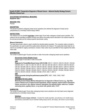
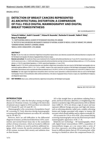
Tetiana M. Babkina et al.Fig.1. Full-field digital mammography RCC, LCC, RMLO, LMLO views.Fig. 2. Full-field digital mammography RCC view, Breast Tomosynthesis RCC view, magnification of architectural distortion in inner quadrant of the rightbreast on Tomo scansthe right breast. Also more BCs associated with AD were detectedin the left breast 10 (66,7%) to 5 (33,3%) in the right breast.As we see in Example 1 (Fig. 1-4) there is architectural distortion in upper inner quadrant of large fatty breast. Due tolarge size, fatty composition of the breast and lack of confidentcorrelation between mammography and ultrasound stereotactic core-needle biopsy was performed. Pathology of suspiciouslesion demonstrated G1 invasive carcinoma of no special type.In another case Example 2 (Fig. 5-7) dense fibroglandularbreast tissue obscured suspicious architectural distortion inupper outer quadrant of the left breast. There was performedexcisional biopsy with preoperative hookwire localization of thelesion which showed at pathology radial scar associated withsclerosing adenosis and atypical ductal hyperplasia.DISCUSSIONDBT is a relatively new modality for Ukrainian breast imaging.First experience of 3D mammography in Ukraine showed superior sensitivity for BC detection by using DBT than FFDM [10].Considering that AD is the most common missed abnormalitywhat caused false-negative mammography results, it’s very important to compare DBT and FFDM in woman with ADs inUkrainian population [11].Our results showed that 17/34 (50,0%) ADs detected by DBTwere missed on FFDM and 7/17 (41,2%) of these FFDM-occult1676AD were malignant. Which are in line with previous study whereAD were visualized better by using DBT than FFDM, moreover73% of them were seen only on the DBT images and 21% of these2D-occult AD were malignant [12].Also our results correlate with one more study showing thatAD is frequently associated with BC. That study showed that 17of 36 (47%) sonographically occult AD, but detected on DBT,were malignant [13]. In our study 35,3% (6/17) sonographicallyoccult but detected on DBT ADs were malignant.According to another study ILCs was presented less frequentlyas masses and more often as AD on DBT [14]. Among 15 malignant lesions associated with AD, in our study, there was onlyone ILC.Aim of earlier study was to determine the management of thelesions that were detected by DBT but were invisible on FFDMor HHUS [15]. Authors point that among 107 patients with DBTpositive but FFDM or HHUS-negative breast lesions there were 48(45%) MRI-negative lesions and none had a suspicious alterationduring the follow-up period [15]. They concluded that breast MRIprior to biopsy may reduce the number of unnecessary biopsiesfor suspicious breast lesions that are DBT positive only [15].Our results are also supported by the results of the recent studythat DBT can detect suspected lesions that are occult in FFDM,especially in women with dense breast [16].After AD is found it’s very important to correlate the findingwith HHUS. Ultrasound-guided CNB has become the first choice
DETECTION OF BREAST CANCERS REPRESENTED AS ARCHITECTURAL DISTORTION: A COMPARISON.Fig. 3. Imaging of navigation during stereotactic core-needle biopsy of suspicious architectural distortion in the right breast.Fig. 4. Pathology revealed breastinvasive carcinoma G1 of no special type (NST). Stained withhematoxylin-eosin, magnification10.0 х.Table III. Breast cancer localization within the breast.Upper outer quadrant9 (61%)Upper inner quadrant1 (7%)Lower outer quadrant1 (7%)Lower inner quadrant3 (18%)Retroareolar region1 (7%)for performing most breast biopsies due to its non-ionising radiation, low cost, full control of the needle in real time, accessibilityin difficult locations, multidirectional punctures and excellentcomfort for patients [17]. In our study each AD was correlatedwith ultrasound images. In most cases 22/34 (64,7%) we foundcorrelation and performed ultrasound-guided CNB. When ADwas occult for HHUS there was performed stereotactic biopsyor excisional biopsy with preoperative mammography-guidedhookwire localization.Two big studies have shown that BC localized more frequentlyin the left breast with a left to right ratio of 1,10 and 1,07 [18,19].In another study authors have shown that the upper outer quadrant was the most frequent site of carcinoma and supportedhypothesis that the high proportion of upper outer quadrantcarcinomas of the breasts is a reflection of the greater amount ofbreast tissue in this quadrant [20, 21]. Our study showed similarresults – 66,7% of BC were detected in the left breast and overall60,0% of all BC were localized in upper outer quadrant.Fifty three percent (8/15) of BC associated with AD were withMCs what correlates with previous research which showed thataround 40% of BCs present with MCs and frequently, serve asthe only mammographic features indicating the presence of amalignant lesion [22]. In other article researches noted that inscreening programs, between 12.7 and 41.2% of women arerecalled with MCs as the only sign of cancer [23].Our study had some major limitations. We made comparisonbetween two methods only for DBT-visible suspicious AD where wehad results of pathology and then tried to identify them on FFDM.Due to its retrospective nature interpreting radiologists knew that ineach case on FFDM there was previously detected suspicion lesion onDBT and this could potentially introduce a bias in their assessment.The number of cancer cases in our sample was limited and wedid not include other findings such as masses, asymmetries andMCs as a main finding in this analysis. Also, when radiologists sawjust MCs on FFDM without association with AD we interpretedit as FFDM-occult AD.CONCLUSIONSThe results of this study suggest that DBT detects morebreast cancers represented as AD which are occult on1677
Tetiana M. Babkina et al.Fig. 5. Full-field digital mammography RCC, LCC, RMLO, LMLO views.Fig. 6. Full-field digital mammography LCC view, Breast TomosynthesisLCC view, preoperative hookwire localization of suspicious architecturaldistortion in upper outer quadrant of the left breast.Fig. 7. Pathology showed radial scarassociated with sclerosing adenosis and atypical ductal hyperplasia.Stained with hematoxylin-eosin,magnification 5.0 х.1678
DETECTION OF BREAST CANCERS REPRESENTED AS ARCHITECTURAL DISTORTION: A COMPARISON.FFDM. Presences of MСs within AD, in the absence ofappropriate history of trauma or surgery, have a highlikelihood of malignancy and obligatorily require biopsy.Our study proposes that combination of DBT and FFDMmay increase detection of BCs represented as AD. We suggestusing HHUS for all ADs detected on FFDM or DBT to correlatethese findings and to perform US-guided biopsy; when MCs arepresent within US-occult AD to perform stereotactic biopsy; ifAD is only seen on DBT to perform excisional biopsy with preoperative mammographic-guided hookwire localization in absenceof possibility to perform tomosynthesis-guided breast biopsy.However, using of DBT in Ukraine is just in the beginning and requires additional investigation in middle-income country settings.REFERENCES1. Fedorenko Z.P. Cancer in Ukraine 2018 to 2019, incidence, mortality, activitiesof oncological service. In: Fedorenko ZP, Michailovich YY, Goulak LO et al. Kyiv:National Cancer Institute of Ukraine; 2020, 42-43 p.2. Perry N., Broeders M., Wolf C. et al. European guidelines for quality assurancein breast cancer screening and diagnosis. Fourth edition. InternationalAgency for Research on Cancer. Lyon. France, 2006, 8 p.3. Broeders M., Moss S., Nyström L. et al. The Impact of MammographicScreening on Breast Cancer Mortality in Europe: A Review of ObservationalStudies. Journal of Medical Screening, J Med Screen. 2012;19(1):14-25. doi:10.1258/jms.2012.012078.4. Rangayyan R.M., Banik S., Desautels J.E. Computer-aided detection ofarchitectural distortion in prior mammograms of interval cancer. J DigitImaging. 2010;23(5):611–631. doi:10.1007/s10278-009-9257-x.5. D’Orsi C.J., Sickles E.A., Mendelson E.B. et al. ACR BI-RADS Atlas, BreastImaging Reporting and Data System. Reston, VA, American College ofRadiology; 2013. 127-128 p.6. Durand M.A.,Wang S., Hooley R.J. et al.Tomosynthesis-detected ArchitecturalDistortion: Management Algorithm with Radiologic-Pathologic Correlation.Radiographics. 2016;36(2):311-21. doi: 10.1148/rg.2016150093.7. Rangayyan R.M., Banik S., Desautels J.E. Computer-aided detection ofarchitectural distortion in prior mammograms of interval cancer. J DigitImaging. 2010;23(5):611–631. doi:10.1007/s10278-009-9257-x.8. Knutzen A.M., Gisvold J.J. Likelihood of malignant disease for variouscategories of mammographically detected, nonpalpable breast lesions.Mayo Clin Proc. 1993;68 :454–460. doi: 10.1016/s0025-6196(12)60194-3.9. Kopans D.B. Digital Breast Tomosynthesis From Concept to Clinical Care.American Journal of Roentgenology. 2014;202(2):299-308. doi: 10.2214/AJR.13.11520.10. Dykan I.M., Bozhok Ye.M., Gurando A.V. Pershyi dosvid vykorystannia 3Dmamohrafii v Ukraini. Luchevaja diagnostika. Luchevaja Terapija [Thefirst experience of using 3D mammography in Ukraine]. 2018;2:40-8. (inUkrainian).11. Burrell H.C., Sibbering D.M., Wilson A.R. et al. Screening interval breastcancers: mammographic features and prognostic factors. Radiology.1996;199(3):811-7. doi:10.1148/radiology.199.3.8638010.12. Partyka L., Lourenco A.P., Mainiero M.B. Detection of mammographicallyoccult architectural distortion on digital breast tomosynthesis screening:initial clinical experience. AJR Am J Roentgenol. 2014;203(1):216-22. doi:10.2214/AJR.13.11047.13. Freer P.E., Niell B., Rafferty E.A. Preoperative tomosynthesis-guided needlelocalization of mammographically and sonographically occult breast lesions.Radiology. 2015;275(2):377-83. doi: 10.1148/radiol.14140515.14. Chamming’s F., Kao E., Aldis A. et al. Imaging features and conspicuity ofinvasive lobular carcinomas on digital breast tomosynthesis. Br J Radiol.2017;90(1073):20170128. doi: 10.1259/bjr.20170128.15. Taskin F., Durum Y., Soyder A., Unsal A. Review and management of breastlesions detected with breast tomosynthesis but not visible on mammographyand ultrasonography. Acta Radiol. 2017. doi: 10.1177/0284185117710681.16. Ray K.M., Turner E., Sickles E.A., Joe B.N. Suspicious Findings at Digital BreastTomosynthesis Occult to Conventional Digital Mammography: ImagingFeatures and Pathology Findings. Breast J. 2015;21(5): 538-42.17. Apesteguía L., Pina L.J. Ultrasound-guided core-needle biopsy of breastlesions. Insights Imaging. 2011;2(4):493–500. doi:10.1007/s13244-0110090-7.18. Amer M.H. Genetic factors and breast cancer laterality. Cancer Manag Res.2014;6:191–203. doi:10.2147/CMAR.S60006.19. Roychoudhuri R., Putcha V., Moller H. Cancer and laterality: a study of thefive major paired organs (UK) Cancer Causes Control. 2006;17(5):655–662.20. Bright C.J., Rea D.W., Francis A. et al. Comparison of quadrant-specific breastcancer incidence trends in the United States and England between 1975 and2013. Cancer Epidemiol. 2016;44:186-194.21. Lee A.H. Why is carcinoma of the breast more frequent in the upper outerquadrant? A case series based on needle core biopsy diagnoses. Breast 2005;14(2):151–2.22. Castronovo V., Bellahcene A. Evidence that breast cancer associatedmicrocalcifications are mineralized malignant cells. Int J Oncol.1998;12(2):305–308. doi.org/10.3892/ijo.12.2.305.23. Mordang J.J., Gubern-Mérida A., Bria A. et al. The importance of earlydetection of calcifications associated with breast cancer in screening. BreastCancer Res Treat. 2018;167(2):451–458. doi:10.1007/s10549-017-4527-7.ORCID and contributionship:Tetiana M. Babkina: 0000-0001-5046-870X AAndrii V. Gurando: 0000-0002-2708-3040 B,DTetiana M. Kozarenko: 0000-0002-0838-9773 EViacheslav R. Gurando: 0000-0001-6303-3799 FVadim V. Telniy: 0000-0001-9860-9663 CDenys V. Pominchuk: 0000-0002-7630-4241 CConflict of interest:The Authors declare no conflict of interest.CORRESPONDING AUTHORViacheslav R. GurandoUzhhorod National University3 Narodna Square 88000 Uzhhorod, Ukrainetel: 380966104524e-mail: vhurando@gmail.comReceived: 2020-06-02Accepted: 2021-06-01A – Work concept and design, B – Data collection and analysis, C – Responsibility for statistical analysis,D – Writing the article, E – Critical review, F – Final approval of the article1679
Results: Overall of 17/34 (50.0%) architectural distortions were identified at digital breast tomosynthesis that were missed at full-field digital mammography what was statistically significant difference ([95% CI, 2.56-7.45]; p 0.00001). Analysis of the results showed that sensitivity of full-field digital mammography for digital breast