Transcription
Lab Manual Exercise # 1Ads by GoogleWayne's WordLil WayneIndexLyricsNoteworthy PlantsLive Album LyricsTriviaLemnaceaeFind Lyrics to a SongBiology 101BotanySearchPhysical Properties & Structure of CellsSee The Cell & Mitosis Crossword PuzzleTable Of Contents:1.2.3.4.5.6.7.8.9.10.11.12.13.14.15.Cells From An Onion SkinDiameter Of Field Of ViewTable Of Cell Size ComparisonsBird Eggs: World's Largest CellsGrains Of Salt & Metric SystemElodea Leaf Cell & PlasmolysisSalt Intake At A Movie TheatreImportant Definitions For Cell ExamPotato Tuber & Banana FruitOak Wood & Specific GravityHuman Cheek Epithelial CellsSome GeneralizationsProkaryotic Cells Of CyanobacteriaIllustration Of Plant & Animal CellsSuccessive Orders Of MagnitudeIf you have difficulty printing out this page, try the PDF version:Click PDF Icon To Read Page In Acrobat Reader. See Text In Arial Font Like In A Book.View Page Off-Line: Right Click On PDF Icon To Save Target File To Your Computer.Click Here To Download Latest Acrobat Reader. Follow The Instructions For Your Computer.Section B. Onion Skin Cell1. The cells of an onion skin are generally rectangular in shape and range in size from 0.25 to 0.4millimeters in length (250-400 micrometers). A millimeter is abbreviated by mm and a micrometer by theGreek letter mu (12th letter of Greek alphabet) followed by an m:file:///C /wayne/lmexer1.htm[4/12/2011 6:27:40 AM]millimeterabout 1/25th of an inchmicrometer1/25,000th of an inch
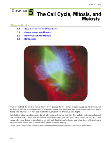
Lab Manual Exercise # 1Left: Microscopic view of an onion skin showing several rectangular cells, each with a small, sphericalnucleus (red arrow). The slide was stained with a drop of yellowish-brown gram's iodine. Right: Highlymagnified view of a cell from the meristematic root tip of an onion showing enlarged nucleus containing16 chromosomes. The cell is in prophase of mitosis, with distinct chromosomes (chromosome doublets)and a disintegrating nuclear membrane.Diameter Of The Field Of ViewCompound microscope showing the 10x ocular (eyepiece) and four objectives (4x, 10x, 40x and 100x).[One objective is not in view.] To calculate the magnification, simply multiply the ocular lens (10x) bythe objective lens. With this microscope you can obtain four different magnifications: 40x, 100x, 400xand 1000x.file:///C /wayne/lmexer1.htm[4/12/2011 6:27:40 AM]
Lab Manual Exercise # 1The field of view when using the 10x objective (100x total magnification) is 2 mm. If 8 plant cellsextend across the field of view (2 mm), then each cell is 2/8 or 0.25 mm long. Remember that thediameter of the field of view changes depending on the power of the objective according to thefollowing table:ObjectiveDiameter Of Field Of ViewMagnification (10x Ocular)4x4.0 mm (4.45)40x10x2.0 mm (1.78)100x40x0.4 mm (0.45)400x100x0.2 mm (0.178)1000xThe original diameters of field of view (fov) were determined with a transparent mm ruler. This is likemeasuring the length of your finger nails with a yard stick. The values in parentheses are more precise.They were determined using a B & L stage micrometer.If you know the diameter of the fov at one magnification, you can determine the diameter of fov atanother magnification with the following formula:diameter of fob#2 diameter of fov#1 x magnification#1 divided by magnification#2For example, if you know the diameter of fov at 100x magnification, the diameter of the fovat 1000x magnification 1.78mm x 100 divided by 1000 0.178 mm or 178 micrometers.file:///C /wayne/lmexer1.htm[4/12/2011 6:27:40 AM]
Lab Manual Exercise # 1Stage micrometer at 1000x magnification with Olympus Compound Microscope. The diameter offield of view (fov) is 0.184 millimeters (184 micrometers). This corresponds to a 0.46 millimeterfov at 400 x magnification.Relative Sizes Of Cells & Viruses 1Diameter or LengthMillimetersMicrometerswater molecule0.0000003850.000385polio virus0.000030.03HIV retrovirus0.00010.1herpes virus0.00020.2Resolving Power of Average Light Microscope: 0.0003 mmsmallpox virus0.00030.3gonorrhea bacterium0.00050.5mimivirus0.00080.8anthrax bacterial spore0.0011.0Staphylococcus bacterium0.0011.0human cheek cell nucleus0.0055.0file:///C /wayne/lmexer1.htm[4/12/2011 6:27:40 AM]
Lab Manual Exercise # 1head of a human sperm0.0055.0puffball Spore0.0055.0human red blood cell0.00757.5snow algae aplanospore0.0330phytolith of crab grass0.03232lichen spore (length)0.03535human cheek cell0.0660fine human hair (width)0.07070Resolving Power of Unaided Human Eye With 20-20 VisionAble to focus on two hairs spaced 70 micrometers apart 8smallest orchid seed0.08585Demodex mite in your nose &Protozoan: Paramecium bursaria0.180180coral-root orchid seed0.20200Wolffia angusta fruit0.30300Amoeba cell0.30300grain of table salt (NaCl)0.30300onionskin cell0.40400Wolffia angusta plant0.60600diameter of pin head1.501500nerve cell (spinal cord)600600,000file:///C /wayne/lmexer1.htm[4/12/2011 6:27:40 AM]
Lab Manual Exercise # 1Eucalyptus regnans100,000100,000,000planet earth (diameter)1.3 x 10101.3 x 10131. The discovery of a virus called "mimivirus" in 1992 complicates the placement of viruses in the overallclassification scheme for living organisms. Prior to this discovery, viruses were generally considerednonliving until they hijack a living cell. Officially, this virus got its name because it mimics bacteria(microbes) in size and complexity. It was placed in a new family of viruses, the Mimiviridae. Mimivirus wasfound inside an amoeba within a cooling tower in Bradford, UK. [The cooling tower was being investigatedas the source of an influenza outbreak.] Mimivirus is the largest known virus, about 0.8 micrometers (800nanometers) across. In fact it is larger than the bacterium causing gonorrhea.Diameter of an Individual Mimivirusmetric unitabbreviation**decimal erµ0.800nanometernm800AngströmÅ8000**html code for abbreviations may not show on all browsers.With the exception of a some bacteriophages, viruses fall into two main morphologicalgroups, those with cubical symmetry and those with helical symmetry. Until 1960, the onlyknown examples of viruses with helical symmetry were those of plant viruses. The beststudied example being the tobacco mosaic virus. The capsid denotes the protein shell thatencloses the nucleic acid. It is composed of numerous repeating structural units. "Linear"viral capsids have RNA genomes that are encased in a helix of identical protein subunits.The length of the helical viral nucleocapsid is determined by the length of the nucleic acid.Cubical viruses have the general molecular symmetry of an icosahedron. Althoughmost viruses are not visible under an ordinary light microscope, mimivirus appearslike a minute spheroid object under a compound microscope using an oilimmersion objective (1000x magnification). An icosahedron has 20 identicalequilateral triangles (facets), each subdivided into more equilateral triagular facets.There are electron micrographs of mimivirus available on the Internet. Simply do asearch for mimivirus using one of the excellent search engines, such asgoogle.com.file:///C /wayne/lmexer1.htm[4/12/2011 6:27:40 AM]
Lab Manual Exercise # 1The mimivirus genome contains 1.2 million bases, more than many bacteria. The bases make up 1,260 genes,which makes it as complex as some bacteria. Most viruses use either DNA or RNA to carry their geneticinformation, but mimivirus has both of these nucleic acids. In addition, mimivirus can make about 150 of itsown proteins, and can even repair its own DNA if it gets damaged. Normal viruses are not capable of proteinsynthesis or DNA repair on their own, they must rely on the organelles of their host cells for these activities.Whether mimivirus should be placed in an existing domain (superkingdom), or in its own domain, remainsto be seen. For more information, see D. Raoult, et al. "The 1.2-Mb Genome Sequence of Mimivirus."Science Published On-line, DOI: 10.1126/Science.1101485 (2004); B. La Scola et al. "A Giant Virus inAmoebae." Science 299 (5615): 2033 (2003).See The Three Domains Of LifeSome scientists have speculated that the evolution of eucaryotic cells involved the merging of two or moregenomes, a phenomenon called symbiogenesis. Eukaryotic cells are characterized by the presence ofmembrane-bound organelles, including chloroplasts, mitochondria and nuclei. The origin of a complex cellhas intrigued scientists for decades, and has been a strongly debated topic between evolutionists andcreationists. According to the endosymbiont hypothesis, bacteria may be the progenitors of cellular organellessuch as chloroplasts and mitochondria. A primitive nucleus (protonucleus) may have evolved from anintracellular virus; however, one weakness of this hypothesis is that viruses generally lack some of the keygenes found in eukaryotes. The complex genome of mimivirus includes these genes, lending support to theevolution of a protonucleus from a virus.See Symbiogenesis & The Endosymbiont HypothesisTypical viruses are not placed in the three major domains of life. They are much smaller and much lesscomplex than cells. They are macromolecular units composed of DNA or RNA surrounded by an outerprotein shell. They have no membrane-bound organelles, no ribosomes (organelle of protein synthesis), nocytoplasm (living contents of a cell), and no source of energy production of their own. They do not exhibitautopoiesis--i.e. they do not have the self-maintenance metabolic reactions of living systems. Viruses lackcellular respiration, ATP-production, gas exchange, etc. However, they do reproduce, but at the expense ofthe host cell. Like obligate parasites, they are only capable of reproduction within living cells. In a sense,viruses hijack the host cell and force it to produce more viruses through DNA replication and proteinsynthesis. Outside of their host cells, viruses can survive as minute macromolecular particles. Viruses mayattack animals and plants. Infectious human viruses can be dispersed though the air (airborne viruses) or bodyfluids (HIV virus). Epidemic viruses (such as HIV) that are passed from person to person via sexualconjugation are remarkably similar to computer viruses. Unfortunately in humans there is no residentantivirus program to alert you of a potential infection, or to quickly scan your body and delete the invaderonce it has entered your system. Humans must rely on their amazing antibody and cell-mediated immuneresponses, some of the most complex and remarkable achievements in the evolution of living systems.See The Five Major Kingdoms Of LifeBiological Viruses vs. Computer VirusesSee The WAYNE'S WORD Virus Article2. This is the width of an oval (elliptical) spore. Spores of this size can easily escape from a folded (closed)file:///C /wayne/lmexer1.htm[4/12/2011 6:27:40 AM]
Lab Manual Exercise # 1paper envelope that is not airtight. Anthrax (Bacillus anthracis) is one of the microorganisms used inbiological warfare because strains have been developed that are extremely infectious through the skin andthrough inhalation. In addition, it forms highly resistant spores that can survive for long periods.Approximately one teaspoon or two grams of anthrax may contain up to 20 billion spores. With an averageinfection rate of 10,000 spores per person, this is theoretically enough spores to infect 2 million people withinhalation anthrax.Left: Highly magnified view (2000x) of human pus showing white blood cells (called neutrophils) withdeeply-lobed purple nuclei. The minute paired dots (red arrow) are diplococcus gonorrhea bacteria(Neisseria gonorrhoeae). Each dot (coccus bacterium) is only about 0.5 micrometers in diameter. Someof the neutrophils have ingested the bacteria through phagocytosis. Right: A culture of rod-shapedanthrax bacteria (Bacillus anthracis). Some of the bacteria have divided by fission (red arrow). [Bothimages came from old (circa 1960) prepared microscope slides enhanced with Adobe PhotoShop by W.P.Armstrong.]Successive Orders Of Magnitude: Galaxy To A Proton3. One human nucleus contains 46 chromosomes (single chromosomes during interphase). These 46chromosomes comprise about 6 feet of DNA (2 meters). This amount of DNA was previously thought tocontain about 100,000 functional genes, but that number has now been reduced to about 30,000 genes (onepercent of the total DNA in the nucleus). In terms of information storage, the genetic information in a cell isroughly equivalent to 500 printed volumes of Encyclopedia Brittanica (12 characters per inch).4. Mature sperm (spermatozoa) are composed of three distinct parts: a head, a middle piece (midpiece), and atail (flagellum). The middle piece and tail contain microtubules in the characteristic 9 2 pattern of cilia andflagella (see animal cell illustration). In the middle piece, mitochondria are packed around the microtubulesfile:///C /wayne/lmexer1.htm[4/12/2011 6:27:40 AM]
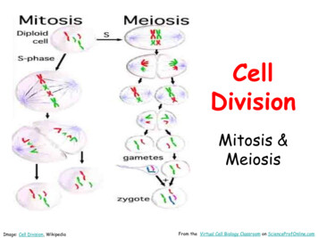
Lab Manual Exercise # 1and provide the energy for movement. The journey of a single sperm in the female reproductive tract isanalogous to a salmon swimming ten miles upstream to spawn. The head contains a nucleus covered by anouter cap called the acrosome which stores enzymes needed to penetrate the cellular and glycoprotein layerssurrounding the egg.Surgical sterilization of a man is called a vasectomy. Each of the two vas deferentia (sperm ducts) from theleft and right testicles are cut and tied in two places, and the portion between the ties removed. Residualsperm may be present in the vas deferentia for up to a month following a vasectomy; therefore, periodicmicroscopic examinations are recommended until sperm are no longer visible in semen samplers.Microscope image of human sperm in semen (1000x). The illustration insert shows the acrosome,head and middle piece of a human spermatozoan.file:///C /wayne/lmexer1.htm[4/12/2011 6:27:40 AM]
Lab Manual Exercise # 15. A baseball-sized puffball (Calvatia gigantea) in San Diego County, California. The inside isfilled with a dark mass of spores intermingled with threadlike hyphae (capillitium). The close-upview of spores (right) was taken at 1,000x. Each spherical spore has a diameter of about 1/200mm (5 µm), slightly smaller than a human red blood cell (7.5 µm). Depending on the species,puffballs range in size from a baseball to a basketball. When they are mature, the puffballreleases millions of spores into the air in a cloud of black dust. This can easily be demonstrated ifyou accidentally kick one. According to David Arora (Mushrooms Demystified, 1986), a largepuffball may contain 7 trillion spores. Lined up single file, this number of spores would extendaround the earth's equator. If each spore produced a puffball the size of a basketball, the resultingpuffballs would extend from the earth to the sun and back!6. Lichens are a symbiotic relationship between algae and fungi. The main bodyof a lichen is composed of fungal tissue (usually class Ascomycota) withphotosynthetic algal cells (usually division Chlorophyta) embedded within thisfungal mass (mycelium). The spores are produced by the fungal component ormycobiont, usually in a cup-shaped body called an ascocarp. Lichen spores canbe 1-2 micrometers in Acarospora chlorophana, or up to 40 micrometers longin Diploschistes scruposus (left). The size, color and shape of spores are usefultraits in separating different species of lichens. Spores up to 40 micrometers orlonger are considered large.Muriform Spore Of Diploschistes muscorumMuriform Spores Of Rhizocarpon disporumTwo-Celled Spores Of Dimaelaena radiatafile:///C /wayne/lmexer1.htm[4/12/2011 6:27:40 AM]
Lab Manual Exercise # 17. The width (diameter) of a human hair rangesfrom very fine (0.017 mm) to very coarse (0.181mm). The right image shows a fine human hair at1,000 x magnification. The hair width is 0.070mm or 70 micrometers. With the unaided eye thesingle hair shaft is visible when placed on a sheetof white paper.8. A simple test for the resolving power of the unaided human eye:A. Two fine human hairs are placed 70 micrometers apart. Each hair is approximately 70micrometers in width (diameter). With 20-20 vision you should be able to differentiatebetween the two hairs at the proper focal distance. B. When placed closer to each other(about 15 micrometers apart), the two hairs appear to blend together and cannot bedifferentiated with the naked eye. A hand lens or magnifying glass is necessary todifferentiate between the two hairs. Although the hairs can be seen on a white background,they cannot be resolved if they are placed close together. The minimum resolution for acomputer monitor is 72 dots per square inch (dpi), otherwise the image becomes pixelated.Printed photos are also composed of dot patterns. Above 150 dpi, the dots generally blendtogether and cannot be resolved. Under a hand lens or microscope the dots can easily beobserved.9. Certain epiphytic orchids of the tropical rain forest produce the world's smallest seeds weighing only onefile:///C /wayne/lmexer1.htm[4/12/2011 6:27:40 AM]
Lab Manual Exercise # 135 millionths of an ounce (1/35,000,000) or 0.81 micrograms. Some seeds are only about 1/300th of an inchlong (85 micrometers). One seed capsule from a single flower may contain up to four million seeds. They aredispersed into the air like minute dust particles or single-celled spores, eventually coming to rest in the uppercanopy of rain forest trees.See The Minute Seed Of A Coral-Root Orchid10. This minute one-seeded fruit is approximately 1/100 of an inch long and weighs about 70 micrograms or1/400,000 of an ounce. Compare this tiny fruit with the most massive pumpkin that weighs over 1000pounds (over 450,000 grams).Wolffia: The World's Smallest Fruit11. The plant body of the Australian species Wolffia angusta is only 0.6 mm long (1/42 of an inch). Itweighs about 150 micrograms or 1/190,000 of an ounce. This is the world's smallest flowering plant, rivaledin minuteness by the Asian species W. globosa.Wolffia: The World's Smallest Flowering PlantStrange Duckweeds From Far Away Lands12. The Australian tree (Eucalyptus regnans) is the world's tallest flowering plant. Some of the largest treesare over 300 feet tall, rivaling in size the California coast redwood (Sequoia sempervirens).See Botanical Record-Breakers13. The planet earth has a diameter of about 8,000 miles (13,000 kilometers) or 13 billion (13,000,000,000)millimeters. It has a volume of about one nonillion (1 X 1030) cubic millimeters. This astonishing volume isroughly equivalent to the volume of one nonillion wolffia plants packed together after 4 months of asexualbudding!If a water molecule is represented by 100, then a wolffiaplant is about 1020 power larger than the water molecule. Theearth is about 1020 power larger than a wolffia plant, or 1040power larger than the water molecule.file:///C /wayne/lmexer1.htm[4/12/2011 6:27:40 AM]
Lab Manual Exercise # 1Successive Orders Of Magnitude: Galaxy To A Proton14. A hair follicle mite (Demodex brevus) in your nose!Hair follicle mites of the genus Demodex are among the smallest multicellularanimals. They were first described in humans in 1841 by Frederick Henle whoreported this minute parasite from "miliary glands" of the ear canal. He wasuncertain about the parasite's taxonomic position in the animal kingdom. [Henle isalso known for the Henle's loop in the vertebrate nephron.] Another scientist fromthis time period by the name of Berger (1845) thought it was a member of thephylum Tardigrada. Tardigrades are minute animals often found on lichens andmosses. According to Desch and Nutting (The Journal of Parasitology 58 (1): 169177, 1972), there are two species of follicle mites on humans. Demodexfolliculorum measures 0.3 to 0.4 mm in length and typically occupies hair follicles.It also called an "eyelash mite" because it commonly occurs in follicles at the baseof eyelashes. Demodex brevis is about half that size (0.15 to 0.2 mm) and typicallylives in sebaceous glands adjacent to hair follicles. The latter mite is only about thesize of a unicellular Paramecium and appears to be the species I found in my nose.Illustration (left) modified from T. Ross and photographs of dorsal side of D.brevus by W.P. Armstrong.file:///C /wayne/lmexer1.htm[4/12/2011 6:27:40 AM]
Lab Manual Exercise # 1Demodex brevis (184 micrometers in length) compared in size witha freshwater protozoan Paramecium bursaria. Magnification 400x.More Information & Images Of The Follicle Mite Demodex brevusBird Eggs: World's Largest Cells By VolumeThe typical bird egg contains an enlarged yolk surrounded by a protein-rich accessory fluid called the eggwhite or albumen. The yolk is essentially the enlarged ovum or egg cell. As this cell undergoes mitosis,the daughter cells become smaller and smaller until they are invisible to the naked eye. Egg-laying(oviparous) birds have large yolks rich in proteins and lipids to sustain the embryo. The developing chickobtains air through minute pores in the calcium carbonate egg shell. Pores in the shell are plugged bycollagen, but are still permeable to air. Chicken egg shells can be brown or white, depending on the breed(variety). For example, Rhode Island Red, New Hampshire and Plymouth Rock varieties lay brown eggs.Most eggs purchased in your local market are unfertilized (haploid). Fertile (diploid) eggs are laid by hensregularly exposed to a rooster. The yolk contains a reddish-yellow carotenoid pigment (astaxanthin) whichproduces its bright color. In fact, chickens throughout the world are fed astaxanthin in their diet tointensify the yolk color.file:///C /wayne/lmexer1.htm[4/12/2011 6:27:40 AM]
Lab Manual Exercise # 1A gigantic bird which lived in southern Madagascar until 900 AD produced the largest egg of any animal.Known as the elephant bird (Aepyornis maximus), it is an extinct member of the Rattae, a clade offlightless birds that includes the ostrich, emu, cassowary, kiwi and rhea, as well as the extinct moa of NewZealand. One enormous egg weighed about 27 pounds (13.6 kg) with a volume of 2.4 gallons (9 liters).This is roughly equivalent in size to 180 chicken eggs or 10,000 hummingbird eggs. The egg was muchlarger than any known dinosaur egg. It has been estimated that the elephant bird was ten feet (3 m) tall andweighed 1,000 pounds (450 kg). An egg preserved at the British Museum of Natural History measures33.7 inches (85.6 cm) around the long axis, with a circumference of 28.5 inches (72.4 cm). The yolk of itsegg was probably the largest single cell that ever existed. Unfortunately, the extinction of this magnificentbird was undoubtedly accelerated by early humans who invaded Madagascar.See New Guinea Hunters & CassowaryThe undisputed largest extant bird egg on earth today is laid by an ostrich. An average egg weighs aboutthree pounds (1.4 kg), and is roughly equivalent to about two dozen chicken eggs. It would takeapproximately 40 minutes to hard-boil an ostrich egg. The yolk of an ostrich egg is the largest cell byvolume; however, nerve cells from the spinal cord of a large hooved mammal may be nearly two feet (0.6m) in length.An egg case from a whale shark (Rhincodon typus) measuring 12 inches (30 cm) by 5.5 inches (14 cm)by 3.5 inches (9 cm) was discovered in the Gulf of Mexico in 1953 at a depth of 186 feet (56.6 m). Theegg contained a perfect embryo of a whale shark 13.7 inches (35 cm) in length. This would certainly be therecord for the largest extant embryo-containing egg; however, it would not be the largest single cellbecause it already divided into a multicellular embryo.Viviparous animals with live birth and embryo development within the mother's uterus have minute eggswith very small yolks. There is no need for a large, nutrient-rich yolk because the embryo receivesnutrients from the mother. Most mammals are viviparous, with the exception of the egg-laying duck-billedplatypus. An unfertilized human egg (ovum) is roughly the size of a printed period at the end of thissentence. After fertilization it contains 46 chromosomes and approximately 30,000 functional genes, all thegenetic information for a complete human organism. In terms of printed information using a 26 letterRoman alphabet and 12 characters per inch, this stored information represents about 500 volumes ofEncyclopedia Brittanica.Reproductive Patterns of Major Animal PhylaEmbryo Development in Major Animal Phylafile:///C /wayne/lmexer1.htm[4/12/2011 6:27:40 AM]
Lab Manual Exercise # 1The ostrich egg is the world's largest cell by volume. The actual cell portion is the inflated yolk(ovum) within the albumen (see chicken egg in following photo).A chicken egg showing the yolk and white accessory fluid (albumen). The yolk is essentially theenlarged ovum or egg cell. The yellow color is enhanced by carotenoid pigments (astaxanthin) inthe chicken feed. As this cell undergoes mitosis, the daughter cells become smaller and smaller untilthey are invisible to the naked eye. Egg-laying (oviparous) birds have large yolks rich in proteinsand lipids to sustain the embryo. The nourishing albumen is also rich in proteins. During itsembryonic development, the chick obtains air through minute pores in the calcium carbonate eggshell. Pores in the shell are plugged by collagen, but are still permeable to air.Read About Carotenoid Pigmentsfile:///C /wayne/lmexer1.htm[4/12/2011 6:27:40 AM]
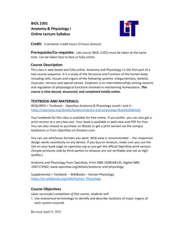
Lab Manual Exercise # 1Section B. Grains Of Salt & Metric SystemMicroscopic view of three cuboidal grains of ordinary table salt (sodium chloride or NaCl). All threegrains are just over one millimeter in length (red bar). Grains of table salt vary slightly in size, but threeaverage grains stacked together adds up to approximately one mm. If three grains equal one millimeter inlength, then a single grain is approximately 0.3 mm or 0.03 cm on a side. Moving the decimal one place tothe left converts millimeters (mm) to centimeters (cm).How Much Does The Grain Weigh?The volume of a single grain in cubic centimeters is (0.03)3 0.000027 cm3.The density of NaCl is 2.165 grams per cubic centimeter. Multiply the density of NaCl by thevolume of a single grain to obtain the weight of the grain:2.165 g/cm3 x 0.000027 cm3 0.000058 g. This value can be rounded off to about 0.00006 g, theweight of one grain.One grain weighs 0.00006 gram 0.06 milligram 60 microgramsAlthough not precise (due to the variability in grains), the size and weight of a grain of table salt makes anice comparison when discussing minute organisms or dosages. For example, the one-seeded fruit ofWolffia angusta and W. globosa (worlds smallest flowering plants) is roughly the size of a grain of tablesalt:file:///C /wayne/lmexer1.htm[4/12/2011 6:27:40 AM]
Lab Manual Exercise # 1The minute one-seeded fruits of Wolffia angusta compared with grains of ordinary table salt (NaCl).The cubical salt grains are about 0.3 mm on a side. The fruits of W. globosa are similar in size.See Metric Conversions In JavaScriptOne of the deadliest seeds on Earth is the castor bean (Ricinus communis). The seeds contain ricin, a verytoxic protein compound known as a lectin. According to the Merck Index: An Encyclopedia ofChemicals, Drugs, and Biologicals (1997), a dose of ricin weighing only 70 micrograms or two millionthsof an ounce (roughly equivalent to the weight of a single grain of table salt from a salt shaker) is sufficientto kill a 150 pound (68 kg) person. The colorful seeds of prayer bead (Abrus precatorius) contain anotherpoisonous lectin called abrin. A dose of abrin equivalent to the weight of about 20 grains of table salt couldbe lethal to an adult human weighing 150 pounds. According to the Merck Index, one thoroughlymasticated seed of prayer bead (A. precatorius) can cause fatal poisoning.In 1978, a Bulgarian dissident, Georgi Markov, was assassinated in London after being pricked by a ricintipped umbrella. Ricin causes a slow and painful death through blood poisoning and a breakdown of thecirculatory system. There is no known antidote for ricin poisoning. Even before the tragic terrorist planecrashes into the Trade Center Twin Towers in New York, some airports hand-inspected umbrellas packed incarry-on luggage. Following the Gulf War, UN investigator teams (UNSCOM) discovered that Iraq waspurifying ricin for possible use in biological warfare, along with anthrax (Bacillus anthracis), botulismtoxin (Clostridium botulinum), gas gangrene (C. perfringens), and aflatoxin (Aspergillus parasiticus).From: Facts on File News Services (23 January and 13 February 1998).According to The Washington Post Online (16 November 2001), Osama bin Ladens's al-Qaida networkalso had working plans for making ricin. Instructions for preparing the poison were found in the cellar of anabandoned house once used as a terrorist training center. According to the article, the ricin may be ingested,injected and inhaled. The article also states that the laxative effect of castor oil is due to ricin, but this isdoubtful. Castor oil is derived from the seeds of the castor bean. It contains 87 percent ricinoleic acid, afatty acid with many industrial uses, along with small amounts of several other fatty acids including oleic(7%), linoleic (3%), palmitic (2%) and stearic (1%). The deadly protein ricin is not a component of purifiedcastor oil.file:///C /wayne/lmexer1.htm[4/12/2011 6:27:40 AM]
Lab Manual Exercise # 1Seeds of prayer bead (left) and castor bean (right).See Article About The Castor Bean ShrubSee Article About Seeds Used For JewelrySee Chemical Compounds In Living SystemsSection C. Elodea Leaf Cell1. Depth of Field: When using a compound microscope this term refers to the thickness of the subject thatis in focus. The Elodea leaf is composed of two layers of cells. Only one layer of cells is in focus whenusing the high power (40x) objective. Therefore, the depth of field is limited to only one layer of cells. Youmust focus u
1. Cells From An Onion Skin 2. Diameter Of Field Of View 3. Table Of Cell Size Comparisons 4. Bird Eggs: World's Largest Cells 5. Grains Of Salt & Metric System 6. Elodea Leaf Cell & Plasmolysis 7. Salt Intake At A Movie Theatre 8. Important Definitions For Cell Exam 9. Potato Tuber & Banana Fruit 10. Oak Wood & Specific Gravity 11. Human Cheek .