Transcription
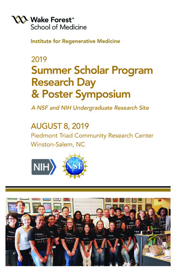
2019Summer Scholar ProgramResearch Day& Poster SymposiumA NSF and NIH Undergraduate Research SiteAUGUST 8, 2019Piedmont Triad Community Research CenterWinston-Salem, NC
WFIRM 2019 Summer Scholars ProgramResearch Day & Poster SymposiumCelebrating Multidisciplinary ResearchExperiences for Undergraduate Scholars inChallenging Areas of Regenerative MedicineThursday, August 8, 2019The Wake Forest Institute for Regenerative Medicine (WFIRM) is aninternational leader in translating scientific discovery into clinicaltherapies. Our mission is to improve patients’ lives through regenerativemedicine.A significant challenge in this promising field is developing the nextgeneration of engineers, scientists, clinicians and entrepreneurscognizant of the challenges and approaches needed to solveregenerative medicine problems and design functional replacementtissues and organs. Here at WFIRM, inherently tied to our mission isthe training of next generation experts to whom we will look towardto continue to advance and deliver upon the promise of this field andmake a lasting impact on health conditions ranging from heart disease,diabetes, injury and aging.Congratulations to our WFIRM Summer Scholars 2019.Today we invite you to join us in celebrating the graduation of 24summer undergraduate scholars, who participated in our annual 10week summer research program. June 3rd, 2019 marked the beginningof WFIRM’s Annual, 10-week, Summer Scholar Program. Today’s finalResearch Day with oral presentations and scientific poster sessionprovides the opportunity for undergraduate researchers who join us fromacross the United States and globe, to present their summer research.We offer congratulations to our talented scholars, their faculty mentorsand near-peer mentors comprised of teams of graduate, postdoctoralfellows and lab technicians. All providing important contributions andsupport. Much success is extended to our 2019 cohort of undergraduatescholars in their future educational and career pursuits. WFIRM isprivileged to be a part of their intellectual and professional growth. Wethank you all for joining us today and the support you have extended tothese fine young adults.Anthony Atala, MDDirector, Wake Forest Institute forRegenerative MedicineJoan F. Schanck, MPASummer Scholars Program Director
Introducing the 2019 WFIRM Summer ScholarsSummer ScholarPrimary Faculty Mentor(s)Honour AdewumiJarvis Christian CollegeChemistry and Biology, JuniorGiuseppe Orlando, MD, PhDAssistant ProfessorJames BennettBucknell UniversityBiomedical Engineering, JuniorAnthony Atala, MDProfessor and Director of WFIRMOlivia CainSpelman CollegeBiology, FreshmanSteve J. Walker, PhDProfessorJames DayNorth Carolina State UniversityChemical Engineering, SophomoreThomas Shupe, PhDAssistant ProfessorJoselyn De Jesus GonzalezUniversity of Puerto Rico – Rio PiedrasCell and Molecular Biology, JuniorGraca Almeida-Porada, MD, PhDProfessor andChristopher Porada, PhDProfessorAnna DealGeorgia Institute of TechnologyBiochemistry, JuniorAnthony Atala, MDProfessor and Director of WFIRMLauren DrakeUniversity of PennsylvaniaBioengineering, JuniorEmmanuel Opara, PhDProfessorAnushka GeraldUniversity of MarylandBioengineering, JuniorSang Jin Lee, PhDAssociate ProfessorCaterina GrassoRice UniversityBioengineering, SophomoreAnthony Atala, MDProfessor and Director of WFIRMAlbert HanRice UniversityBioengineering, JuniorJames Yoo, MD, PhDProfessorJada JacksonTuskegee UniversityChemical Engineering, SophomoreGiuseppe Orlando, MD, PhDAssistant ProfessorBrandon KassoufGeorgia Institute of TechnologyBiomedical Engineering, FreshmanYuanyuan Zhang, MD, PhDAssociate ProfessorEmma KoukosSaint Michael’s CollegeBiology, JuniorSteve J. Walker, PhDProfessorDaniel LeeWinston-Salem State UniversityExercise Science, SeniorDavid McGuirtElon UniversityBiology, JuniorSang Jin Lee, PhDAssociate ProfessorKhalil Bitar, PhDProfessorSamuel MossUniversity of Wisconsin – MadisonBiomedical Engineering, JuniorAleks Skardal, PhDAssistant ProfessorAlexandra SaldanaLaTourneau UniversityBiomedical Engineering, JuniorAnthony Atala, MDProfessor and Director of WFIRM
David McGuirtElon UniversityBiology, JuniorKhalil Bitar, PhDProfessorIntroducing the 2019 WFIRM Summer ScholarsSamuel MossUniversity of Wisconsin – MadisonSummerScholarBiomedical Engineering,JuniorAleks Skardal, PhDAssistant ProfessorPrimary Faculty ring,JuniorChemistryBiology, JuniorAnthony Atala,MDMD, PhDGiuseppeOrlando,ProfessorProfessorand Director of WFIRMAssistantMacaiahSheffieldJamesBennettGeorgia MilitaryCollegeBucknellUniversitySports y Criswell,AnthonyAtala,PhDMDAssistant ProfessorProfessorand Director of WFIRMOlivia CainEmma Carin StattSpelman CollegeUniversity of DaytonBiology, FreshmanPre-Med, JuniorJames DayNorth Carolina State UniversityMallory ThomasChemicalEngineering, SophomoreNorth Carolina State UniversityBiologicalFreshmanJoselyn DeSciences,Jesus GonzalezUniversity of Puerto Rico – Rio PiedrasJacobThompsonCellandMolecular Biology, JuniorUniversity of IowaBiomedicalAnna Deal Engineering, JuniorGeorgiaInstitute of TechnologyNikhil VettikattuBiochemistry,Junior CaliforniaUniversity of SouthernHumanBiology, JuniorLauren DrakeUniversityof PennsylvaniaOlivia ZyniewiczBioengineering,JuniorUniversity of NotreDameNeuroscienceand Behavior, JuniorAnushkaGeraldUniversity of MarylandBioengineering, JuniorEmmanuel Opara, PhDSteve J. Walker, PhDProfessor andProfessorYuanyuan Zhang, MD, PhDAssociate ProfessorThomas Shupe, PhDGracaAlmeida-Porada,MD, PhDAssistantProfessorProfessor andChristopherPorada, PhDMD, PhDGracaAlmeida-Porada,ProfessorProfessor andChristopher Porada, PhDShay Soker, PhDProfessorProfessorAnthony Atala, MDProfessorand Directorof WFIRMVijay Gorantla,MD, PhDAssociate ProfessorEmmanuel Opara, PhDProfessorYoung Min Ju, PhDAssistant ProfessorSang Jin Lee, PhDAssociate ProfessorCaterina GrassoRice UniversityBioengineering, SophomoreAnthony Atala, MDProfessor and Director of WFIRMAlbert HanRice UniversityBioengineering, JuniorJames Yoo, MD, PhDProfessorJada JacksonTuskegee UniversityChemical Engineering, SophomoreGiuseppe Orlando, MD, PhDAssistant ProfessorBrandon KassoufGeorgia Institute of TechnologyBiomedical Engineering, FreshmanYuanyuan Zhang, MD, PhDAssociate ProfessorEmma KoukosSaint Michael’s CollegeBiology, JuniorSteve J. Walker, PhDProfessorDaniel LeeWinston-Salem State UniversityExercise Science, SeniorSang Jin Lee, PhDAssociate Professor
WFIRM 2019 Summer Scholars ProgramResearch Day & Poster SymposiumThursday, August 8, 2019WFIRM 2019 SUMMER SCHOLARS FINAL RESEARCH DAYOral Presentation Venue: PiedmontResearch Center (PTCRC)Thursday, TriadAugustCommunity8, 2019115 S. Chestnut St., Winston-Salem, NC 27101Oral Presentation Venue: Piedmont Triad Community Research Center (PTCRC)Poster Session and LuncheonVenue:Wake ForestInstitute for115 S. ChestnutStreet, Winston‐Salem,NC 27101Regenerative MedicinePoster Session and Luncheon Venue: Wake Forest Institute for Regenerative Medicine,391 TechnologyWay, Winston-Salem,NC 27101391 Technology Way, Winston‐Salem, NC 27101ScheduleSCHEDULE7:45 am to 8:00 am7:45am-8:00amSummerScholars Arrivalat PTCRC:Photo SessionSummerScholarsarrivalat PTCRC:photo session8:00am-8:30am8:00 am to 8:30 amGuestarrivalat RegistrationPTCRC: registrationwithGuestArrivals:w/coffee, biscuitsand refreshmentsmuffins at PTCRC8:30am-8:45am8:30 am to 8:45 ,Director,WFIRMandAnthony Atala, MD,MD, Director,WFIRMandSchanck, MPA,MPA, SummerScholarsProgram tor8:45am-10:00am8:45 am to 10:00 amSummerScholars’PresentationsSummerScholars’ Presentations– Part -I Part I(Note:GroupQ&AQ&A ession 11 Nikhil VettikattuUniversity of Southern California2 Joselyn De Jesus GonzalezUniversity of Puerto Rico – RioPiedras3 Macaiah SheffieldGeorgia Military College4 Olivia ZyniewiczNotre Dame UniversitySession 25 Emma KoukosSaint Michael’s College6 Olivia CainSpelman College7 Mallory ThomasNorth Carolina State University8 Anna DealGeorgia Institute of TechnologySession 39 Honour AdewumiJarvis Christian College10 Jacob ThompsonUniversity of Iowa11 James BennettBucknell UniversityNONINVASIVE NEAR INFRARED FLUORESCENCE IMAGING ORMACROPHAGE MEDIATED INFLAMMATIONDEVELOPMENT OF IN VITRO ASSAYS TO VALIDATE IMMUNOLOGICALOUTCOMES IN SHEEP UNDERGOING CELL AND GENE THERAPY FORHEMOPHILIA ATHE EFFECT OF MUSCADINE GRAPE EXTRACT (MGE) TREATMENT ONMACROPHAGE ACTIVITY IN SKELETAL MUSCLE AFTER INJURYTARGETED ANGIOGENESISINTEGRATIVE MICRORNA‐MRNA CO‐EXPRESSION ANALYSIS OF RIGHT‐SIDEDCOLONIC HYPOMOTILITY IN CHILDREN WITH AUTISMCOMPARING MIRNA AND GENE EXPRESSION PROFILES OF BLADDERMUCOSA IN IC/BPS PATIENTSTHE EFFECTS OF SPACE RADIATION ON GASTROINTESTINAL TISSUESHUMAN PLACENTAL‐DERIVED STEM CELLS ATTENUATE EXPERIMENTALNECROTIZING ENTEROCOLITISREMODELING THE EXTRACELLULAR MATRIX OF THE LUNGS CREATES A PRE‐METASTATIC NICHEEFFECTS OF MICROBIOTA METABOLITES ON COLON CANCER AND ITSIMMUNE MICROENVIRONMENT IN AN ORGANOID MODELDIGITAL ANALYSIS OF FULL‐THICKNESS WOUNDS CONFIRMS ENHANCEDEPIDERMAL REMODELING THROUGH THE APPLICATION OF BIOPRINTEDSKINPage 4
12 Samuel Moss12 Samuel MossUniversity of Wisconsin ‐University of Wisconsin ‐MadisonMadison13 Davis McGuirt13 Davis McGuirtElon UniversityElon UniversityBIOPRINTING OF MULTIPLE MYELOMA MICROENVIRONMENT TOBIOPRINTING OF MULTIPLE MYELOMA MICROENVIRONMENT TOEVALUATE PATIENT‐DERIVED ORGANOIDSEVALUATE PATIENT‐DERIVED ORGANOIDSFABRICATION OF ELECTROSPUN PCL SCAFFOLDS FOR REPLICATION OFFABRICATION OF ELECTROSPUN PCL SCAFFOLDS FOR REPLICATION OFSMALL INTESTINE SMOOTH MUSCLE TISSUESMALL INTESTINE SMOOTH MUSCLE TISSUE10:00 am to 10:15 am10:00am-10:15am10:00 am to 10:15 amCoffeeBreakCoffeeBreakCoffeeBreak10:15 am to 11:20 am10:15am-11:20am10:15 am to 11:20 amSummerScholars’ Presentations– Part II- Part IISummerScholarsPresentationsSummerScholars’ Presentations– Part II(Note: Group Q&A follows each session)(Note:GroupQ&Afollowseach(Note: Group Q&A follows each session) session)Session 4Session 414 Albert Han14 Albert HanRice UniversityRice UniversityEFFECTS OF PRINTING CONDITIONS ON PRINTABILITY FOR EXTRUSION‐EFFECTS OF PRINTING CONDITIONS ON PRINTABILITY FOR EXTRUSION‐BASED BIOPRINTINGBASED BIOPRINTING15 Caterina Grasso15 Caterina GrassoRice UniversityRice UniversityA NOVEL ORGANOID THAT IS STRUCTURALLY AND FUNCTIONALLYA NOVEL ORGANOID THAT IS STRUCTURALLY AND FUNCTIONALLYREPRESENTATIVE OF HUMAN SKINREPRESENTATIVE OF HUMAN SKIN16 James Day16 James DayNorth Carolina State UniversityNorth Carolina State UniversityCHARACTERIZATION OF HUMAN PRIMARY CELLS GROWN IN REMDO’SCHARACTERIZATION OF HUMAN PRIMARY CELLS GROWN IN REMDO’SCHEMICALLY‐DEFINED AND XENO‐FREE MEDIUMCHEMICALLY‐DEFINED AND XENO‐FREE MEDIUM17 Daniel Lee17 Daniel LeeWinston‐Salem State UniversityWinston‐Salem State UniversityUTILIZATION OF DECELLULARIZED EXTRACELLULAR MATRIX FOR THEUTILIZATION OF DECELLULARIZED EXTRACELLULAR MATRIX FOR THEDEVELOPMENT OF BONE TISSUE‐SPECIFIC BIOINK FOR CELL‐BASED 3DDEVELOPMENT OF BONE TISSUE‐SPECIFIC BIOINK FOR CELL‐BASED 3DBIOPRINTINGBIOPRINTINGMATHEMATICAL AND PHYSICAL MODELING OF GELMA BIOPRINTING FORMATHEMATICAL AND PHYSICAL MODELING OF GELMA BIOPRINTING FOROPTIMIZED URETHRA BIOPRINTINGOPTIMIZED URETHRA BIOPRINTINGIN VITRO LIVER MODELS USING PEG BASED HYDROGELSIN VITRO LIVER MODELS USING PEG BASED HYDROGELS18 Alexandra Saldana18 Alexandra SaldanaLaTourneau UniversityLaTourneau University19 Anushka Gerald19 Anushka GeraldUniversity of MarylandUniversity of MarylandSession 5Session 520 Lauren Drake20 Lauren DrakeUniversity of PennsylvaniaUniversity of Pennsylvania21 Emma Carin Statt21 Emma Carin StattUniversity of DaytonUniversity of Dayton22 Brandon Kassouf22 Brandon KassoufGeorgia Institute of TechnologyGeorgia Institute of Technology23 Jada Jackson23 Jada JacksonTuskegee UniversityTuskegee University24 Anna Jones24 Anna JonesUniversity of North CarolinaUniversity of North CarolinaENGINEERING AN ALGINATE MATRIX TO MIMIC THE PANCREATICENGINEERING AN ALGINATE MATRIX TO MIMIC THE PANCREATICMICROENVIRONMENT TO ENHANCE ENCAPSULATED ISLET CELL FUNCTIONMICROENVIRONMENT TO ENHANCE ENCAPSULATED ISLET CELL FUNCTIONCONTROLLED DELIVER OF IGF1 USING ALGINATE MICROBEADSCONTROLLED DELIVER OF IGF1 USING ALGINATE MICROBEADSTHE REGENERATIVE POTENTIAL OF EXOSOMES FROM URINE‐DERIVEDTHE REGENERATIVE POTENTIAL OF EXOSOMES FROM URINE‐DERIVEDSTEM CELLSSTEM CELLSEXTRACELLULAR MATRIX AS A CELL CULTURE MEDIA SUPPLEMENT FOREXTRACELLULAR MATRIX AS A CELL CULTURE MEDIA SUPPLEMENT FORLONG‐TERM ISLET CULTURELONG‐TERM ISLET CULTURETHE BIOLOGICAL EFFECTS OF PANCREAS EXTRACELLULAR MATRIXTHE BIOLOGICAL EFFECTS OF PANCREAS EXTRACELLULAR MATRIXHYDROGEL IN RECAPITULATING HUMAN ISLETS' BIOLOGICAL NICHEHYDROGEL IN RECAPITULATING HUMAN ISLETS' BIOLOGICAL NICHE11:20 am to 11:35 pmWrap‐Up/Certificates of Completion11:20 am to 11:35 pmWrap‐Up/Certificates of Completion(Note: Walk to WFIRM for Poster Session & Luncheon)(Note: Walk to WFIRM for PosterSession & Luncheon)11:20am-11:35amWrap-Up/Certificationsof Completion11:45pmtoto12:30 forpmPosterSession at WFIRM (2nd Floor Collaboration Area)Note:WalkWFIRM& Luncheon11:45pm to12:30 pmPoster SessionPosterSession at WFIRM (2 Floor Collaboration Area)nd12:30 pm to 1:45 pm12:30 pm to 1:45 pm11:45am-12:30pm1:50 pm to 2:30 pmLunch with Scholars at WFIRM (2ndnd Floor Collaboration Area)Lunchwith Scholarsat WFIRMFloor CollaborationArea) Area)PosterSessionat WFIRM(2(2nd FloorCollaborationLab Tours at WFIRM1:50 pm to 2:30 pmLabat rea)(Note: Guests sign‐up during registration.Scholarsassist withwithdemos,Scholarsoverview of theirin lab atCollaborationstations)(Note: Guests sign‐up during registration. Scholars assist with demos, overview of their work in lab at stations)1:50pm-2:30pmLab Tours at WFIRMNote: Guests sign-up during registration. Scholars assist with demos, overview of their work in lab at stations)Page 5Page 5
1Noninvasive Near Infrared Fluorescence Imaging Surveillance of Macrophage MediatedInflammationN. T. Vettikattu*, F. Selek, H. Kapucu, H. Karagoz, Lu Liu, F. Zor, J.M. Janjic, V. S. Gorantla*Summer Scholar, Wake Forest Institute of Regenerative MedicineAims and Objectives: Macrophages (MΦ) play key roles in innate and adaptive immunity during sterileand non-sterile inflammation. Recent studies have highlighted their role in acute rejection ( AR), graftvasculopathy, nerve regeneration and autoimmune disease. The central role of macrophages (MΦ) in theimmune response makes them an attractive target for noninvasive monitoring of inflammation.Recent development of MΦ-targeted, perfluorocarbon (PFC) nanoemusions (NE) with near infra-redfluorescence (NIRF) probes enable tracking of peripheral macrophages to sites of inflammation. OtherNIRF probes like, MMPSense 680 are selectively cleaved by proteases from activated MΦ in inflamedtissues. The aim of this study was to develop an in-vivo, non-invasive strategy for real-time, sequential,monitoring of MΦ migration, trafficking and activation that could help guide and determineresponsiveness to treatment in acute or chronic inflammatory states.Methods: Complete Freund’s Adjuvant (CFA) (InvivoGen, San Diego, CA) was used to cause a lastingimmune response and induce localized sterile inflammation in the rat hind limb. 100 µl of CFA wasinjected in the right leg of each rat and 100 µl of PBS was injected in the left leg (control). 50 µl ofMMPSense 680 (Perkin Elmer, Akron, OH) was injected alone or co-injected with CFA in the right leg(or with PBS in opposite control legs) according to study group. PFC-NE (500 µl) was systemicallyadministered on day -1 via penile vein. NIRF imaging was performed on a LI-COR Pearl (LICORBiosciences, Lincoln, NE) in both legs at 700 nm (MMPSense 680) and 800 nm (PFC-NE) on Day 0[0,1,3 6 hours] and POD 1, 2, 3, and every third day till end point of day 30. NIRF signal intensity wasquantified and correlated with histology of skin samples to determine tissue inflammatory cell infiltration.Results: The time course of signal changes on imaging showed that MMPSense signal started as early1 day and peaked between 1 and 3 days. The PFC-NE signal began to increase between 1 and 2 days andpeaked between 3 and 6 days after CFA. Signal intensity was significantly different at these time pointsbetween experimental versus control limbs. Both signals co-localized to the site of CFA injection,indicating both the migration and subsequent activation of systemic MΦ at the site of inflammation . Thedelayed time to peaking of NIRF signal on PFC-NE indicates the time for peripheral macrophages totraffic to the inflamed limb tissues (24-48 hours post- injury), whereas the earlier peaking of signal onMMPSense 680 indicates the activation of in situ macrophages in the immediate proximity of CFAinjection. The co- localization of the MMP and NE signal between days 1 and 6 demonstrate that MΦfrom the periphery are homing to the site of inflammation and becoming activated, working inconjunction with the resident MΦ of the tissue.Conclusion: Our non-invasive real-time, surveillance strategy for imaging peripheral infiltrating andlocally activated MΦ could help preventive or personalized treatment of acute or chronic inflammatoryconditions and improve the safety and efficacy of therapies. Since inflammation is an early precursor ofthe immune response, monitoring inflammation can provide valuable information for the regulation ofimmunosuppressants in transplant patients, ultimately reducing the substantial toxicity burden of thesedrugs. Correlation with histology can increase the positive predictive value of these strategies.Acknowledgement: This summer research position was made possible by NSF REU grant #1659663entitled, “Engineering New REU Approaches to Challenges in MultiTERM” (Atala, Schanck).
2DEVELOPMENT OF IN VITRO ASSAYS TO VALIDATE IMMUNOLOGICAL OUTCOMES INSHEEP UNDERGOING CELL AND GENE THERAPY FOR HEMOPHILIA A*J. De Jesús González, M. Rodríguez**, A. Atala, C.Porada, G. Almeida-Porada* Summer Scholar, Wake Forest Institute for Regenerative MedicineHemophilia A (HA) is an X-linked disorder caused by the lack, or the reduced activity, of functional clottingfactor VIII (FVIII) and is one of the most common severe bleeding disorders. It may be inherited or arisefrom a spontaneous mutation, affecting 1 in 5000 males (1). The most common treatments for HA patientsare based in replacement or on-demand therapy with plasma derived- or recombinant- FVIII intravenousinfusions. Although HA patients have several therapeutic options available, treatment is limited by highcost, and lifelong treatment. In addition, HA patients develop immunological responses against the infusedFVIII protein. Cell and/or gene therapy is expected to become a therapeutic alternative to FVIII factorconcentrates (2).The overall goal of this research is to provide a lifelong cure to HA using a prenatal cell and gene therapydelivery approach, to allow long-term engraftment of FVIII secreting cells and induction of central immunetolerance to both, FVIII and the gene modified donor cells.In order to test prenatal stem cell and gene delivery (PNTx) platforms to treat HA, we use sheep, as a largeanimal model. Thus, in order to validate these therapies and evaluate immunological outcomes, reliable andrigorous tests have to be developed for this animal model.We hypothesize that prenatal exposure to FVIII–transduced donor cells in sheep fetuses induces centralimmune tolerance postnatally and deletion of T cell FVIII specific clones, thus here we aim to determinethe normal levels of T cells in our animals, and develop and validate methods to determine antigenspecific T cells.Lymphocytes and monocyte-derived dendritic cells (MDDC) were isolated from peripheral bloodmononuclear cells (PBMC) of sheep. For the characterization of the T cell subsets, the cells were labeledwith antibodies that bind specific cell markers for the identification of CD4, γδ, and FoxP3 T cells via flowcytometry. Next, a protocol was designed, and optimized to detect antigen-specific T cells against the FVIIItransgene product. In parallel, we also developed a universal positive read out for the experiment, since itwould be possible that FVIII-specific T cells were deleted after PNTx due to induction of central tolerance.Thus, we examined whether we were able to detect the presence of keyhole limpet hemocyanin (KLH)specific T cells in sheep. To this end, MDDC (3) were cultured from PBMC in AIM V, cytokines andgrowth factors. CD4 T and γδ T-cells were isolated through negative selection. T cells and MDDC werethen co-cultured with keyhole limpet hemocyanin (KLH) for a duration of 14 days on a growth media of10% fetal bovine serum with cytokines to stimulate T cell differentiation and proliferation (4). The CD4 Tcells that became activated in response to KLH were identified by activation markers expressed on the cellsurface via flow cytometry.Data analysis shows that the baseline of T cells in sheep is predominantly composed of CD4 T cells(17.82%) and γδ T cells (23.71%). In addition, our results demonstrated the detection of KLH-specificCD4 T cells via presence of key activation markers. Based on the current data in our animal model, weconclude that 1) frequencies of T cell subsets can be identified and 2) antigen-specific T cells can bedetected, both serving as fundamental findings for subsequent identification of FVIII-specific T cells inPNTx recipients.
Acknowledgement: This summer research position was made possible by NSF REU grant #1659663entitled, “Engineering New REU Approaches to Challenges in MultiTERM” (Atala, Schanck).References:1. Graw, J.et al. Haemophilia A: from mutation analysis to new therapies. Nat. Rev. Genet. 6, 488501 (2005)2. High, K.A. Gene transfer as an approach to treating hemophilia. Circ. Res. 88, 137-144 (2001)3. da Silva Simoneti G, Saad ST, and Gilli SC (2014) An efficient protocol for the generation ofmonocyte derived dendritic cells using serum-free media for clinical applications in postremission AML patients. Ann Clin Lab Sci 44:180–188.4. Jacquemin, M. (2002). CD4 T-cell clones specific for wild-type factor VIII: a molecularmechanism responsible for a higher incidence of inhibitor formation in mild/moderate hemophiliaA. Blood, 101(4), 1351–1358.
3THE EFFECT OF MUSCADINE GRAPE EXTRACT (MGE) TREATMENTON MACROPHAGE ACTIVITY IN SKELETAL MUSCLE AFTER INJURY*M. Sheffield, J. Poteracki, Y. Zhou, T. Criswell*Summer Scholar, Wake Forest Institute for Regenerative MedicineBackground:Muscadine Grape Extract (MGE) is a nutraceutical derived from grapes that contains polyphenols andother compounds. Macrophages are vital for inflammation. They activate inflammatory responses, fightpathogens, and initiate tissue repair at the site of injury. We tested the effect of MGE to differentiatemonocytes (M0) to M1 pro-inflammatory and/or M2 anti-inflammatory macrophages, which are involvedin the healing process.Objectives:Our goal for the project was to see if MGE enhanced macrophage conversion from M1 to M2 in vitro andin skeletal muscle in vivo after injury.Methods:In vitro, we treated a monocyte cell line (M0) with LPS to for induction of M1 macrophages and IL4 forinduction of M2 macrophages. We cultured the different macrophage populations with MGE 24hr prior todifferentiation. qPCR was used to determine the expression of markers specific for either M1 or M2macrophage16 hours after treatment. In vivo experiments consisted of providing MGE in the drinkingwater to rats 2 weeks prior to injury. Muscles were harvested from the rats 3, 7 and 14 days post-injuryand stained for the pan-macrophage marker CD68.Results:Using qPCR, we determined that MGE treatment resulted in a significant decrease in the M1 marker Il6,but not the M1 marker Il1b. Furthermore, MGE had no effect on any of the M2 markers examined. Invivo, MGE decreased macrophage count early after injury (day 3 – acute inflammatory phase) andincreased macrophage count at day 7 after injury (remodeling inflammatory phase).Conclusions:This data suggests that MGE may prove to be an effective anti-inflammatory treatment for injuredskeletal muscle.Acknowledgement:This summer research position was made possible by NSF REU grant #1659663 entitled, “EngineeringNew REU Approaches to Challenges in MultiTERM” (Atala, Schanck).
4TARGETED ANGIOGENESIS*O.M. Zyniewicz, J. H. Park, Y. M. Ju*Summer Scholar, Wake Forest Institute for Regenerative MedicineRegenerative medicine has made many advancements in the production of regenerative tissue; however,these approaches are limited in size and physiological relevance due to the tissues’ need to maintain aconstant supply of nutrients and oxygen (1, 2, 3). The delivery of these resources occurs via angiogenesis,or the growth of blood vessels (1, 2, 3). As the natural process of angiogenesis is relatively timeconsuming (5 µm/hour), methods have been developed to enhance and control the angiogenic process (2).These methods include the introduction of natural and artificial pro-angiogenic growth factors,implantation of prevascularized constructs, induced vascularization of natural and synthetic scaffolds, andproduction of synthetic vascular architecture. However, these methods are limited with regard torobustness, viable longevity, and replication of physiological sizing (1, 2).The complexity of the human body adds a level of difficulty to angiogenic process; natural vasculaturedevelops with specific hierarchy, geometry and exerts paracrine activity to the parenchyma (5). Theproximity of nonvascular and vascular tissue necessitates meticulous control over the location ofangiogenesis, thus angiogenesis of regenerating tissue must be controlled (4, 5).In this study, we hypothesized that simultaneous delivery of pro- and anti-angiogenic factors via ahydrogel scaffold will allow for control of angiogenesis and promotion of vasculature in the targeted area.Vascular endothelial growth factor, VEGF, was the pro-angiogenic factor investigated in this experiment.Bevacizumab is an FDA approved anti-cancer drug that was used as the anti-angiogenic factor in thisexperiment.An in vitro study was performed to determine the efficacy of the pro- and anti-angiogenic factors bymeasuring the effects of varying concentrations of VEGF and bevacizumab on cell cytotoxicity,proliferation and migration. All concentrations of bevacizumab and VEGF showed no cytotoxic effects oncells. Cell proliferation testing under fetal bovine serum supplementation conditions showed a clear trendwhere increased concentrations of bevacizumab correlated with low cell proliferation.To evaluate angiogenic properties in ovo, a chorioallantoic membrane assay was performed. Egg shellwindows were made in fertilized eggs after three days of incubation. VEGF and bevacizumab weretransferred onto the egg membrane via a silicon rubber plate. Blood vessel growth measured on Day 10showed decreased blood vessel growth after treatment with bevacizumab compared to treatment withVEGF. For future studies, a drug-eluting scaffold was designed on an IOP2 printer with 80 kpa at a feedrate of 200mm/min, using F-127 DA biomaterial. The proposed model will contain two channels to eluteVEGF and bevacizumab respectively in order to direct angiogenesis to the targeted area.Acknowledgement: This summer research position was made possible by NSF REU grant #1659663entitled, “Engineering New REU Approaches to Challenges in MultiTERM” (Atala, Schanck).References:1. Silva, E.A.; Mooney, D.J. Spatiotemporal control of vascular endothelial growth factor delivery2. Laschke,M.W. hydrogelsVascularizationin angiogenesis.Tissue Engineering:Angiogenesisvs Inosculation.from injectableenhancesJ ThrombHaemost 2007;5: 590-8. Eur SurgRes 2012; 48: 85-92.3. Dew, L.; MacNeil, S.; Chong, C.H. Vascularization strategies for tissue engineers. Regen Med2015; 10: 211-224.4. Baranski et al. Geometric control of vascular networks to enhance engineered tissue integrationand function. PNAS 2013; 110:75886-7591.5. Post, M. J.; Rahimi, N.; Caolo, V. Update on vascularization in tissue engineering. Regen Med2013; 5: 757-770.
5INTEGRATIVE miRNA-mRNA CO-EXPRESSION ANALYSIS OF RIGHT-SIDED COLONICHYPOMOTILITY IN CHILDREN WITH AUTISM*E. Koukos, A. Krigsman, T. Simon, S. J. Walker*Summer Scholar, Wake Forest Institute for Regenerative MedicineGastrointestinal (GI) symptoms are more common and contribute to defects of cognition and behavior inchildren with autism spectrum disorder (ASD) compared to typically developing (TD) children.1-2Furthermore, of the GI symptoms most commonly observed in children with ASD chronic constipation isreported by parents as being especially problematic.2 We have observed children with ASD who havesought medical assistance for chronic constipation on a background of colonic inflammation. Two clinicaltrends (phenotypes) were observed based on the children’s response to anti-inflammatory therapy: (1)patients who experience remission from constipation while undergoing anti-inflammatory therapy (fastresponders), and (2) patients who experience recurrent right-side fecal loading while undergoing antiinflammatory therapy (slow responders).3 Total gene expression derived from right colon biopsies of 35patients (15 fast responders, 20 slow responders), distinguished ASD fast responders from ASD slowresponders.3 The objective of this study is to further compare the two ASD subgroups through microRNA(miRNA) and total gene (mRNA) co-expression analysis to identify potential molecular regulators of theatypical constipation.Expression profiles for miRNA were assessed
Emmanuel Opara, PhD Professor Anushka Gerald University of Maryland Bioengineering, Junior Sang Jin Lee, PhD Associate Professor Caterina Grasso Rice University Bioengineering, Sophomore Anthony Atala, MD Professor and Director of WFIRM Albert Han Rice University Bioengineering, Junior James Yoo, MD, PhD Professor Jada Jackson