Transcription
Kim et al. BMC Cancer (2015) 15:1015DOI 10.1186/s12885-015-2015-1RESEARCH ARTICLEOpen AccessClinical significance of OCT4 and SOX2protein expression in cervical cancerBo Wook Kim1,4, Hanbyoul Cho2, Chel Hun Choi1,3, Kris Ylaya1, Joon-Yong Chung1, Jae-Hoon Kim2*and Stephen M. Hewitt1*AbstractsBackground: Cancer stem cell markers have become a major research focus because of their relationship withradiation or chemotherapy resistance in cancer therapy. Cancer stem cell markers including OCT4 and SOX2 havebeen found in various solid tumors. Here, we investigate the expression and clinical significance of OCT4 and SOX2in cervical cancer.Methods: To define the clinical significance of OCT4 and SOX2 expression, we performed immunohistochemistryfor OCT4 and SOX2 on 305 normal cervical epithelium samples, 289 cervical intraepithelial neoplasia samples, and161 cervical cancer cases and compared the data with clinicopathologic factors, including survival rates of patientswith cervical cancer.Results: OCT4 and SOX2 expression was higher in cervical cancer than normal cervix (both p 0.001). OCT4overexpression was associated with lymphovascular space invasion (p 0.045), whereas loss of SOX2 expressionwas correlated with large tumor size (p 0.015). Notably, OCT4 and SOX2 were significantly co-expressed inpremalignant cervical lesions, but not in malignant cervical tumor. OCT4 overexpression showed worse 5-yeardisease-free and overall survival rates (p 0.012 and p 0.021, respectively) when compared to the low-expression group,while SOX2 expression showed favorable overall survival (p 0.025). Cox regression analysis showed that OCT4 was anindependent risk factor (hazard ratio 11.23, 95 % CI, 1.31 - 95.6; p 0.027) for overall survival while SOX2 overexpressionshowed low hazard ratio for death (hazard ratio 0.220, 95 % CI, 0.06–0.72; p 0.013).Conclusions: These results suggest that OCT4 overexpression and loss of SOX2 expression are strongly associated withpoor prognosis in patients with cervical cancer.Keywords: Neoplastic stem cells, OCT4, SOX2, Prognosis, Survival, Uterine cervical neoplasmsBackgroundCervical cancer is one of the most common gynecologicmalignancies worldwide and remains a leading cause ofcancer-related death for women in developing countries[1]. Such high mortality rates are ascribed to disease recurrence despite cervical resection as well as ineffectivetreatment options for advanced disease. Radiation therapy is widely employed for advanced cervical cancer, but* Correspondence: jaehoonkim@yuhs.ac; genejock@helix.nih.gov2Department of Obstetrics and Gynecology, Gangnam Severance Hospital,Yonsei University College of Medicine, 146-92 Dogok-Dong, Gangnam-Gu,Seoul 135-720, South Korea1Experimental Pathology Lab, Laboratory of Pathology, Center for CancerResearch, National Cancer Institute, National Institutes of Health, MSC 1500,Bethesda, MD 20892, USAFull list of author information is available at the end of the articleradiation resistance is an obstacle for cancer eradication.Radiation resistance of cancer cells is acquired by intrinsic and extrinsic factors including tumor hypoxia, cellcycle and DNA repair and radiation resistance in cancerstem cells (CSCs) [2, 3].CSCs are a small subpopulation of cancer cells thathave stem cell features such as self-renewal and the ability to differentiate into multiple cell types. Radiationtherapy or chemotherapy largely eliminates cancer cells,including cervical cancer, but some tumor cells surviveand acquire radiation or chemotherapy resistance [4, 5].These resistant cancer cells are difficult to eradicate, andthey show properties of CSCs.Among CSC markers, octamer-binding transcriptionfactor 4 (OCT4) and sex determining region Y-box 2 2015 Kim et al. Open Access This article is distributed under the terms of the Creative Commons Attribution 4.0International License (http://creativecommons.org/licenses/by/4.0/), which permits unrestricted use, distribution, andreproduction in any medium, provided you give appropriate credit to the original author(s) and the source, provide a link tothe Creative Commons license, and indicate if changes were made. The Creative Commons Public Domain Dedication o/1.0/) applies to the data made available in this article, unless otherwise stated.
Kim et al. BMC Cancer (2015) 15:1015(SOX2) are transcriptional factors involved in the regulation of several target genes including NANOG, Fgf4 andUtf1, as well as OCT4 and SOX2 [6–10]. OCT4 belongsto the POU (Pit-Oct-Unc) transcriptional factor familyand plays a key role in stem cell pluripotency and differentiation by determining the fate of embryonic stemcells [11]. OCT4 expression in cancer stem-like cells isassociated with self-renewal and tumorigenesis via regulation of its target genes [12]. OCT4 expression has beenshown to be correlated with poor tumor differentiationand metastasis, as well as poor prognosis in colon, pancreas and lung cancer [13–15]. SOX2, a member of theSRY-related HMG-box (SOX) family of transcription factors, stimulates the reprogramming of adult cells into induced pluripotent stem cells and maintains stem cell-likeproperties in cancer by complexing with other stem cellmarkers such as NANOG and OCT4 [9]. SOX2 expression was reported to be correlated with tumorigenesis,chemoresistance and maintenance of stem cell-like phenotype in cancer cells [16, 17]. In addition, SOX2 has beenshown to be highly expressed in premalignant lesions suchas squamous dysplasia and carcinoma in situ in lung [18].Prior studies suggest OCT4 and SOX2 have a key role oftumorigenesis and prognosis of cancer. However, theprognostic significance of OCT4 and SOX2 is not clearlydefined in cervical premalignant and malignant lesion. Inthis study, we investigated the clinical significance ofOCT4 and SOX2 expression in cervical neoplasia.MethodsPatient selectionA total of 450 patients with cervical cancer and cervicalintraepithelial neoplasia (CIN) were collected from patients who enrolled at Gangnam Severance Hospital,Yonsei University College of Medicine in Seoul, Koreaand the Korea Gynecologic Cancer Bank through Bio &Medical Technology Development Program of theMinistry of Education, Science and Technology, Koreabetween 1996 and 2010. One hundred sixty-oneparaffin-embedded specimens of cervical cancer, 289CIN and 305 matched normal tissues were included inthe study. Medical records were obtained to review patient data including age, cancer stage, tumor differentiation, cell type, tumor size, lymphovascular spaceinvasion (LVSI) and lymph node (LN) metastasis. Cervical cancer was staged according to the InternationalFederation of Gynecology and Obstetrics (FIGO) stageand histologically classified and graded according toWorld Health Organization (WHO) grade. Patients withsurgical indications underwent radical hysterectomy withpelvic and aortic lymph node dissection. Concurrentchemoradiation therapy was added in cases with risk factors such as LN metastasis, parametrial invasion andpositive resection margin. Inoperable patients underwentPage 2 of 8radiation or chemoradiation therapy. Tissue samples andmedical records were obtained with informed consent ofall patients and approval of the local research ethicscommittee (approval no. 3-2010-0030; Seoul, SouthKorea). This study was additionally approved by the Office of Human Subjects Research at the National Institute of Health.Tissue microarray construction and immunohistochemistryTissue microarrays (TMAs) were constructed from 450 patients with primary invasive cervical cancer or CIN, as wellas 305 matched non-adjacent normal epithelial tissues.After hematoxylin and eosin slides were reviewed by apathologist, areas containing each category were indicatedby marking them. Four 1-mm punches were then takenfrom the corresponding regions of the paraffin blocks andtransplanted into a recipient paraffin block using a tissuearrayer (Pathology Devices, Westminster, MD).For immunohistochemical staining, all paraffinembedded sections were cut at 5-μm thickness followedby deparaffinization through xylene and dehydration withgraded ethanols. Antigen recovery was performed in heatactivated antigen retrieval pH 6 (Dako, Carpinteria, CA)for OCT4 and SOX2, and then specimens were incubatedwith 3 % H2O2 for 10 min. Non-specific binding wasblocked with protein block (Dako) for 20 min at roomtemperature. The sections were incubated with rabbitpolyclonal anti-OCT4 antibodies (Abcam, Cambridge,MA; Cat. #ab19857) at 1:250 for 30 min or with rabbitmonoclonal anti-SOX2 antibodies (Cell Signaling,Danvers, MA; Cat. #3579) at 1:500 for 2 h, respectively.Subsequently, antigen-antibody reaction was detectedwith EnVision Dual Link System-HRP (Dako) and visualized with DAB (3, 3’-Diaminobenzidine; Dako). Tissuesections were lightly counterstained with hematoxylin andthen examined by light microscopy. Negative controls(substitution of primary antibody with TBS) were run simultaneously. Positive controls included testicular seminoma and lung squamous cell carcinoma for OCT4 andSOX2 [19] antibodies, respectively.Quantitative evaluation of immunostainingImmunohistochemically stained slides were digitized at 20 magnification utilizing an Aperio Scanscope CS(Aperio, Vista, CA, USA). Images were reviewed usingan online software application, Digital Image Hub (SlidePath, Dublin, Ireland). Once the areas were annotated,they were sent for automated image analysis utilizingTissueIA (SlidePath’s Tissue IA system, version 3.0,Dublin, Ireland). Within Tissue IA, an algorithm was developed to quantify OCT4 and SOX2 expression levels.The staining intensity of OCT4 and SOX2 wascategorized as 0 (no staining), 1 (weak), 2 (moderate)and 3 (strong). The overall immunohistochemical score
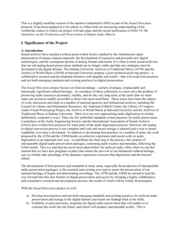
Kim et al. BMC Cancer (2015) 15:1015(histoscore) was expressed as the percentage of positivecells multiplied by their staining intensity (possiblerange, 0–300) [20].Page 3 of 8Table 1 Patient clinicopathologic characteristicsFrequency%43.3aAgeDiagnostic categoryStatistical analysisHistoscores were compared using one-way ANOVA testand independent t-test. The immunohistochemical cutoff for high expression of tumor markers was determined through receiver operating characteristic (ROC)curve analysis. The sensitivity and (1 - specificity) fordiscrimination of dead and alive was determined foreach immunohistochemistry (IHC) score and plotted,thus generating a ROC curve. The cut-off value wasestablished to be the point on the ROC curve wheresum of sensitivity and specificity was maximized.Kaplan-Meier survival analysis was performed to determine the association of OCT4 and SOX2 expressionwith survival, and the survival curves were compared between groups using log-rank tests. Multivariate analysesof hazard ratio for death were performed using Cox proportional hazards regression. Chi-square test was usedto evaluate the association between OCT4 and SOX2.Statistical analyses were performed using SPSS version21.0 (SPSS Inc., Chicago, IL). A value of p 0.05 wasconsidered statistically significant.Normal30540.4Low grade CIN597.8High grade CIN23030.5Cancer16121.3 IIA11873.3 .4AD169.9Other148.7 4 cm11269.6 4 e2114.2Positive12785.8FIGO stageTumor differentiationbCell typeTumor sizecLVSIResultsClinicopathologic characteristics of casesTable 1 presents the patients’ clinicopathologic characteristics. Of 161 patients with cervical cancer, 118 patients were stage IIA or less and 43 patients were stageIIB or higher. The mean age was 43.3 years (range, 19–83 years). The tumor sizes ranged from 0.2 to 12.0 cm(mean, 2.8 cm). The histopathology included 131 squamous cell carcinoma, 16 adenocarcinoma, 7 adenosquamous and 7 other types (3 small cell carcinomas, 2neuroendocrine and 2 mixed cell types). Patients withcervical cancer were evaluated for survival analysis andthe mean follow-up time of surviving patients was54.3 months (range, 1–179). Fifteen patients (9.3 %) diedduring the follow-up period.LN metastasisdeHPV test in CINCIN cervical intraepithelial neoplasia, FIGO International Federation of Gynecologyand Obstetrics, SCC squamous cell carcinoma, AD adenocarcinoma, LVSIlymphovascular space invasion, LN lymph node, HPV human papillomavirusamean valuebcalculated based on 156 cases with available tumordifferentiation informationccalculated based on 153 cases with available LV invasion informationdcalculated based on 155 cases with available LN metastasis informationecalculated based on only 148 cases of CIN with available HPV test dataOCT4 and SOX2 protein expressionExpression of OCT4 and SOX2 was evaluated by IHCin cervical neoplasia and cancer specimens. Subsequently, we performed analysis of both markers usingquantitative image analysis software. RepresentativeIHC images of OCT4 and SOX2 are presented inFig. 1. OCT4 expression was observed primarily inthe nucleus with limited cytoplasm expression, whileSOX2 was restricted to the nucleus (Fig. 1). Only nuclear staining was considered OCT4- and SOX2positive.Of the cancer specimens, 92 of 161 cancers(57.1 %) had high expression of OCT4 (histoscore 200) and 125 of 161 cancers (77.6 %) had high expression of SOX2 (histoscore 30). Association ofOCT4 and SOX2 expression with clinicopathologiccharacteristics in cervical cancer is summarized inTable 2. OCT4 and SOX2 expression was significantlydifferent depending on diagnostic category (p 0.001).OCT4 overexpression was associated with lymphovascular space invasion (p 0.045), whereas loss of SOX2
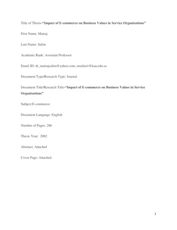
Kim et al. BMC Cancer (2015) 15:1015Page 4 of 8abcdFig. 1 OCT4 and SOX2 expression in formalin-fixed, paraffin-embedded cervical cancer tissues. Representative immunohistochemical image ofOCT4 negative (a) and positive (b), SOX2 negative (c) and positive (d). Insets show high magnification of areas indicated with boxes. Scale bar: 100 μmexpression was correlated with large tumor size (p 0.015).There were no other correlations between OCT and SOX2expression and clinicopathologic characteristics.We next examined the association between OCT4 andSOX2 expression, Chi-squared distribution was used inmalignant and premalignant lesions. In premalignantcervical lesions, SOX2 expression presented a significantcorrelation with OCT4 (p 0.004), while there was noassociation between OCT4 and SOX2 in malignanttumors (p 0.543; Table 3).Prognostic significance of OCT4 and SOX2 expressionFive-year disease-free and overall survival rates were analyzed through the Kaplan-Meier plots as shown inFig. 2. In survival analysis with OCT4 expression, 22 recurrences and 13 deaths occurred in 83 cases of OCT4high expression, while 7 recurrences and 3 deaths wereobserved in 65 cases of low expression. The 5-yeardisease-free and overall survival rates were 89.2 and95.4 % in low OCT4 expression and 73.5 and 84.3 % inhigh OCT4 expression. OCT4 overexpression was associated with shorter disease-free and overall survival thanthe low expression group (p 0.012 and p 0.021, respectively) (Fig. 2a and d). In survival analysis of SOX2,there were 20 recurrences and 8 deaths in 125 highexpression patients, while 8 recurrences and 7 deathsoccurred in 36 low-expression patients during the 5-yearfollow-up period. The 5-year disease-free and overallsurvival rates were 77.8 and 80.6 % in cases with lowSOX2 expression and 84.0 and 93.6 % in case with highSOX2 expression cases. High expression of SOX2 was associated with better overall survival than low expression(p 0.025) (Fig. 2e). When survival of patients with expression of high OCT4/low SOX2 was compared withsurvival of patients with low OCT4/high SOX2, KaplanMeier analysis revealed a significant difference in diseasefree and overall survival (p 0.016 and p 0.001, respectively; Fig. 2c and f).Cox proportional multivariate analysis of relationshipsbetween prognostic variables and survival are shown inTable 4. FIGO stage was an independent survival factor forboth disease-free and overall survival analysis (p 0.001and p 0.041, respectively). OCT4 overexpression showedindependent poor overall survival with a hazard ratio of11.23 (p 0.027), while high expression of SOX2 presentedbetter disease-free and overall survival compared to lowexpression, as shown in Table 4 (p 0.019 and p 0.013,respectively).DiscussionOCT4 and SOX2 are important transcriptional factorsinvolved in maintenance of pluripotency and selfrenewal in cancer stem cells, aberrant expression ofOCT4 and SOX2 might contribute to carcinogenesis invarious cancers [15, 21, 22]. Radioresistance is importantin the treatment and prognosis of cervical cancer and itis known to be associated with cancer stem cells [3].This study examined the clinical correlation and prognostic significance of stemness-related OCT4 and SOX2protein expression assessed by IHC in premalignant andmalignant cervical tumors. The results demonstrate thatOCT4 and SOX2 protein expression is elevated in premalignant and malignant cervical tumors compared tonormal cervix and this finding is consistent with a
Kim et al. BMC Cancer (2015) 15:1015Page 5 of 8Table 2 Association between clinicopathologic characteristics and OCT4 or SOX2 expressionOCT4SOX2p valueMean Histoscore (95 % CI)Diagnostic categoryMean Histoscore (95 % CI) 0.001 0.001Normal113.3 (105.1–121.4)36.5 (32.3–40.8)Low-grade CIN197.6 (177.4–217.7)40.0 (28.8–51.2)High-grade CIN219.0 (211.3–226.8)91.7 (79.9–103.5)Cancer208.5 (196.7–220.3)105.4 (91.8–119.1)FIGO stage0.498 IIA205.7 (191.6–219.7) IIB215.1 (192.6–237.6)0.529108.1 (92.1–124.2)98.3 (71.5–125.1)Tumor differentiation0.4380.112Well moderate203.8 (187.7–219.8)112.8 (94.4–131.1)Poor213.4 (195.3–231.4)90.1 (69.0–111.3)Cell type0.450SCC205.8 (192.4–219.2)Other217.2 (190.8–243.6)0.060111.7 (96.4–127.0)78.3 (69.0–111.3)Tumor size0.8680.015 4 cm208.7 (194.2–223.2)116.5 (100.1–132.8) 4 cm206.5 (185.5–227.6)80.3 (56.3–104.2)LVSI0.045No195.3 (175.8–214.8)Yes220.1 (205.3–234.9)p value0.106115.5 (95.4–135.6)91.8 (71.2–112.5)LN metastasis0.2060.879No202.6 (187.1–218.0)105.6 (88.6–122.7)Yes221.0 (199.7–242.3)103.1 (75.6–130.6)HPV test in CIN0.2920.907Negative246.8 (223.7–215.7)124.9 (81.5–168.7)Positive227.9 (215.7–240.1)127.6 (110.5–144.6)SCC squamous cell carcinoma, AD adenocarcinoma, LVSI lymphovascular space invasion, LN lymph node, HPV human papillomavirusprevious study [23]. OCT4 was an independent poor survival factor but SOX2 showed as a favorable prognosticfactor.In this study, OCT4 protein was observed clearly inthe nucleus and partially in the cytoplasm. Similar toTable 3 Association of OCT4 and SOX2 expression in CIN andcervical cancer patientsOCT4 expressionNo.Low (%)High (%)SOX2 Low ( )5529 (53.1)26 (46.9)SOX2 High ( )23462 (26.3)173 (73.1)CINp value0.004Cancer0.543SOX2 Low ( )3615 (41.7)21 (58.3)SOX2 High ( )12554 (43.1)71 (56.9)CIN cervical intraepithelial neoplasiaour findings, OCT4 has been reported in the cytoplasm as well as in the nucleus in previous studies[24, 25]. This staining pattern may arise from thepresence of an OCT4 isoform. OCT4 is known tohave two isoforms, OCTA and OCTB. OCT4A is observed in the nucleus and OCT4B is observed in thecytoplasm in prostate and cervical cancer [24, 26].Because OCT4 is a transcriptional regulator, the active form of OCT4 is always located in the nucleus.For this reason, we focused our automated digitalimage analysis on OCT4 protein expression in thenucleus only. Notably, OCT4 expression increasedduring cancer progression but within cancers, it wasnot correlated with known prognostic factors, such asstage, LN metastasis or tumor size. Nonetheless, itshowed high hazard ratio of death in multivariateanalysis. In previous published results, OCT4 expression was associated with unfavorable prognosis
Kim et al. BMC Cancer (2015) 15:1015Page 6 of 8Fig. 2 Kaplan-Meier survival curves of OCT4 and SOX2 expression in cervical cancer. Cervical cancer patients with high OCT4 expression had shorter 5-yeardisease-free survival (a, P 0.012) and worse 5-year overall survival (b, P 0.021) than those with low expression. Patients with high SOX2 expression hadlonger 5-year overall survival than those with low expression (e, P 0.025). The patients with low SOX2/high OCT4 expression had shorter5-year disease-free survival (c, P 0.016) and worse 5-year overall survival (f, P 0.001) than those with high SOX2/low OCT4 expressionshowing poor tumor differentiation, tumor invasionand metastasis in the lungs, stomach, esophagus andoral cavity [13, 25, 27, 28]. Although there have beenlimited reports on the association between OCT4 andprognosis in cervical cancer, Shen et al. reported thatOCT4 expression was associated with radiationresistance and unfavorable survival in locally advancedsquamous cell carcinoma [29]. That study furthershowed OCT4 overexpression in the radiation resistance group, but it was not associated with high riskprognostic factors including FIGO stage and tumorsize, which is similar to our results. It is interestingthat OCT4 is associated with poor survival withoutcorrelation to known prognostic factors and evendisease-free survival. As a stem cell related protein,OCT4 expression can be related more with overallsurvival than with disease-free survival which is possibly more related with residual tumor after resection.Further research is required to clarify the associationbetween OCT4 and high-risk prognostic factors.SOX2 is known to play an important role in regulatingthe cell cycle, DNA repair and self-renewal in stem cells[30]. It is associated with tumorigenesis, chemoresistanceand maintenance of stem cell-like property in cancer cells,Table 4 Multivariate survival analysis of the association between prognostic variables and survival in cervical cancer patientsVariablesDisease-free survivalOverall survivalHR [95 % CI]P valueHR [95 % CI]FIGO stage ( IIB)8.67 [2.77–27.11] 0.0014.33 [1.06–17.72]0.041Tumor size ( 4 cm)1.16 [0.47–2.84]0.7391.79 [0.54–5.92]0.340LN metastasis1.72 [0.57–5.23]0.3341.60 [0.40–6.41]0.500OCT4 3.75 [1.24–11.55]0.11711.23 [1.31–95.64]0.027SOX2 0.47 [0.18–1.20]0.0190.22 [0.06–0.72]0.013FIGO International Federation of Gynecology and Obstetrics, HR hazard ratio, LN lymph node, CI confidence intervalP value
Kim et al. BMC Cancer (2015) 15:1015which suggests poor overall survival [16, 31, 32]. High expression of SOX2 was reported to be associated with alack of cell differentiation and to contribute cell migrationand invasion in cervical cancer cell line [33]. In addition,Shen et al. showed that SOX2 is highly expressed in patients with radiation resistance and predicts poor survival[29]. In contrast, SOX2 expression was associated withprolonged survival in the current study. These discrepancies might be explained by the lack of standardized methodology, different standards of interpretation ordifferences in studies’ patient populations. Similar to ourstudy, Wilbertz et al. reported that SOX2 gene amplification and protein expression are associated with favorablesurvival outcomes in squamous cell lung cancer [34]. Inaddition, a recent meta-analysis reported that SOX2 expression presents a positive prognosis in non-small celllung cancer [35]. Previously, SOX2 overexpression was reported to be associated with favorable prognosis in squamous cell lung carcinoma, but was correlated with poorsurvival in adenocarcinoma [34–36]. Notably, the poorsurvival associated with SOX2 expression that was reported in GI tract cancer mostly pertained to adenocarcinoma [37–39]. Cervical cancer consists of squamous cellcarcinoma followed by adenocarcinoma and our datacomprised 80.7 % squamous cell carcinoma and 14.8 %adenocarcinoma. Further research is required to clarifythe prognostic significance of SOX2 in cervical cancer andvariation in prognosis according to cell type.Premalignant cervical lesion demonstrated significantcorrelation between OCT4 and SOX2, while malignantlesion did not present an association between OCT4 andSOX2. The lack of a correlation between OCT4 andSOX2 in malignant lesions has not been explainedclearly because OCT4 and SOX2 are known to work cooperatively and self-regulate themselves via the OCT4/SOX2 complex in embryonic stem cells [6, 8]. However,in the current cancer tissue samples, OCT4 and SOX2were associated with opposite effects on survival andlose their association in cervical cancer, as well. Similarto our results, no correlation between OCT4 and SOX2was reported in cervical cancer [23]. In addition, Li et al.also reported that OCT4 and SOX2 were not coexpressed and also showed different survival outcomesin lung cancer tissue samples [40]. Furthermore, overexpression of SOX2 inhibited the activity of OCT4 promotor in embryonal carcinoma cells [41]. OCT4 and SOX2are known to function cooperatively through the OCT4/SOX2 complex, but OCT4, SOX2 and Nanog have beenreported to form individual complexes with nucleophosmin to control stem cell fate determination [42]. In previous study, we also observed a similar phenomenonthat Nanog expression in precancerous cervical tissuewas correlated with Tcl1a and pAkt but this relationshiplost in cancerous tissue [43]. Considering previousPage 7 of 8results and our contradictory survival data, OCT4 andSOX2 might function independently or inhibit activityduring tumor progression, and eventually lose their connection in cervical cancer.ConclusionsIn conclusion, this study investigated the immunohistochemical expression of OCT4 and SOX2 in large number of cervical cancer patients by means of imageanalysis for IHC scoring. OCT4 and SOX2 showed highexpression in premalignant and malignant cervicaltumors. Co-expression of OCT4 and SOX2 was observed in premalignant tumors, but no association wasobserved in malignant cervical tumors. OCT4 high expression showed poor disease-free survival and overallsurvival while SOX2 high expression showed favorableoverall survival in patients with cervical cancer. Cox regression analysis confirmed that OCT4, and SOX2 expression was an important prognostic indicator incervical cancer. OCT4 was associated with poor prognosis, while SOX2 showed favorable prognosis. Our findings suggest further investigation into OCT4 and SOX2as biomarkers in cervical cancer.AbbreviationsAD: adenocarcinoma; CI: confidence interval; CIN: cervical intraepithelialneoplasia; CSC: cancer stem cell; FIGO: International Federation ofGynecology and Obstetrics; H&E: hematoxylin and eosin; HPV: humanpapillomavirus; HR: hazard ration; IHC: immunohistochemistry; LN: lymphnode; LVSI: lymphovascular space invasion; OCT-4: octamer-bindingtranscription factor 4: SOX2, sex determining region Y-box 2; ROC: receiveroperating characteristic; SCC: squamous cell carcinoma; TMA: tissuemicroarray; WHO: World Health Organization.Competing interestsThe authors declare that there is no conflict of interest.Authors’ contributionsBWK, J-YC, J-HK and SMH conceived of the study and devised the experimentaldesign. SMH designed and build the tissuemicroarrays. BWK, HC, CHC and KYperformed experiments. BWK, HC, CHC, J-YC, J-HK and SMH performed dataanalysis for experiments or clinical records. BWK, HC and J-YC drafted the finalversion of the manuscript and figure legends. J-HK and SMH revised the figures,added critical content to the discussion and were responsible in revisingall portions of the submitted portion of the manuscript. All authors readand approved the final manuscript.AcknowledgmentsThis research was supported by the Intramural Research Program of theNational Institutes of Health National Cancer Institute, Center for CancerResearch.Author details1Experimental Pathology Lab, Laboratory of Pathology, Center for CancerResearch, National Cancer Institute, National Institutes of Health, MSC 1500,Bethesda, MD 20892, USA. 2Department of Obstetrics and Gynecology,Gangnam Severance Hospital, Yonsei University College of Medicine, 146-92Dogok-Dong, Gangnam-Gu, Seoul 135-720, South Korea. 3Department ofObstetrics and Gynecology, Samsung Medical Center, SungkyunkwanUniversity School of Medicine, Seoul 135-710, Republic of Korea.4Department of Obstetrics and Gynecology, Kangdong Sacred Heart Hospital,Hallym University, Seoul 135-701, South Korea.
Kim et al. BMC Cancer (2015) 15:1015Received: 1 May 2015 Accepted: 15 December 2015References1. Ferlay J, Shin HR, Bray F, Forman D, Mathers C, Parkin DM. Estimates ofworldwide burden of cancer in 2008: GLOBOCAN 2008. Int J Cancer. 2010;127:2893–917.2. Feng D, Peng C, Li C, Zhou Y, Li M, Ling B, et al. Identification andcharacterization of cancer stem-like cells from primary carcinoma of thecervix uteri. Oncol Rep. 2009;22:1129–34.3. Krause M, Yaromina A, Eicheler W, Koch U, Baumann M. Cancer stem cells:targets and potential biomarkers for radiotherapy. Clin Cancer Res. 2011;17:7224–9.4. Bao S, Wu Q, McLendon RE, Hao Y, Shi Q, Hjelmeland AB, et al. Glioma stemcells promote radioresistance by preferential activation of the DNA damageresponse. Nature. 2006;444:756–60.5. Yin T, Wei H, Gou S, Shi P, Yang Z, Zhao G, et al. Cancer stem-like cellsenriched in Panc-1 spheres possess increased migration ability andresistance to gemcitabine. Int J Mol Sci. 2011;12:1595–604.6. Chew JL, Loh YH, Zhang W, Chen X, Tam WL, Yeap LS, et al. Reciprocaltranscriptional regulation of Pou5f1 and Sox2 via the Oct4/Sox2 complex inembryonic stem cells. Mol Cell Biol. 2005;25:6031–46.7. Nishimoto M, Fukushima A, Okuda A, Muramatsu M. The gene for theembryonic stem cell coactivator UTF1 carries a regulatory element whichselectively interacts with a complex composed of Oct-3/4 and Sox-2. MolCell Biol. 1999;19:5453–65.8. Okumura-Nakanishi S, Saito M, Niwa H, Ishikawa F. Oct-3/4 and Sox2 regulateOct-3/4 gene in embryonic stem cells. J Biol Chem. 2005;280:5307–17.9. Rodda D
Kaplan-Meier survival analysis was performed to deter-mine the association of OCT4 and SOX2 expression with survival, and the survival curves were compared be-tween groups using log-rank tests. Multivariate analyses of hazard ratio for death were performed using Cox pro-portional hazards regression. Chi-square test was used