Transcription
Improving the Intercellular Uptake and Osteogenic Potency of CalciumPhosphate via Nanocomplexation with the RALA PeptideO’Doherty, M., Mulholland, E. J., Chambers, P., Pentlavalli, S., Ziminska, M., Chalanqui, M. J., Pauly, H. M.,Sathy, B. N., Donahue, T. H., Kelly, D. J., Dunne, N., & McCarthy, H. O. (2020). Improving the IntercellularUptake and Osteogenic Potency of Calcium Phosphate via Nanocomplexation with the RALA Peptide.Nanomaterials, 10(12), [2442]. https://doi.org/10.3390/nano10122442Published in:NanomaterialsDocument Version:Publisher's PDF, also known as Version of recordQueen's University Belfast - Research Portal:Link to publication record in Queen's University Belfast Research PortalPublisher rightsCopyright 2020 the authors.This is an open access article published under a Creative Commons Attribution License ch permits unrestricted use, distribution and reproduction in any medium, provided the author and source are cited.General rightsCopyright for the publications made accessible via the Queen's University Belfast Research Portal is retained by the author(s) and / or othercopyright owners and it is a condition of accessing these publications that users recognise and abide by the legal requirements associatedwith these rights.Take down policyThe Research Portal is Queen's institutional repository that provides access to Queen's research output. Every effort has been made toensure that content in the Research Portal does not infringe any person's rights, or applicable UK laws. If you discover content in theResearch Portal that you believe breaches copyright or violates any law, please contact openaccess@qub.ac.uk.Download date:08. Jul. 2022
nanomaterialsArticleImproving the Intercellular Uptake and OsteogenicPotency of Calcium Phosphate via Nanocomplexationwith the RALA PeptideMichelle O’Doherty 1,† , Eoghan J. Mulholland 1,† , Philip Chambers 1 , Sreekanth Pentlavalli 1 ,Monika Ziminska 1 , Marine J. Chalanqui 1 , Hannah M. Pauly 2 , Binulal N. Sathy 3 ,Tammy H. Donahue 2,4 , Daniel J. Kelly 3,5,6,7 , Nicholas Dunne 1,3,5,7,8,9,10,11, * andHelen O. McCarthy 1,12, *123456789101112*†School of Pharmacy, Queen’s University Belfast, Belfast BT9 7BL, UK; modoherty04@qub.ac.uk (M.O.);emulholland11@qub.ac.uk (E.J.M.); philip.chambers@qub.ac.uk (P.C.); s.pentlavalli@qub.ac.uk (S.P.);m.ziminska@qub.ac.uk (M.Z.); mchalanqui01@qub.ac.uk (M.J.C.)Department of Biomedical Engineering, University Colorado State University, 1374 Campus Delivery,Fort Collins, CO 80523, USA; hmpauly1@gmail.com (H.M.P.); thautdonahue@umass.edu (T.H.D.)Trinity Centre for Bioengineering, Trinity Biomedical Sciences Institute, Trinity College Dublin,Dublin 2, Ireland; binulalns@gmail.com (B.N.S.); kellyd9@tcd.ie (D.J.K.)School of Biomedical Engineering, University of Massachusetts Amherst, 130 Natural Resources Road,Amherst, MA 01003, USADepartment of Mechanical and Manufacturing Engineering, School of Engineering, Trinity College Dublin,Dublin 2, IrelandDepartment of Anatomy, Royal College of Surgeons in Ireland, Dublin 2, IrelandAdvanced Materials and Bioengineering Research Centre (AMBER),Royal College of Surgeons in Ireland and Trinity College Dublin, Dublin 2, IrelandSchool of Mechanical and Manufacturing Engineering, Dublin City University, Dublin 9, IrelandCentre for Medical Engineering Research, School of Mechanical and Manufacturing Engineering,Dublin City University, Dublin 9, IrelandAdvanced Manufacturing Research Centre (I-Form), School of Mechanical and Manufacturing Engineering,Dublin City University, Glasnevin, Dublin 9, IrelandAdvanced Processing Technology Research Centre, Dublin City University, Dublin 9, IrelandSchool of Chemical Sciences, Dublin City University, Dublin 9, IrelandCorrespondence: nicholas.dunne@dcu.ie (N.D.); h.mccarthy@qub.ac.uk (H.O.M.)These authors contributed equally to this work.Received: 3 November 2020; Accepted: 27 November 2020; Published: 7 December 2020 Abstract: Calcium phosphate-base materials (e.g., alpha tri-calcium phosphate (α–TCP)) havebeen shown to promote osteogenic differentiation of stem/progenitor cells, enhance osteoblastosteogenic activity and mediate in vivo bone tissue formation. However, variable particle sizeand hydrophilicity of the calcium phosphate result in an extremely low bioavailability. Therefore,an effective delivery system is required that can encapsulate the calcium phosphate, improvecellular entry and, consequently, elicit a potent osteogenic response in osteoblasts. In this study,collagenous matrix deposition and extracellular matrix mineralization of osteoblast lineage cellswere assessed to investigate osteogenesis following intracellular delivery of α-TCP nanoparticles.The nanoparticles were formed via condensation with a novel, cationic 30 mer amphipathic peptide(RALA). Nanoparticles prepared at a mass ratio of 5:1 demonstrated an average particle size of 43 nmwith a zeta potential of 26 mV. The average particle size and zeta potential remained stable for up to28 days at room temperature and across a range of temperatures (4–37 C). Cell viability decreased24 h post-transfection following RALA/α-TCP nanoparticle treatment; however, recovery ensuedby Day 7. Immunocytochemistry staining for Type I collagen up to Day 21 post-transfection withRALA/α-TCP nanoparticles (NPs) in MG-63 cells exhibited a significant enhancement in collagenNanomaterials 2020, 10, 2442; aterials
Nanomaterials 2020, 10, 24422 of 16expression and deposition compared to an untreated control. Furthermore, in porcine mesenchymalstem cells (pMSCs), there was enhanced mineralization compared to α–TCP alone. Taken togetherthese data demonstrate that internalization of RALA/α-TCP NPs elicits a potent osteogenic responsein both MG-63 and pMSCs.Keywords: calcium phosphate; peptide; RALA; intercellular; osteogenic; bone engineering1. IntroductionDevelopments in biomaterial, stem cell and nanomedicine technologies are continually improvingthe quality of tissue-engineered scaffolds [1–6]. However, these applications are limited and donot replicate the innate bone environment [3]. The focus is now on developing strategies aimed atenhancing the intrinsic repair mechanisms of the body to stimulate new, biologically functional bonetissue regrowth to replace tissue that has been lost as a result of trauma or disease [6]. Promotingosteogenic differentiation and bone forming potential of osteoprogenitor/osteoblast cells is an emergingapproach for improving bone tissue engineering [7–10]. Osteoprogenitors and osteoblasts, derivedfrom bone marrow stromal cells, are found at the advancing surface of repairing bone and play akey role in bone production, maintenance and remodeling; producing, depositing and mineralizingthe extracellular collagen matrix [11,12]. Therefore, the modulation of the osteogenic properties ofthese cells represents a potential target to promote soft tissue attachment to bone. Bergemann et al.,(2015) have demonstrated the influence of implant material surface topography on the behaviorof primary human osteoblasts [13]. This investigation demonstrated that in vitro, micro-structuredzirconia surfaces can modulate key bone-related gene expression in human primary osteoblasts,enhancing osseointegration of oral implants. Furthermore, Hu and Olsen (2016) have shown in vitrothat osteoblast-derived vascular endothelial growth factor functions as a paracrine factor on osteoblasticlineage cells promoting osteoblastic maturation and mineralization [14].Calcium phosphate-based materials exhibit similar chemical and structural properties to naturalbone minerals and so are widely used in a range of biomedical applications for bone repair: As cements tofill bone defects, coating materials to improve orthopedic implant biocompatibility and in biodegradablebioceramics and composites [15–18]. Biomaterials containing a calcium phosphate component havebeen shown to promote osteogenic differentiation of progenitor and stem cells and can facilitate in vivobone tissue formation [19,20]. In particular, alpha-tricalcium phosphate (α-TCP) has been frequentlyutilized as a bone substitute material as it is biocompatible, osteoconductive, osteoinductive andbiodegradable and elicits no immunogenic effect [21]. Furthermore, implants coated with α-TCP havebeen shown to improve fixation and integration as the calcium phosphate can form chemical bondswith native bone [22]. Lui et al., (2015) demonstrated that elevated extracellular α-TCP levels enhancedosteogenic differentiation in rat bone mesenchymal stem cells (MSCs) by upregulating a number ofosteogenic related-genes [23].Variable particle size and hydrophilicity of α-TCP limits its bioavailability. Yet the role ofintracellular calcium phosphate in the mineralization of the extracellular matrix (ECM) has beendemonstrated [24]. Therefore, a more efficacious delivery system is required that can encapsulate thecalcium phosphate, improve cellular entry and, consequently, elicit a potent osteogenic response inosteoblasts. To this end, cationic 30 mer amphipathic peptide (RALA) is a novel, non-viral, 30-aminoacid peptide gene delivery vehicle, comprised of repeating arginine/alanine/leucine/alanine units [25].The amphipathic nature of RALA facilitates interaction with the lipid bi-layers, enabling transportacross cellular membranes. Furthermore, the pH-sensitive α-helicity of the RALA peptide facilitatesendosomal disruption and escape, preventing degradation of the cargo with delivery to the cytosol.Consequently, the RALA delivery system can significantly improve the bioavailability of hydrophilictherapeutics [26–28].
Nanomaterials 2020, 10, 24423 of 16In this study, we postulate that the intracellular uptake of α-TCP in the form of nanoparticles(NPs), facilitated by condensation with intracellular delivery peptide RALA, will improve its long-termbioavailability and consequently stimulate the osteogenic potential and bone forming properties ofosteoblasts, which could have a key role in bone repair and regeneration.2. Materials and Methods2.1. Materials2.1.1. RALA PeptideThe RALA peptide was synthesized as previously described [25–28]. Briefly, the peptidewas produced by 9-fluorenylmethyloxycarbonyl (FMOC solid-state peptide synthesis (Biomatik,Washington, DE, USA) and supplied as a desalted, lyophilized powder. The product was purified andvalidated by reversed-phase high-performance liquid chromatography; molecular mass was confirmedas 3327.98 Da.2.1.2. Synthesis of Alpha-Tri Calcium PhosphateThe α-TCP (Ca3 (PO)4 ) powder was synthesized according to the following solid-statereaction [29,30] (Equation (1)):2CaHPO4 (s) CaCO3 (s) Ca3 (PO)4 (s) CO2 (g) H2 O(g)(1)Calcium phosphate (CaHPO4 ) and calcium carbonate (CaCO3 ) precursors were mixed at a molarratio of 2:1, which was turbo-blended and sintered in a furnace (Elite BRF15/5, Elite Thermal SystemsLtd., Harborough, UK) at 1400 C for 6 h. The sintered specimens were then removed and placedinto a stainless steel bowl and rapidly cooled using compressed air, reducing the possible phasetransformation of α-TCP to beta-TCP (β-TCP). The cooled powder was subsequently ground in ethanol(99.5%) using a planetary mill (Pulverisette 6, Frisch, Idar-Oberstein, Germany) at 5 min intervals for atotal of 30 min at 600 5 RPM, which produced an average D10 particle size of 1.04 0.1 µm, D50 of3.51 0.4 µm and D90 of 10.86 0.6 µm as measured by laser diffraction. The milled α-TCP was storedin a desiccator until required.2.1.3. PlasmidpEGFP-N1 (GFP) expression plasmid, purchased from Clontech (Mountain View, CA, USA),was amplified in MAX Efficiency DH5α Competent Cells (Life Technologies, Inchinnan, UK),purified using PureLink HiPure Plasmid Filter Maxiprep Kit (Life Technologies, Inchinnan, UK) andquantified by UV absorption at 260 nm using a NanoDrop 2000c spectrophotometer (Thermo Scientific,Loughborough, UK).2.2. Methods2.2.1. X-ray DiffractionCrystalline phase purity was analyzed by X-ray diffraction using an X’Pert PRO MPD (PANalyticalLtd., Malvern, UK) (CuKα radiation, at 40 kV on a rotating stage, Range (2q): 5–6). The JCPDS filefrom the X’Pert High Score V 2.2b software (PANalytical Ltd., Malvern, UK) was used to identify theα-TCP phase (#09-0348).2.2.2. Fourier Transform Infra-Red SpectroscopyStructural information of the α-TCP powder at the molecular level was determined by Fouriertransform infrared spectroscopy (FTIR) (FT-IR 4100, Jasco Ltd., Dunmow, UK). For this using KBrpellets of α-TCP powder was prepared and scanned in the wavelength range of 500–4000 cm 1 at256 scans with a resolution of 4 cm 1 .
Nanomaterials 2020, 10, 24424 of 162.2.3. Scanning Electron MicroscopyMorphological analysis of the α-TCP powder was performed by scanning electronmicroscopy (SEM) using the Hitachi Analytical Tabletop Microscope/Benchtop SEM TM3030 system(Hitachi High-Technologies Europe GmbH, Warrington, UK). An acceleration voltage of 5 kV wasused. All powders were mounted on aluminum discs and sputter coated with gold under vacuum toinduce conductively.2.2.4. Particle Size and Zeta Potential AnalysisRALA/α-TCP NPs were prepared at mass ratios of 1, 5, 10 and 15 to a final volume of 1 mL; e.g.,to achieve a ratio of 10:1, 10 µg of RALA was added to 1 µg of α-TCP in aqueous solution. NPs wereallowed to incubate at room temperature for approximately 30 min. In a separate set of experiments,RALA/α-TCP NPs were prepared at a mass ratio of 5 and incubated at: (i) room temperature for 0, 1, 7,14, 21 and 28 days and (ii) a range of temperatures from 4 to 37 C. Particle size and zeta potentialwere subsequently analyzed using the Nano ZS Zetasizer and DTS software (Malvern Instruments,Malvern, UK).2.2.5. Cell CultureHuman osteosarcoma cells MG-63 (ATCC CRL-1427 ) were purchased from ATCC (Manassas,VA, USA). Cells were maintained and expanded in Eagle’s minimum essential medium (MEM) (Gibco ,Life Technologies, Inchinnan, UK) supplemented with 10% fetal bovine serum (FBS) (Life Technologies,Inchinnan, UK) in a humidified incubator at 37 C and 5% CO2 . Prior to use, cells were trypsinized,using trypsin/EDTA in phosphate buffered solution (PBS) (2X) (Life Technologies, Inchinnan, UK),centrifuged, and re-suspended in MEM. Osteosarcoma MG-63 cells represent a cell type commonlyassociated with initial cell material characterization. This cell line is known for the high proliferationpotential as they have the ability to continuously divide and grow and are therefore utilized innumerous in vitro biocompatibility studies. However, porcine mesenchymal stem cells (pMSCs) wereused to support any conclusions made about biocompatibility and osteogenic functionality of theRALA/α-TCP nanoparticles. pMSCs were isolated from the femora of freshly sacrificed 3–8 month-oldpigs, obtained from a slaughterhouse (Agri-Food and Biosciences Institute, Belfast, UK). Bone marrowwas harvested and homogenized in Dulbecco’s Modified Eagle Medium (DMEM) 10% FBS usinga 16-gauge needle. Bone marrow suspension was centrifuged at 650 g for 5 min. After removingsupernatant cells were counted and seeded at a density of 10 106 cells/T75 flask in MSC expansionmedia, DMEM, 1X GlutaMAX, 10% FBS and 1% pen/strep (Gibco , Life Technologies, Inchinnan, UK).Media was replaced every three days and flasks monitored day for emergence of MSCs.2.2.6. Nanoparticle TransfectionMG-63 cells were seeded in a 96-well flat bottom plate (VWR International, Lutterworth, UK) at adensity of 1 104 cells per well and incubated in complete culture medium for 24 h. Two hours priorto transfection, the cell culture media was replaced with OptiMEM medium (Invitrogen, Inchinnan,UK) optimized for transfection. The RALA/α-TCP NPs were prepared using a mass ratio of 5 such thatthe final amount of α-TCP/well was 1 µg, and 5 µg RALA. Untreated wells were also prepared and ablank RALA NP control group (RALA—GFP NPs) such that the amount of RALA was the same asthat used for the RALA/α-TCP NPs. Cells were incubated with nanoparticles at 37 C/5% CO2 for 4 hbefore medium was replaced with complete culture medium. The following day, culture media wasreplaced with complete medium supplemented with dexamethasone (50 nM), ascorbic acid (50 µg/mL)and β-glycerophate (10 mM). In a separate set of experiments, MG-63 cells were seeded in a 24-well flatbottom plate (VWR International, Lutterworth, UK) at a density of 3 104 cells and transfected withRALA/α-TCP NPs prepared using a mass ratio of 5 such that the final amount per well was 6.7 µg, and33.3 µg for α-TCP and RALA, respectively.
Nanomaterials 2020, 10, 24425 of 162.2.7. Transmission Electron MicroscopyThe RALA/α-TCP NP structure was examined using a transmission electron microscope (TEM,JEOL-100CXII, Welwyn Garden City, UK). NPs were prepared at a mass ratio of 5. A formvar/carbonmesh grid (Agar Scientific, Stansted, UK) was then placed face down on a 10 µL sample of theRALA/α-TCP NPs for 10 min. The grid was dried overnight and stained for 5 min with 5% aqueousuranyl acetate at room temperature.2.2.8. Cell Viability: MTS Cell Proliferation AssayMG-63 cell viability post-transfection with RALA/α-TCP NPs was assessed using the CellTiter96 Aqueous One Solution Cell Proliferation Assay (MTS) (Promega, Southampton, UK) according tothe manufacturer’s protocol. At various time points (i.e., 1, 7, 14, 21 and 28 days) post-transfection,20 µL of CellTiter 96 Aqueous One Solution Reagent was added per well of a 96-well plate containing100 µL of fresh culture medium and incubated at 37 C/5% CO2 for 2–3 h. The absorbance of each wellwas measured at 490 nm using microplate reader (PowerWave XS, BIO-TEK , Winooski, VT, USA).Results were expressed as mean relative absorbance (%) compared to the control samples.2.2.9. Cell Toxicity: Lactate Dehydrogenase AssayThe cytotoxicity of RALA NP transfection was assessed using Pierce lactate dehydrogenase (LDH)cytotoxicity assay kit (Thermo Fisher Scientific, Loughborough, UK) according to manufacturer’sprotocol. Briefly, at time points 1, 7, 14, 21 and 28 days post-transfection, 50 µL of media was removedand transferred to a fresh 96-well plate and 50 µL of reaction mixture added. After incubationat room temperature for 30 min, the reaction was stopped and LDH activity was determined byspectrophotometric absorbance at 490 nm and 680 nm (PowerWave XS, BIO-TEK , Winooski, VT,USA). Results were expressed as mean relative absorbance (%) compared to the control samples.2.2.10. ImmunocytochemistryTransfected and control MG-63 cells were fixed in 4% PFA for 30 min at room temperature andblocked for 1 h in PBS containing 1% bovine serum albumin (BSA, Sigma Aldrich, Gillingham, UK)and 0.01% Tween 20 (Sigma Aldrich, Gillingham, UK). Thereafter cells were incubated for 1 h at roomtemperature with primary antibodies against collagen (Abcam, Cambridge, UK). After washing in PBS,cells were incubated in appropriate secondary antibodies for 1 h at room temperature and observedunder a fluorescence microscope (EVOS FL, Life Technologies, Loughborough, UK). Anti-rabbit andAlexa Fluor 568 IgG (Abcam, Cambridge, UK) was used as secondary antibodies. ImageJ image analysissoftware (NIH, Bethesda, MD, USA) was subsequently used to quantify the average fluorescenceintensity of the cells in each treatment group and made relative to the untreated control cells.2.2.11. Mineralization: Alizarin Red Stain AssayThe presence of calcium in the cell cultures was determined by Alizarin Red Staining (ARS)(Sigma–Aldrich, UK) at Day 7, 14 and 21 post-transfection with RALA only, RALA/GFP andRALA/α-TCP NPs. Briefly, the pMSCs were rinsed with cold PBS and then fixed in 4% PFA for30 min. Following a rinse with distilled water, the cells were stained with 40 mM alizarin red (pH 4.2)for 30 min at room temperature.2.2.12. Alizarin Red CPC ExtractionTo quantify the level of matrix mineralization, ARS monolayers were washed in triplicate withdH2 O. ARS was extracted from the monolayer by incubation in 500 µL 100 mM cetylpyridiniumchloride (CPC) for 30 min. The dye was subsequently removed and 200 µL aliquots were transferredto a 96-well plate prior to reading at 540 nm (FLUOstar Omega BMG LabTech, Aylesbury, UK).
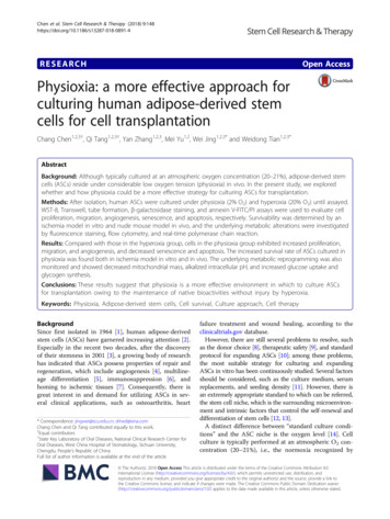
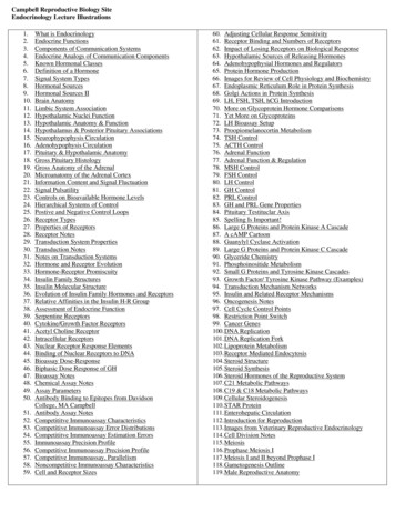
Nanomaterials 2020, 10, 24426 of 162.2.13. Statistical AnalysisStatistical analysis was performed using GraphPad Prism 6 (GraphPad Software Inc., San Diego,CA, USA). Statistical significance of differences between groups was determined by using one-wayANOVA with the Student-Newman–Keuls post-test. A p-value less than 0.05 was consideredstatistically significant.3. Results3.1. Physiochemical Properties of α-TCP ParticlesX-ray diffraction analysis of α-TCP powder showed a peak profile typical of and comparable toα-TCP of a similar standard (JCPDS # 09-0348) (Figure 1A). Characteristic peaks are highlighted by anasterix (*). FTIR analysis confirmed that calcium and phosphate were present in the α-TCP powder(Figure 1B). Formation of tri-calcium phosphate was also confirmed on FTIR analysis as the bandspresent on α-TCP spectrum (Figure 1C) are characteristic of α-TCP, as previously described [31,32].SEM showed that the synthesis and milling process produced smooth granules of α-TCP withirregular-shaped morphology (Figure 1B).120,000100,00080,00060,00040,00020,000Figure 1. Chemical and morphological characterization of alpha-tricalcium phosphate (α-TCP) powder.(A) representative XRD pattern of α-TCP exhibiting characteristic peaks; (B) FTIR spectra of α-TCP;and (C) SEM micrographs of the surface of α-TCP after final milling step show that the granules ofα-TCP have an irregular-shaped morphology. (Scale bar 100 µm; scale bar 30 µm), (N 3 SEM).3.2. Characterization of RALA/α-TCP NanoparticlesEffective intracellular uptake is dependent on the size and net charge of the nanoparticle.Therefore, following the formulation of RALA/α-TCP NPs by incubation at room temperature for30 min (Figure 2A), particle size, charge and shape of RALA/α-TCP were assessed (Figure 2A,B).Results indicated that RALA condensed the α-TCP micro-sized particles into NPs 100 nm at eachmass ratio ranging from 1 to 15. At mass ratio of 5 the average size of the RALA/α-TCP NPs was 43 nmwith a zeta potential of 26 mV (Figure 2B). TEM analysis showed that particles exhibited uniformshape and indicated no aggregation (Figure 2A). These biophysical characteristics indicate that theparticles are within the ideal size and charge range for receptor mediated endocytosis.
Nanomaterials 2020, 10, x FOR PEER REVIEW7 of 16and indicated no aggregation (Figure 2A). These biophysical characteristics indicate that the particlesare within the ideal size and charge range for receptor mediated endocytosis.Nanomaterials 2020, 10, 24427 of 16Figure 2. Formulation and characterization of cationic 30 mer amphipathic peptide (RALA)/α-TCP NP.Figure 2. Formulation and characterization of cationic 30 mer amphipathic peptide (RALA)/α-TCP(A)RALA/α-TCPnanoparticlesNP. redpreparedbybymixingmixing 11 µgµgα-TCPα-TCP withwith reconstitutedreconstituted thenincubatedatroomtemperatureRALA (lyophilized) at varying mass ratios. Particles were then incubated at room temperature forfor3030 min.Formulation and morphology of the NPs was confirmed by TEM. (Scale bar 50 nm). (B) Particle sizeand zeta potential of RALA/α-TCP NPs after formulation at mass ratios (RALA:α-TCP) 1, 5, 10 and15. The RALA peptide effectively condenses α-TCP into positively charged nanoparticles 100 nmin size up to mass ratio 15. Significant differences are denoted by the symbols (*) for hydrodynamicsize and (#) for zeta potential, compared to α-TCP Only (N 3 SEM) *** p 0.001, ### p 0.001,#### p 0.0001. (C) representative size distribution by number of RALA/ α-TCP 7:1.
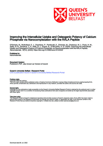
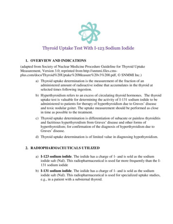
size and zeta potential of RALA/α-TCP NPs after formulation at mass ratios (RALA:α-TCP) 1, 5, 10and 15. The RALA peptide effectively condenses α-TCP into positively charged nanoparticles 100nm in size up to mass ratio 15. Significant differences are denoted by the symbols (*) forhydrodynamic size and (#) for zeta potential, compared to α-TCP Only (N 3 SEM) *** p 0.001,Nanomaterials2020, 10,24428 of 16### p 0.001,#### p 0.0001. (C) representative size distribution by number of RALA/ α-TCP 7:1.3.3. Stability of RALA/α-TCP Nanoparticles3.3. Stability of RALA/α-TCP NanoparticlesThe stability of RALA/α-TCP NP with time and over a temperature range was also investigatedThe stability of RALA/α-TCP NP with time and over a temperature range was also investigated(Figure 3). For a mass ratio of 5:1, the particle size remained consistent when exposed to temperatures(Figure 3). For a mass ratio of 5:1, the particle size remained consistent when exposed to temperaturesranging from 4–37 C (Figure 3A). NPs were also stable when stored at room temperature over aranging from 4–37 C (Figure 3A). NPs were also stable when stored at room temperature over aperiodof 28(Figure3B),consistentlyparticlesizesizeofof 50 50nmnmininsizesizeandperiodof days28 days(Figure3B),consistentlymeasuringmeasuring anan averageaverage particleand 25 25mV mVin CPintostableNPsthatretainin charge. These results suggest that RALA condensed the α-TCP into stable NPs that retainthe theidealcharacteristicsforforintercellularsize andand chargechargeatat4 4 C, C,roomroomandideal threspectrespect toto sizeandbodytemperature.bodytemperature.Figure 3. Characterization of RALA/α-TCP NP stability (mass ratio 5). (A) particle size remained stableFigure 3. Characterization of RALA/α-TCP NP stability (massratio 5). (A) particle size remainedwhen NPs were incubated over a temperature range of 4–37 C. (B) particle size and zeta potential ofstable when NPs were incubated over a temperature range of 4–37 C. (B) particle size and zetaRALA/α-TCP NPs were consistent at room temperature over 28 days, i.e., the average particle size waspotentialof RALA/α-TCP NPs were consistent at room temperature over 28 days, i.e., the average 50 nm and 25 mV in charge. (N 3 SEM).particle size was 50 nm and 25 mV in charge. (N 3 SEM).3.4. Cytotoxicity of RALA/α-TCP NanoparticlesThe transfection concentration and cytotoxic effects of the NPs was assessed in vitro using MG-63cells, a pre-osteoblast-like-cell (Figure 4A–D). At 24 h post-transfection (addition of RALA/α-TCPNPs so that 1 µg of α-TCP was added to each well of a 96-well plate) cell morphology was observed
3.4. Cytotoxicity of RALA/α-TCP NanoparticlesThe transfection concentration and cytotoxic effects of the NPs was assessed in vitro using MG63 cells, a pre-osteoblast-like-cell (Figure 4A–D). At 24 h post-transfection (addition of RALA/α-TCPNanomaterials 2020, 10, 24429 of 16NPs so that 1 µg of α-TCP was added to each well of a 96-well plate) cell morphology was observed(Figure 4A). No significant morphological changes were found between the RALA/α-TCP treated(Figure4A).significantmorphologicalfound betweenRALA/α-TCPtreatedthecells andtheNocontroluntreatedcells. Dark changesdepositswereof RALA/αTCPNPsthewereobserved sofRALA/αTCPNPswereobservedwithinthecultures and were not removed upon washing with PBS, suggesting these particles were associatedcultureswerenot removedwashing withsuggestingtheseparticles werewith theandcellseitherintra- or uponextracellularly.In PBS,a separateset ofexperiments,cellsassociatedwere nsfected with varying amounts of RALA/α-TCP NPs so that 0.5, 1.0 and 5.0 µg of α-TCP werewithvaryingamountsof RALA/α-TCPNPsfromso that0.5,1.0 and5.0indicatedµg of α-TCPperofwelladdedper wellof 96-wellplate. ResultstheMTSassaythatwerethe addedviabilitycellsof96-well plate.Results from theassay indicatedthedecreasedviability oftocellswithtransfectedwith RALA/α-TCPNPMTScontaining5.0 µg of thatα-TCP71%transfectedafter 24 h (FigureRALA/α-TCPNP containing5.0 µg of α-TCPdecreasedto 71%exhibitedafter 24 hcomparable(Figure 4B).cellCellstreated to4B). Cells treatedwith NPs containing0.5 and1.0 µg α-TCPviabilitywithNPscontaining0.5 andµg α-TCPcomparablecell viabilitycontrolcells.controluntreatedcells.Cell 1.0viabilitywas exhibitedthen assessedover a longerperiodtoviaMTSuntreated(Figure dviaMTS(Figure4C)andLDHassays(Figure4D)LDH assays (Figure 4D) comparing RALA/α-TCP NP (with α-TCP content 1 µg/well 96-well plate)comparingRALA/α-TCPα-TCP1 µg/wellgene96-wellplate) withRALA/GFP(controlwith geniccontent),free α-TCPsupplementedNPgroupwithandnon-osteogenicgene content),free α-TCPsupplementedmedia theand24controlculturemediacontrol untreatedcells. Despitean initialincrease inculturecytotoxicity,h nsfection (Figure 4D) cell viability of RALA/α-TCP NP treated cells returned to control tocontrollevelsbyDay7.ThiswassupportedbyaDay 7. This was supported by a similar finding using the MTS assay (Figure 4C).similar finding using the MTS assay (Figure 4C).Figure 4. In vitro characterization of RALA/α-TCP transfection and cytotoxicity. (A) representativeFigurecontrast4. In vitrocharacterizationof RALA/α-TCPand cytotoxicity.(A) representativephaseimagesof MG-63 osteoblast-likecells 24 transfectionh after transfectionwith 1 µg RALA/α-TCPNPsphasecontrastmassimagesof5.MG-6324 h after(Scaletransfectionwith1 µgatformulationratioWhiteosteo
zirconia surfaces can modulate key bone-related gene expression in human primary osteoblasts, enhancing osseointegration of oral implants. Furthermore, Hu and Olsen (2016) have shown in vitro . (FMOC solid-state peptide synthesis (Biomatik, Washington, DE, USA) and supplied as a desalted, lyophilized powder. The product was purified and


![Clinical significance of incidental [18 F]FDG uptake in the .](/img/32/s12876-016-0545-x.jpg)