Transcription
RESEARCH ARTICLEA Novel DNA Vaccine Coding For H5 and N1 Genes ofHighly Pathogenic Avian Influenza H5N1 SubtypeMS Elnagar Eman1, AN Gamal Maha2, FF Zaki2, MA Saad1, YA Soliman2A b s t r ac tControl of avian influenza infection requires a good vaccine that could induce both humoral and cell-mediated immune response,specifically IFN-ɣ production, to maintain a high level of protection along with the minimal level of viral shedding after the infectionto prevent secondary epidemics. In the current study, deoxyribonucleic Acid (DNA) vaccine coding for full-length H5 and N1 geneshave been produced and evaluated in SPF-chicken. Humoral immune response estimated by haemagglutination inhibition (HI) assayrevealed that the DNA vaccine gave a high titer of antibodies at the day 28-post vaccination and 14 days post-challenge. However, theshedding level was minimal with the DNA vaccine (0.1 Log 10 EID50). The IFN-ɣ transcript was upregulated at a higher level in the DNAvaccinated group. The results revealed that the DNA vaccine could induce a high level of humoral and IFN-ɣ level that maintains a highlevel of protection (92%) with the advantage of limiting the shedding level and thus, prevent secondary epidemics.Keywords: Avian influenza, DNA vaccine, Haemagglutinin gene (H5), IFN-ɣ, Neuraminidase gene (N1).Ind J of Vet Sci and Biotech (2020): 10.21887/ijvsbt.15.4.1IntroductionAvian influenza infection is caused by a group of virusesin the family Orthomyxoviridae, genus influenza virus A.Diagnosis of avian influenza based on virus isolation followedby molecular characterization of the haemagglutinincleavage site to identify its pathogenicity. Infections inbirds can give rise to a wide variety of symptoms that mayvary depending on the host species, strains of virus, thehost’s immune response, environmental conditions, and thepresence of secondary exacerbating organisms (OIE 2012).Vaccination programs, along with biosafety andbiosecurity measures, would be a powerful tool to supportthe eradication of the infectious virus from the environmentthrough increase resistance to field challenge, eliminatingviral shedding from infected birds and thus reduce viraltransmission and secondary epidemics (Capua et al., 2004).Although vaccination is the primary method for control ofthe influenza disease, the lack of proofreading activity by theRNA polymerase leads to the recurrent appearance of severalmutations in the virus progeny (Deng et al., 2006).The most widely used completely inactivated influenzavirus, vaccine, only confer optimized protection to thecirculating strain of the virus (Xu et al., 2011). This type ofvaccine cannot induce a cellular immune response, and thusviral shedding will not be hindered. Therefore due to thereasons mentioned above and due to logistical, surveillanceand monetary constraints, current vaccination approachesare not sufficient. Furthermore, due to the antigenic variantcharacteristics of the virus, vaccines may need to be updatedwith frequency to new circulating strains.DNA vaccines provide an alternative approach tomaintain both humoral and cell-mediated immune responses1Vet. Serum and vaccine research Institute, Abbasia, Cairo.Central Laboratory for Evaluation of Veterinary Biologics, Abbasia,Cairo. 131, Elsekka Elbeida , St. Abbasia, Cairo. Egypt.Corresponding Author: Y.A. Soliman, Central Laboratory for Evaluation of Veterinary Biologics, Abbasia, Cairo. 131, Elsekka Elbeidast,Abbasia, Cairo. Egypt., e-mail: dryousefadel@gamil.comHow to cite this article: Eman, M.S.E., Maha, A.N.G., Zaki, F.F., Saad,M.A., Soliman, Y.A. (2020). A novel DNA vaccine coding for H5 andN1 genes of highly pathogenic avian influenza H5N1 subtype. IndJ Vet Sci and Biotech, 15(4): 1-11.Source of support: Nil2Conflict of interest: None.Submitted: 24/03/2020 Accepted: 30/03/2020 Published: 30/04/2020to a higher degree. DNA vaccine, when administrated I/M orS/C, would give both humoral and cellular immune responsespresented to the MHC-I and MHC-II via the resident APC suchas Langerhans cells, dendritic cells and even by myocytes.The importance of induction cellular immune response maybe the fundamental advantage of the use of DNA vaccine inthe elimination of infection and eradication of the disease asit will prevent the shedding of the pathogen after infection(Shedlock and Weiner 2000).In the current study, DNA vaccine based on theexpression of H5 and N1 genes from locally circulatingfield isolate was produced and tested for its protectiveefficacy in chickens. Also, its ability to reduce viral sheddingfrom dead or diseased birds was under investigationto maintain the environmental load of the virus at theminimal. The Author(s). 2020 Open Access This article is distributed under the terms of the Creative Commons Attribution 4.0 International License (http://creativecommons.org/licenses/by/4.0/), which permits unrestricted use, distribution, and non-commercial reproduction in any medium, provided you give appropriate credit tothe original author(s) and the source, provide a link to the Creative Commons license, and indicate if changes were made. The Creative Commons Public DomainDedication waiver ) applies to the data made available in this article, unless otherwise stated.
A novel DNA vaccine coding for H5 and N1 genes of highly pathogenic avian influenza H5N1 subtypeM at e r ia l sandMethodsAll applicable international, national, and/or institutionalguidelines for the care and use of animals were followed. Inorder to develop a novel DNA vaccine coding for H5 and N1genes of highly pathogenic avian influenza H5N1 subtypefollowing steps were undertaken. Amplification of the full-length H5 and N1 genes of theH5N1 isolate by RT-PCR amplification Sequencing of the full-length H5 and N1 genes Cloning and expression vector (DNA vaccine) coding thefull H5 and N1 genes Preparation of the DNA vaccine coding for H5 and N1genes Vaccination of the chickens with DNA vaccine carryingFull H5 and N1 genes Evaluation of the protective efficacy of the DNA vaccine:Four parameters viz.antibody titer, protection percentage,shedding test, and measurement of the IFN-ɣ transcriptlevel using qRT-PCR.Avian influenza virus: (A/chicken/Qalubia/ch1.12.61/2017(H5N1))highly pathogenic Egyptian field strain clade2.2 wasused in the current study for both DNA vaccine preparationand challenge test. The strain was propagated on 9 days oldSPF.ECE and the allantoic fluid was collected and clarifiedand stored at -20 CAmplification of the full-length H5 and N1 genes:Viral RNA was extracted from the clarified Allantoic fluidaccording to the method of Sambrook et al. (1989) usingQIAamp Viral RNA Mini Kit (Qiagen Germany, cat #52904), asper the manufacturer’s instructions. The full-length H5 andN1 genes of the H5N1 isolate were amplified by two stepsRT-PCR. First, the cDNA was synthesized using M-MuLV FirstStrand cDNA Synthesis Kit (Biomatik cat # K5147) accordingto the manufacture instruction. The second step PCR wasdone using PfuUltra II Hotstart PCR Master Mix (AgilentUSA cat # 600850). Primers that target full orf of H5 and N1genes (Table 1) were designed and verified by LasergeneDNAStare software V 15 using the full-length orf of H5 andN1 sequences of avian influenza H5N1 Egyptian isolatesretrieved from the gene bank database. The ampliconswere electrophoresed on a 1% agarose and the size of theamplicons were determine using SynGene tool softwareV4.01 (SynGen Corporation, Cambridge, England)Sequencing of the full-length H5 and N1 genes:For the preparation of the full H5 and N1 genes forsequencing, the PCR products were separated on 1% lowmelting agarose. The bands were sliced off and purified withthe biospin PCR purification kit (Biobasic cat # BSC03S1), asdescribed by the manufacturer. Sequencing reactions wereperformed in a MJ Research PTC-225 Peltier Thermal Cyclerusing ABI PRISM 3730XL Analyzer BigDyeTM Terminator CycleSequencing Kits with AmpliTaq DNA polymerase (FS enzymeApplied Biosystems), following the protocols supplied by themanufacturer. Single-pass sequencing was performed oneach template using the primer used for PCR amplification.The fluorescent-labeled fragments were purified from theunincorporated terminators with an ethanol precipitationprotocol. The samples were resuspended in distilled waterand subjected to electrophoresis in an ABI 3730xl sequencer(Applied Biosystems).Cloning of full H5 and N1 genesThe amplified full-length H5 and N1 genes were clonedseparately in the donor vector using pENTR Directional(pENTR/SD/D-TOPO) Cloning Kits (life technologies USACat # K2420-20) according to the manufacture instructions,followed by transformation in TOPO 10 E.coli and inoculatedon LB-Kanamycin agar and incubated overnight at 37 C.Transformants were subcultured on LB broth-kanamycin (50ng/mL) at 37 C in a shaker incubator at a speed of 150 rpmfor 24 h. (Soliman et al., 2016)The transformants were analyzed by QPCR for thepresence of the gene of interest. Briefly, the plasmid DNATable 1: Sequences of primers used in the current studyTarget genePrimer Sequence (5’- 3’)full-length orf of H5 gene AI-H5- f5’ – CACCATGGAGAAAATAGTGCTTCTTCTT- 3’AI-H5- r5’ –AATGCAAATTCTGCATTGTAACGA- 3’full length orf of N1 gene AI-N1- f5’ – CACCATGAATCCAAATCAGAAGATAAT- 3’AI-N1- r5’ – CTTGTCAATGGTGAATGGCAACT - 3’QPCR for H5 geneHA5-f5’ – CAAGACTCTATCAAAACCCAACCAC- 3’HA5-f5’ – CACCCTCTCACYATCGGRGAATGCC- 3’QPCR for N1 geneNA-f5’ – GTCTTTGACAGTCCCRTTGGAGTGC- 3’NA-r5’ – ACTACCKGTTCCATCATTGGGGCGT- 3’QPCR IFN-ɣIFN-ɣ-f5’ –GTGAAGAAGGTGAAAGATATCATGGA- 3’IFN-ɣ -r5’ –GCTTTGCGCTGGATTCTCA- 3’IFN-ɣ pFam- 5’ –TGGCCAAGCTCCCGATGAACGA- 3’ -TamraQPCRβ- actin f 5’ –CTCCATCATGAAGTGTGACGTT- 3’β- actinβ- actin –r 5’ –ATCTCCTTCTGCATCCTGTCAG- 3’β- actin-p Fam-5’ –CAAGGACCTCTATGCCAACACAGTGCT-- 3’Tamra2The Indian Journal of Veterinary Sciences and Biotechnology, Volume 15 Issue 4 (April-June 2020)RefThis studyThis studyNaksupan et al., 2008Naksupan et al., 2008Kaiser et al., 2003Hong et al., 2006
A novel DNA vaccine coding for H5 and N1 genes of highly pathogenic avian influenza H5N1 subtypewas extracted from the overnight culture using Pure Link. HQMini Plasmid Purification Kit (Invitrogen Cat. # K2100-01). Tenng of the DNA plasmid was mixed with 12.5μl of the BrilliantII QPCR Master Mix (Agilent Cat # 600804), 100nM of each ofthe forward and reverse primers (Table 1) and 200 nM of thedual-labeled probe. The program was adjusted at 95 C for10 min initial denature then 40 cycles at 95 C for 30 seconds,annealing at 58 C for 30 sec and extension at 72 C for 30 sec.Transformants that give positive Ct ( indicating thepresence of H5 or N1 insert ) were further sub-culturedon LB-broth-kanamycin, and plasmids were purified andsubjected to homologous recombination with the GatewaypcDNA-DEST40 vector (life technologies USA Cat # 12274-015)according to the manufacturer’s instructions. Recombinantdestination vector carrying either H5 (pDEAST40/H5) orN1 (pDEAST40/N1) genes were transformed in TOPO 10E.coli, cultured on LB-ampicillin (100IU/ml) overnight at37 C. Growing colonies were sub-cultured overnight onLB-ampicillin broth in shaker incubator and tested for thepresence of H5 or N1 genes using QPCR as before.Blood samples were taken from the jugular vein of allbirds before vaccination, 15, 21, and 28 days post-vaccination(before challenge) and after 7 and 14 days post-challengefrom live birds. Heparinized blood samples were taken tomeasure the level of the IFN-ɣ transcript at days 3, 5, 7, 15and 28 post vaccination (before challenge), and 3,7 and 14days post-challenge from live birds. Cloacal swaps were takenfrom life birds 14 days post-challenge and, when possible,from the recently died chickens.Preparation of the DNA vaccine coding for H5 and N1genesA single colony of E.coli harboring the destination vectorcarrying either H5 or N1 genes was picked up in 10 mL ofLB broth containing 100 IU/mL ampicillin and incubatedovernight at 37 C with agitation (200 rpm) in shakerincubator. The whole culture was added to 1 litter LB brothwith antibiotics and incubated as before. The culture wasthen cooled down on the ice and subjected to plasmidpurification using Maxi prep kit (BioBasic cat # BS466). Theplasmid preparation (DNA vaccine) was quantified usingQubit dsDNA BR Assay Kit (Life technologies Cat# Q32850).The concentration was adjusted at 1 µg/µL and stored at-20 C till used for vaccination.Shedding testCloacal swaps were swirled in 1 mL of sterile saline andthe titer of the viral shedding was measured using EID50following the standard method in routine practice.Vaccination of the chickens with DNA vaccine carryingFull H5 and N1 genesThree weeks old, White Leghorn SPF chickens (Nile-SPF-eggsfarm, Koom Oshiem, Fayom, Egypt) were housed in closedsystem Bio-isolators with sterilized food and water supply.The chickens were divided into 3 groups 25 chicken eachand were vaccinated as in Table (2). At day 28 of vaccination,chicken challenged with 0.2 mL (virus titer 6 Log 10EID50) of the challenging virus strain (A/chicken/Qalubia/ch1.12.61/2017 (H5N1)) via eye drop route.Evaluation of the vaccine potential of the DNA vaccinecoding for H5 and N1 gene.Protection rateThe protection rate was calculated 14 days post challengeusing the following equation:(total number of bird in agroup– number of dead birdsProtection percentage �––––––––––––– X 100total number of birds)IFN-ɣ transcript quantitation using qRT-PCR Purification of Avian influenza protein antigen:Viral proteins of the strain (A/chicken/Qalubia/ch1.12.61/2017(H5N1) was purified using Trizol Reagent (Invitrogen cat No15596026) according to the manufacturer’s instructions.The purified protein was resuspended in CAHPS and theconcentration of the protein was measured using Qubit IIassay (Invitrogen Cat # Q33211) and adjusted at 5 µg/ml.The protein preparation kept in aliquots in low bind polypropelling tubes at -80 C till used. Purification of peripheral blood mononuclear cells (PBMCs)from chicken groups:PBMCs were separated by density-gradient centrifugationusing Ficoll-Paque (biowest cat# L0560-500) followingthe method of Harrington et al. ( 2007). To induce theIFN-γ transcript, the cells were pulsed with either 10 µl ofavian influenza protein (specific stimulant) or with PBS(unstimulated negative control cells). The cells incubated at37 C for 20 h in a 5% CO2 incubator. After 20 h, the cells wereTable 2: Vaccination program and doses of the experimental infection studyGroupGroup one(25 birds)Group two(25 birds)Group three(25 birds)SymbolG1G2Vaccine type and dose0.5mL of DNA vaccine containing50µg (pDEAST40/H5) and 50µg (pDEAST40/N1)Commercially inactivated oil adjuvanted avian influenza vaccineG30.5 mL saline as a non-vaccinated group (control)The Indian Journal of Veterinary Sciences and Biotechnology, Volume 15 Issue 4 (April-June 2020)3
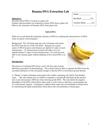
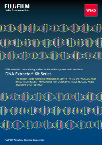
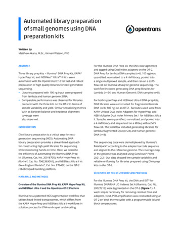
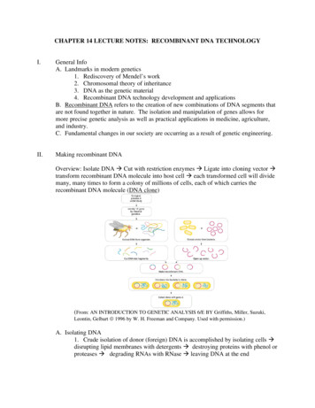
A novel DNA vaccine coding for H5 and N1 genes of highly pathogenic avian influenza H5N1 subtypeharvested and the total cytoplasmic RNA was purified usingTrizol reagent according to the manufacturer’s instructions.All RNA samples were treated with RNase-free DNase I(Qiagen cat# 79254) to remove any traces of genomic DNAcontamination. Quantitation of the IFN- γ transcript:The level of IFN-γ mRNA from cultured PBMCs either pulsedwith avian influenza proteins or the un-pulsed culture wasmeasured using Q-PCR assay (Yeong et al., 2014: Pete et al.,2003 ).Total RNA was reverse transcribed to cDNA with theOmniscript cDNA Synthesis kit (Qiagen Cat # 205111). Thereal-time PCR reaction was performed from resultant cDNAfollowing the method of Harrington et al. ( 2007) using 200nM of the probe and 100 nM each of forward and reverseprimers (sequence shown in table 1), and 1X of the brilliant IIQPCR master mix (Agilent cat # 600804).β actin genes were used as housekeeping non-regulatedreference genes for normalization of target gene expression.The results were analyzed using Livak method. Relativetranscript abundance of the IFN-γ gene equals ΔCt values (ΔCt Ct target – Ct reference). Relative changes in the transcriptwere expressed as 2 -ΔΔCt values (Schmittgen and Livak 2008).Statistical analysisThe statistical analysis was done using SPSS V21. ANOVA testwith LSD as post hook analysis was used to calculate thesignificance of value at 95% confidence interval for both HItiter and IFN-γ assay.R e s u ltsandCloning of the full –length H5 and N1 genes in thedonor vectorTo produce the DNA vaccine coding for full-length H5 andN1 genes, gateway technology was adopted. First, thefull-length H5 and N1 amplicons were cloned in the donorvector (pENTR /SD/D-TOPO). After the transformation of thecompetent E. coli Topo 10 cells with the recombinant donorvector, many colonies were grown after overnight incubationat 37 C. (Figures 2.A and B)Fig. 1: The PCR amplification of the full-length N1 and H5 genes. (A)Clear visible fragment migration about 1400 bp corresponding to thefull-length NA1 gene. (B) Fragment migration of about 1700 bp wasseen (B). M is DNA ladderDiscussionAmplification of the full length of H5 and N1 genes ofHPAI H5N1 subtypeAvian influenza virus (A/chicken/Qalubia/ch1.12.61/2017(H5N1)) was used to amplify the full-length orf of both H5 andN1 gens to be used in the subsequent cloning procedures,as seen in Fig. 1, clear visible fragments with a molecular sizeof 1700 bp and1400bp corresponding to the full-length H5and N1genes respectively. The specific amplified fragmentswere sliced off and subjected to purification for cloningSequencing analysis of the full H5 and N1 genes:Nucleotide sequence and the phylogenetic tree for the fulllength H5 gene and the full-length N1 gene (Supplementaryfile), was created by MegAlin suit of DNASTARE software. Thephylogenetic tree was built using the maximum likelihoodmethod using the sequence of Egyptian isolates found onthe gene bank and it was found that the strain used in thisstudy has a maximum identity 98-99% with most of them.The phylogenetic tree revealed that there was minimumsubstitution with all test retrieved genes that make the strainsuitable for DNA vaccine production4Fig. 2: .E.coliTopo 10 transformed with recombinant pENTR/SD/DTOPO / H5 (A), pENTR/SD/D-TOPO / N1 (B), pDEST40/ H5 (C) andpDEST40/N1 (D). Note the growth of many colonies with nearly thesame size after overnight culture.The Indian Journal of Veterinary Sciences and Biotechnology, Volume 15 Issue 4 (April-June 2020)
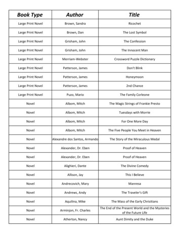
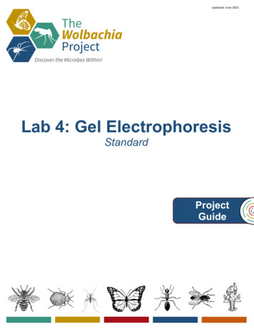
A novel DNA vaccine coding for H5 and N1 genes of highly pathogenic avian influenza H5N1 subtypeAnalyzing the transformantsAnalyzing the transformants with miniprepThe preparation of the plasmids from the overnight culturewas done to verify the size of the recombinant entry vector.The donor vector, pENT R /SD/D-TOPO, is a 2601bp, so theexpected size or the recombinant vector should be 4301bpfor the H5 and 4001bp for the N1 recombinant vectorsrespectively. As seen in Fig. 3 a very clear band migratingabout 4.3 and 4.1 Kbp were seen with all the tested 8 colonies.Bands with molecular size longer also observe, whichrepresents plasmid dimmers or trimmers. The supercoiledplasmid also has seen at the much lower size (about 3Kbp).After analyzing the transformants with miniprep QPCRTo verify the presence of the H5 or N1 inserts in thecorresponding donor vector, a real-time PCR assay wasperformed using miniprep as a templet and primer sets thattarget either H5 or N1 gens. As seen in Figures 4 A and B, thepENTR /SD/D-TOPO carrying the full-length H5 gene gave apositive Ct range from 14.18 -15.89, and pENTR /SD/D-TOPOcarry the full-length N1 gene gave a positive Ct range from22.25-23.14.Homologous recombination between the entry vectorand destination vectorIn order to produce a mammalian expression vector, agateway destination vector pDEST 40 was used for thehomologous recombination with the entry vector. Theresulted from recombinant destination vector (pDEST 40/H5) and (pDEST 40/N1) were transformed into one shotE.coli Topo 10 competent cells. After overnight culture withselective antibiotics (ampicillin), few colonies have beenobserved (Fig. 2 C, D).The maxi prep of the destination plasmid was done usingMaxi prep kit (Bio Basic cat # BS466), and the concentrationwas measured using Qubit II Fluorometer. The concentrationwas ranging from 80-100ng/µl (in 4mL elution volume) from 4litter overnight culture (the total concentration thus is 320µg/litter). The DNA vaccines (pDEST 40/H5) and (pDEST 40/N1)preparations were concentrated, combined and adjusted in aconcentration of 50 µg of each vaccine per 0.5 ml sterile salineEvaluation of the protective efficacy of the DNAvaccineFour parameters viz.antibody titer, protection percentage,shedding test, and measurement the IFN-ɣ transcript level.The antibody titer as measured by HIDynamic changes of antibody titers in chickens as measuredby HI assays (Table 3) revealed a significant difference(p 0.05) between G1 (DNA vaccine) and G2 (availablecommercial vaccine) at 28 days post-vaccination and 14 dayspost-challenge, where there is a great difference (p 0.05)when G1 or G2 when compared with G3 (non- vaccinatedgroup) at all time points.Fig. 3: Electropherogram of the miniprep from E. coli Topo10 coloniestransformed with recombinant entry clone pENTR /SD/D-TOPOcarrying the full-length H5 (A) and N1 (B). Note the migration ofthe 4.3 Kbp recombinant vector carrying the H5 gene and 4.1 Kbprecombinant vector carrying the N1 gene. Other bands migrating at asize over 9Kbp also observed, which represents plasmid dimmers.Fig. 4: Real time qPCR amplification of the H5 gene (A) and N1 gene (B) from the pENTER SD/D/H5 miniprep. All the tested miniprep gavepositive Ct, indicating successful cloning.The Indian Journal of Veterinary Sciences and Biotechnology, Volume 15 Issue 4 (April-June 2020)5
A novel DNA vaccine coding for H5 and N1 genes of highly pathogenic avian influenza H5N1 subtypeTable 3: Mean antibody responses to pDEAST40/H5 , pDEAST40/N1, and the commercially available inactivated vaccine, in comparison withthe non-vaccinated control SPF chickens as measured by HI assay. Results expressed as mean HI titer (Log 2) SEGroupsGroup 1 (G1)pDEAT40/H5 and (pDEST 40/N1) DNA vaccinesGroup 2 (G2)Commercially available inactivated vaccineGroup 3 (G3)Control non vaccinatedMean HI titer ( Log2) SEDays post-vaccination0152128Days post-challenge71508.88 0.4419.5 0.2511.63 0.428.5 0.42313.63 0.4205.75 0.4128.25 0.1649.5 0.3277.5 0.32710.13 0.3980000No bird surviveProtection percentageAfter the challenge, DNA vaccinated chickens (G1) showed nosign of disease. The birds remained healthy, active with eatingand drinking rates normal. The survival rate in this group was92%, and the dead birds (deaths were at day 10-12) showedvery mild post mortem signs not more than slight cyanosedcombs (Table 4).Birds in G2 (vaccinated with the commercially availablevaccine) showed depression with loss of appetite, roughenfeather. This vaccine could not provide more than 80% survivalrate with extreme PM lesions externally (the cyanosed combsand wattles, hemorrhage on the chunk) or internally (bloodspots on the coronary fat, hemorrhagic tracheas)However, after challenge, G1 group showed an increasedlevel of IFN-γ transcript by the 3rd days post-challenge(912838), which reach its maximum by the 7th -day postchallenge (4E 06) then begin to decline (558037), yetit remains significantly higher than the G2 (p 0.05). G2showed a moderate increase of the transcript level by the3rd-day post-challenge that lasts till the 14th days postchallenge. All birds in G3 group were dead by the 3rd daypost-challenge.The shedding as measured by EID50:Cloacal swaps were taken from all birds survived after thechallenge and from dead birds within 24hours of death. Thecloacal swaps were kept in sterile saline at -20C until the EID50test was performed. As shown in Table 4, the DNA vaccinemanaged to decrease the shedding level to as minimal 10 incomparison to the inactivated commercially available vaccine( 3.25 Log 10 ) . In contrast, the level of shedding in the nonvaccinated group were as high as 5.24 Log 10.Measurement the IFN-ɣ transcript level:After vaccination, G1 showed slight elevation of the transcriptlevel by the 5th day and seen till the 7th days post-vaccinationonly while G2 and G3 showed no detectable level during the28 days post-vaccination (Table 5 and Fig. 5)Fig. 5: Mean fold change in IFN- transcript of chicken vaccinated witheither DNA vaccine or inactivated vaccine in comparison with thenon-vaccinated chickenTable 4: The protection rate, the lesions and the shedding level recorded with each group after 14 days of challenge (- No lesions: Very mildlesions (one sign only externally or internally): Moderate lesions (more than one sign): sever lesions (typical for natural infection)GroupG1G2G3Vaccine typepDEAST40/H5 and pDEAST40/N1Commercially available vaccineControl non vaccinated#of deaths2/255/2525/25Survival rate %92800Presence of signs ondead birds Presence of P/Mlesions Mean EID 50 of theshedding03.255.24Table 5: Transcriptional level of the target IFN-γ gene expressed as fold change post-vaccination and post-challenge.6GroupsTranscriptional level of the target IFN-γ gene expressed as fold changeDays 65E-07Days 7The Indian Journal of Veterinary Sciences and Biotechnology, Volume 15 Issue 4 (April-June 2020)3d7d9128384E 069.189659.302All birds were died14d5580372241.1
A novel DNA vaccine coding for H5 and N1 genes of highly pathogenic avian influenza H5N1 subtypeDiscussionVaccination programs against avian influenza would be apowerful tool for viral eradication if used along with biosafetymeasures. Two main parameters should be established witha good vaccine candidate. First, the vaccine should give ahigh percentage level; second, the vaccine should decreaseand limit viral shedding to prevent secondary epidemicsand could eliminate the virus from the environment. In thecurrent study, DNA vaccine coding for full-length H5 and N1have fulfilled these two main parameters.The full-length H5 and N1 genes (orf) were amplifiedfrom the selected strain (A/chicken/Qalubia/ch2.2.64/2017(H5N1)). The specific amplicon migrating about 1700 and1400bp corresponding to H5 and N1 respectively, were seen;many nonspecific amplification fragments have also seen,which may result from the relatively lower annealing tempand long extension time changing these condition resultedin negative results. After cloning in the entry vector, theminiprep revealed the 4 Kbp product corresponding to therecombinant pENTR /SD/D-TOPO/N1 and 4.3Kbp productcorresponding to the recombinant pENTR /SD/D-TOPO/H5with other bands at 9Kbp representing polymer forms of theplasmids. This phenomenon always is seen in the high copynumber plasmids. qPCR quantitation of the miniprep gavepositive Ct values (Ct range 14.18-15.86) for pENTR /SD/DTOPO/H5 and Ct values (Ct range 22.25-23.14) for pENTR /SD/D-TOPO/N1, this difference in the concentration ofplasmids may be due to difference in the efficiency of cloning.The mammalian expression vector (pDEST40) has a molecularsize of 7.1kb. This vector derived from the pcDNA 3.1/V5-His vector. It is designed to allow high-level, constitutiveexpression of the gene of interest (e.g. haemagglutinin H5and neuraminidase N1) in a variety of mammalian hosts.PDEST40 contains the essential elements for controllingthe selective and high-level expression of the gene insertsin the mammalian cells, including Human cytomegalovirusimmediate-early (CMV) promoter/enhancer for high-levelexpression (Boshart, et al., 1985 and Southern and Berg1982).The HI titer revealed that DNA vaccine in a single dose(which was significant higher than inactivated vaccine) couldelicit a powerful humoral immune response that prevent theviral cell entry through binding to the HA protein antigenwith subsequent reducing both mortalities and the clinicalsigns observed with infected birds (Angeletti et al., 2017).One of the very important outcomes of the use of the DNAvaccine is the control of the shedding. The shedding seenin the group vaccinated with the pcDEST40/H5, and N1 waszero, which is significantly different (p 0.05) than that of thegroup received the inactivated vaccine(3.25 Log 10 EID50) andwith the control non-vaccinated chicks (5.24 Log 10 EID50).In the current study, the level of IFN-γ transcript was veryhigh even after 14 days post-challenge, which explains thezero shedding level in the group received the DNA vaccine.The fold change in the transcriptional level of IFN-γ wassignificantly higher (p 0.05 ) in the DNA vaccinated groupwhen compared with either the group vaccinated with theinactivated commercial vaccine or the negative control groupat all time point tested.Based on the current study, the DNA vaccine codingfor H5 and N1 genes would be a suitable alternative to thetraditional oil adjuvanted inactivated vaccines mainly due toits inexpensive, rapidly produced, stable, and safe properties(Kutzler, and Weiner 2008) along with its capability to produceboth humoral and cellular immune response that gaveboth high protection level with zero viral shedding afterchallenge. Other advantages, including the ability to expressdiverse antigens, rapid production, and inability to revertinto virulent forms, easy storage, long shelf life, and possibleavoidance of the cold chain, also have much consideration inthe commercialization of this type of vaccines.C o n c lu s i o nIn conclusion, the pDEST 40/H5 and pDEST 40/N1 gave avery high percentage of protection (92%) and very high HItiter and IFN-γ level, this DNA vaccine also give a zero level ofviral shedd
Strand cDNA Synthesis Kit (Biomatik cat # K5147) according to the manufacture instruction. The second step PCR was done using PfuUltra II Hotstart PCR Master Mix (Agilent USA cat # 600850). Primers that target full orf of H5 and N1 genes (Table 1) were designed and verified by Lasergene DNAStare software V 15 using the full-length orf of H5 and