Transcription
Flapless SurgeryDeploying Alpha-Bio Tec’s NeO forCombined Immediate Post-extractionImplant and Flapless ImplantationDr. Paolo BorelliDDS, ItalyDr. Paolo Borelli graduated in dentistry from the Universityof Turin, Italy. In 2006, he obtained a Masters in Prostheticsfrom the University of Turin. Since 2004, Dr. Borelli has beena member of the Order of Doctors, Turin. He co-authoredtwo books, "Prosthetic Rehabilitation" Vol. 3 (UTET, 2004)and "Biological Approach to Edentulous Patient Treatment"(Quintessence, 2008). Dr. Borelli is a co-founder of the StudyClub of Genoa, Milan and Turin, which focuses on guidedsurgery techniques. He is a teaching assistant in oral surgeryin Koeszeg, Hungary under the direction of Professor Dr. P.Famà. Dr. Borelli has been a guest speaker at seminars andconferences in Italy and abroad and he manages a privatepractice in Turin, Italy.Dr. Massimiliano FavettiDDS, ItalyDr. Massimiliano Favetti graduated with honors from theUniversity of L'Aquila, Italy in 1995, where he collaboratedwith the ENEA Research Center on the study ofbiocompatibility of metals in dentistry. Dr. Favetti specializesin prosthetics, implantology and oral surgery. Since 2008, hehas been on the Board of Experts, Italian Civil Court, Rome.His main interests are piezoelectric surgical techniques andCAD/CAM systems for prosthetics and implantology; he hasbeen a guest speaker on these topics at various conferencesand courses. Dr. Favetti has used the Alpha-Bio Tec. implantsystem since 2005. He is currently the owner of DentamedClinics, Rome.75
Deploying Alpha-Bio Tec’s NeO for Combined ImmediatePost-extraction Implant and Flapless ImplantationAbstractThe upper molar area often presents challenges for immediateimplantation. In addition to favorable anatomical conditions,such as divergent roots and a barely pneumatized maxillarysinus, it is necessary to have high performance implantsystems available, able (despite the limited availability ofbone typical of these conditions) to achieve high primarystability.This case study presents a 41-year old patient who, following,the failure of a fixed prosthesis on her natural teeth, wasrehabilitated using two Alpha-Bio Tec's NeO implants. Aflapless implant was selected to be inserted in area 15 andan immediate post-extraction implant in area 16.BackgroundAn immediate post-extraction implant presents tremendousadvantages for the patient in reducing the edentulous phaseand the number of surgical steps. In order to be placedsuccessfully, such an implant requires careful planning,optimal site preparation and the utilization of suitableimplants by the clinician [1] .The utilization of immediate implants is a viable alternativeto replacing missing teeth in cases of severe periodontaldisease, periapical pathology, extensive cavities or incurablefractures [2] .In extreme conditions, such as poor bone density, it isrecommended to utilize spiral implants, with which it ispossible to obtain adequate primary stability [3] .76The new Alpha-Bio Tec's NeO implant features a very refineddesign, allowing for easily obtained high torque values asa result of its ability to stabilize bone tissue. This featurebecomes even more important when operating in complexpost-extraction sites, such as in multi-rooted teeth, wherethe scarce bone availability needs to be optimized. Anotherfeature of this new implant system is its versatility – itsability to be used in any bone density and for any surgicaltechnique, from flapless implants to those combined withregenerative procedures.OverviewThe patient is a 41-year old woman, moderate smoker (5-6cigarettes per day), with no meaningfully adverse healthhistory. The patient reports pain around an old implantedprosthesis in the maxillary right quadrant. Clinical examinationof the area reveals inflammation and gingival bleedingaround tooth 16, while a radiographic evaluation of the areashows good bone availability. The recommended approachis to remove the existing bridge (14 – pontic – 16), place anew crown on tooth 14, place an implant using a flaplesstechnique in the area of the missing tooth 15, extract tooth16, and place an immediate post-extraction implant as areplacement of tooth 16.Extraoral ExaminationPatient presents toned perioral muscles and a high smileline that permits full exposure of the front teeth, also due toprotrusion of the maxillary central and lateral incisors.
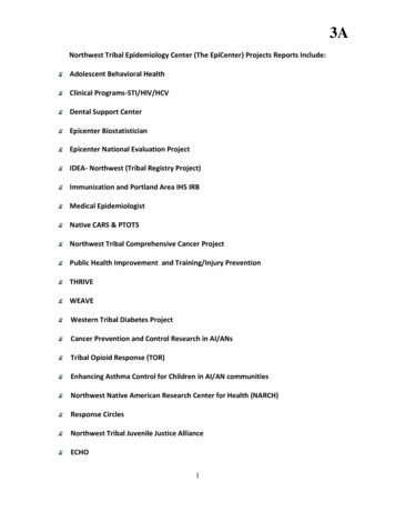
Flapless SurgeryIntraoral ExaminationGood level of oral hygiene and absence of tooth mobility.Thick mucosal biotype with no evidence of lesions. All teethshow signs of wear and tear as a result of parafunctionalactivity, which may also be the cause of the widespreadgingival recession. Mucosal swelling in evident in the areaof tooth 16. Some incongruous prosthetic artifacts exist.Radiographic ExaminationThe initial ortho-panoramic radiography (Fig. 1) showssufficient bone availability to enable the implant placement inareas 15 and 16 without adopting regenerative techniques.1Initial orthopantomographyMaterials UsedØ 3.75 x 11.5 mm NeO implant (Alpha-Bio Tec., Israel) inarea 15Ø 4.2 x 10 mm NeO implant (Alpha-Bio Tec., Israel) in area 16Temporary TLAC-AR abutment (Alpha-Bio Tec., Israel) onimplant in area 15Non-absorbable polyamide suture (Supramid; B. BraunMelsungen, Germany)Temporary polycarbonate crown (InLine, BM. Dental,Italy) on implant in area 15Final crown in IPS e-max CAD (Ivoclar Vivadent, Italy) ontooth 14Final crowns with Prettau CAD zirconium structure(Zirkonzahn, Italy) and ZirPress veneering (IvoclarVivadent, Italy) on implant areas 15 and 16Treatment Objectives and Work PlanThe treatment plan includes the removal of the existingprosthesis in the maxillary right quadrant and the placementof two implants: in area 15 using a flapless techniqueand in area 16 as an immediate post-extraction implant.Immediate screw retained prosthetic rehabilitation in area15 is scheduled after the end of the surgical phase to reduceany imperfections resulting from missing teeth. The finalprosthesis, expected to be placed approximately 3 monthsafter surgery, will be constructed by creating a ceramiccrown with chair side CAD/CAM technique on tooth 14,and zirconium-ceramic crowns on the abutments in areas15 and 16.Surgical PhaseThe old bridge was removed after administering plexusanesthesia. Impairment of tooth 16 (unsalvageable) wasevidenced (Fig. 2).HS6-5 healing screw (Alpha-Bio Tec., Israel)Final TLAO-2 abutments (Alpha-Bio Tec., Israel) on implantsin areas 15 and 162Initial situation after removalof the old prosthesisAdditional MaterialsAbsorbable haemostatic sponges (Cutanplast Dental;Ogna Lab, Italy)77
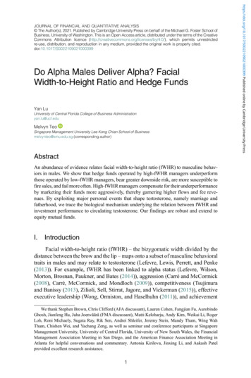
The extraction of the root residues revealed a very wellrepresented inter-radicular septum, enabling implant placement(Fig. 3).5NeO implant insertion ininter-root septum of tooth163Inter-root septum afterextraction of tooth 16The progression of the implant within the site is gradual, andthe steep rise in the insertion torque occurs only in the lastfew millimeters, easily reaching values of 50 Ncm (Fig. 6).A mucosal operculum in area 15 was performed whilesimultaneously preparing the two implant sites. Thepassage of a 2 mm pilot drill revealed low bone density (D3),and therefore under-preparation of the sites was decidedupon in order to obtain the necessary primary stability.For the site in area 15, which received an Ø3.75 x 11.5 mmimplant (NeO, Alpha-Bio Tec., Israel), it was sufficient to usea 2 mm drill up to 11.5 mm depth. Area 16 was preparedto receive the Ø4.2 x 10 mm implant (NeO, Alpha-Bio Tec.,Israel) with a 2 mm drill to 10 mm depth; a crest housing wascreated for implant installation with a 2.8 mm drill to 4 mmdepth (Fig. 4).4Under-preparation of theimplant sites6Tightening of NeO implantwith dynamometric ratchet;high insertion torque (50 Ncm)At directly accessible sites, it is advisable to use a straightmanual driver that allows, where enough bone density ispresent, altering the implant placement trajectory in orderto optimize the prosthetic axis. In fact, the NeO implantfeatures such a powerful apical thread that it is possible touse it as an actual osteotome (Fig. 7).7NeO implant insertion withmanual driver in area 15The geometric characteristics of the NeO implant, makingit self-tapping and self-compacting, allows it to reach hightorque values even in compromised sites (Fig. 5).78
Flapless SurgeryThe surgical procedure was completed by filling the postextraction alveoli of area 16 with absorbable hemostaticsponges (Cutanplast Dental, Ogna Lab, Italy), applying ahealing screw on the implant (HS6-5, Alpha-Bio Tec., Israel)and suturing the area with non-absorbable polyamidepseudo-monofilament (Supramid, B. Braun Melsungen,Germany (Fig. 8).8Placement of healingabutment and suture inarea 1610Placement of temporarycrown on abutmentThe provisional crown was bonded to the abutment usinga flowable composite and then the screwed-on crownwas removed from the patient’s mouth. This procedureallowed adjustment of the screwed-on provisional outsideof the oral cavity (Fig. 11), thus achieving a high degree ofaccuracy in the finishing and polishing of the emergenceprofile (Fig. 12).11Immediate loading of the implant in area 15 was accomplishedby modifying a temporary abutment (TLAC-AR, Alpha-BioTec. , Israel) (Fig. 9).Realization of screwed onprovisional9Grinding of temporaryTLAC-AR abutment12Finished and polishedtemporary crownTo avoid clogging the opening passage during the provisionalfitting procedures, a long transfer screw was used to holdthe temporary abutment in place and then the suitably preconstructed crown, pre-molded in polycarbonate (InLine, BM.Dental, Italy), was fitted over it (Fig. 10).The provisional crown was attached to the implant bytightening the screw to 20 Ncm and closing the hole withanother flowable composite (Fig. 13).79
1315Application of temporaryabutment and closingthe hole with flowablecompositeDamaged provisional at 35days after surgeryTo limit the risk of overload on the implant, the provisionalwas adjusted to eliminate contacts in both in centric occlusionand in lateral and protrusive movements (Fig. 14).The decision was made to remove the provisional and(to avoid additional stress that could effect the implantstability) to apply a HS6-5 healing screw instead (Fig. 16).1416Provisional withoutocclusal loadApplication of healingscrew in place ofprovisionalThe patient was discharged with a recommendation to adhereto the following drug regimen: Amoxicillin Clavulanic Acid:1 g every 12 hours for the following three days, Ketoprofen1 g every 8 hours on the first day and as needed in thefollowing days, Chlorhexidine 0.2% spray at least 3 times aday for the next 7 days.The intraoral radiography did not show any evidence ofbone loss around the implants (Fig. 17).17Intraoral radiography at35 days after surgeryAdditional Check-UpsA week after surgery, the sutures were checked and removed.As the patient reported no discomfort, her follow up checkup was planned a month after surgery.At 35 days after surgery, despite all the recommendationsprovided to the patient about the diet to be followed duringthe healing period, she showed up at the follow-up visit witha damaged screwed-on provisional on 15, evidently due tosome masticatory overload (Fig.15).80Prosthodontics PhaseDuring the osseointegration phase, the old crown was replacedon tooth 14 with AIPS e-max CAD integral ceramic (IvoclarVivadent, Italy) produced directly in the dental clinic in a single
Flapless Surgerysession with the CAD/CAM Cerec system (Sirona, Germany),(Fig. 18).1821TLAO-2 abutmentsprepared on modelCrown in IPS e-max CADon tooth 14 made withSirona CerecAt 90 days after surgery the final impressions were taken witha single-phase individual open tray procedure, positioningthe HTLO impression transfers (Fig. 19) on the implantsutilizing VPES (Vinyl Polyether Silicone) EXA'lence GC (GCEUROPE, Belgium), (Fig. 20).19Alpha-Bio Tec. HTLOtransfer placed onimplantsIt was decided to adopt a fully digital work flow that, inaddition to maintaining accuracy of the details of theimpressions, also allows for optimizing execution times,reducing costs and achieving remarkable aesthetics. TheCAD/CAM (Zirkonzahn, Italy) system first allowed us toperform scans of the prepared models (Fig. 22), followed bythe design of the two crowns of 15 and 16 with the pressedzirconium technique (Fig. 23) and finally, milling of theprosthetics.22CAD/CAM scannedmodels20Dental impression in VPESwith open tray technique23CAD design of teeth 15and 16 for press techniqueon zirconiumTwo TLAO-2 (Alpha-Bio Tec., Israel) abutments were providedto the laboratory. After pouring plaster models, the abutmentswere modified by grinding them to 0 (Fig. 21).81
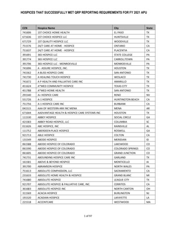
The structures were milled from hard Prettau zirconium(Fig. 24), while the anatomical occlusal details were milledfrom hard castable resin (Fig. 25).27Intraoral occlusalfunctionalization24Milled structures fromhard Prettau zirconiumdisks25Anatomical details milledfrom hard castable resinOnce sent back to the laboratory, the crowns were finalizedwith structural ceramization techniques by means of diecasting, utilizing ZirPress Ivoclar ceramic (Ivoclar Vivadent,Italy), characterized by saturating the surface of the color(Fig. 28).28Crowns designed in thelaboratory with ceramicdie casting techniqueAfter sintering the structures in zirconium and controls onthe model (Fig. 26), the crowns were sent for fitting tryingin the patient’s mouth.26Controls on the modelIn the final session, the abutments were positioned bytightening them to 30 Ncm (Fig. 29) and crown shape, colorand contacts were crosschecked (Figs. 30-31) prior tocementation.29Abutment 30 Ncmtightening torqueThe intraoral test was carried out without difficulty andbasically consisted of the optimization of occlusal contacts(Fig. 27) using articulating paper of 40 microns thickness.
Flapless Surgery30Cemented crowns31Control of occlusalcontacts aftercementationIn addition to extremely thorough planning, it is essential thatsuitable implants are available in order to proceed to theirimmediate placement and, if appropriate, to their immediateprosthetization. The Alpha-Bio Tec. NeO implant representsthe ultimate expression of the versatile features of animplant, as it can be implanted in virtually all conditions, fromconventional implants to immediate implant surgery, anddeploying all techniques, from flapless surgery to immediateloading. The predictability of a prosthetic implant treatmentdepends on many factors. Consequently, in addition to highquality implants and prosthetic components, it is essentialto achieve a high level of prosthesis. The new CAD/CAMtechnologies, new materials and new laboratory techniques [5]can help in this endeavor, while also minimizing technicalexecution time as described in this case.ReferencesThe final radiographic control (Fig. 32) was performed toensure not to leave any residual cement, and highlights thefit of all the prosthetic structures.32Final X-raySummaryState-of-the-art techniques and technologies applicable toimplant prosthetics make it possible to recommend quicksolutions to a patient, such as the immediate insertion ofimplants post-extraction and flapless surgery interventions,wherever possible.1.Romanos GE. Wound healing in immediately loaded implants.Periodontol 2000. 2015 Jun; 68(1): 153-672.Tarazona B, Tarazona-Álvarez P, Peñarrocha-Oltra D, PeñarrochaDiago M. Relationship between indication for tooth extractionand outcome of immediate implants: A retrospective study with5 years of follow-up. J Clin Exp Dent. 2014 Oct 1; 6(4): e384-83.Danza M, Zollino I, Paracchini L, Riccardo G, Fanali S, CarinciF. 3D finite tooth number analysis to detect stress distribution:Spiral family implants. J Maxillofac Oral Surg. 2009 Dec;8(4): 334-94.Kapos T, Evans C. CAD/CAM technology for implant abutments,crowns, and superstructures. Int J Oral Maxillofac Implants.2014; 29 Suppl: 117-365.Abduo J, Lyons K. Rationale for the use of CAD/CAM technologyin implant prosthodontics. Int J Dent. 2013
new crown on tooth 14, place an implant using a flapless technique in the area of the missing tooth 15, extract tooth 16, and place an immediate post-extraction implant as a replacement of tooth 16. Extraoral Examination Patient presents toned perioral muscles and a high smile line that permits full exposure of the front teeth, also due to