Transcription
6-204 OA1 11/28/17 5:19 PM Page 36Malaysian Orthopaedic Journal 2017 Vol 11 No 3Kumar M, et aldoi: http://dx.doi.org/10.5704/MOJ.1711.014A Prospective Study to Evaluate the Management ofSub-trochanteric Femur Fractures with Long ProximalFemoral NailKumar M, MS Orth, Akshat V*, MS Orth, Kanwariya A**, MD Anaest, Gandhi M***, MDS Paediatr DentDepartment of Orthopaedics, Dr SN Medical College and Hospital, Jodhpur, India*Department of Orthopaedics, Jhalawar Hospital and Medical College, Jhalawar, India**Department of Anaesthesia, Dr SN Medical College and Hospital, Jodhpur, India***Department of Paediatric Dentistry, Government Dental College, Jaipur, IndiaThis is an open-access article distributed under the terms of the Creative Commons Attribution License, which permits unrestricted use,distribution, and reproduction in any medium, provided the original work is properly citedDate of submission: 9th November 2016Date of acceptance: 21st June 2017ABSTRACTIntroduction: Sub-trochanteric fractures of the femurremains one of the most challenging fractures faced byorthopaedic surgeons. This study was done to analyse themanagement and complications of sub-trochanteric fracturesusing long proximal femoral nail (PFN).Materials and Methods: This was a prospective study of 50patients with sub-trochanteric fractures of femur who weretreated with long PFN at a tertiary care center from July 2012to June 2016. The fractures were classified according toSeinsheimer classification. All patients were assessedfunctionally by Harris Hip Score.Results: Average duration of union was 17.08 weeks (range13 to 32 weeks), union was achieved in 92% cases. Closedreduction was achieved in 68% cases and open reduction wasrequired in 32% cases. Various intraoperative complicationswere seen in 12% and delayed complications in 26% ofcases. Good anatomical results were achieved in 86% ofcases and 14% were fair. As per Harris Hip score, excellentresults were noted in 28% cases, good in 56% cases and fairin 16% cases.Conclusion: The long PFN is a reliable implant for subtrochanteric femur fractures, with high rate of bone unionand minimal soft tissue damage. Intramedullary fixation hasbiological and biomechanical advantages, but the surgery istechnically demanding.Key Words:sub-trochanteric fractures, Seinsheimer classification, longProximal Femoral NailINTRODUCTIONSub-trochanteric fractures are femoral fractures that involvethe lesser trochanter and extend distally up to 5cm1. Thesefractures account for 10% to 34% of all hip fractures2. Thesub-trochanteric region is usually exposed to high stressesduring routine activities. Axial loading forces through the hipjoint create a large moment arm, with significant lateraltensile stresses and medial compressive loads. In addition tothe bending forces, muscle forces at the hip also createtorsional effects that lead to significant rotational shearforces. In the sub-trochanteric region thickness of corticalbone is more and vascularity is less which produce healingdisturbances.High compressive and tensile forces of muscles separate thefracture fragments and cause instability of the fracture.Hence this fracture is difficult to manage and is associatedwith many complications including mal-union, delayedunion, non-union and implant failure3. Due to theseanatomical features conservative treatment is not preferred,and if there are no absolute contra indications and the patientcan tolerate surgery, surgery is the treatment of choice4. Thegoal of operative treatment is restoration of normal length,anatomical alignment and angulation to restore adequatetension to the abductors. Early mobilization and weightbearing are possible with advances in implants and fixationtechnology. The two primary options for treatment of subtrochanteric fractures are intramedullary fixation andextramedullary fixation5.Extramedullary implants including condylar blades platesand proximal femoral locking plates have been used to treatsub-trochanteric fractures, but they were associated withcomplications of high rate of reduction loss, fixation failureand the need for reoperation6. Compared with extramedullaryimplants, intra-medullary implants have severalbiomechanical advantages with benefits, including less softCorresponding Author: Akshat Vijay, Department of Orthopaedics, Jhalawar Hospital and Medical College, Jhalawar, IndiaEmail: akshat.vijay15@gmail.com36
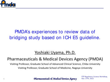
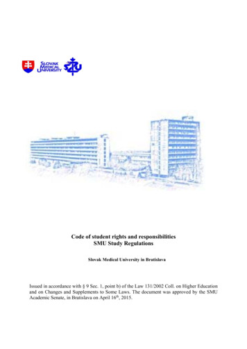
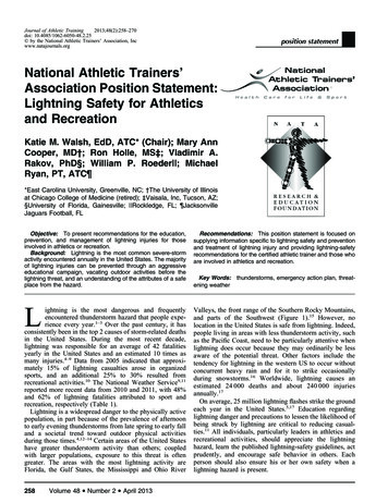
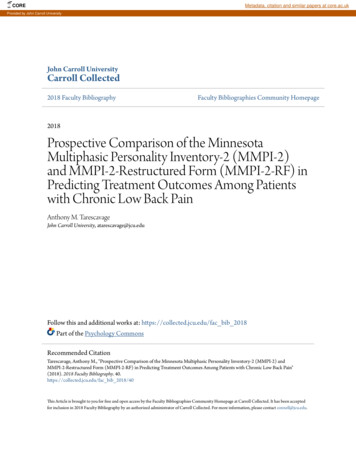
6-204 OA1 11/28/17 5:19 PM Page 37Management of Sub-trochanteric Femur Fracturestissue dissection, dynamic locking, ease of insertion,potentially less blood loss, restoration of the mechanical axisand, most importantly, allowance for immediate weightbearing after fixation7. There have few studies to compareresults of these two modalities. The purpose of our study wasto evaluate the results, complications and functionaloutcomes of long PFN in the management of subtrochanteric femur fractures.MATERIALS AND METHODSThis prospective study was conducted on 50 patients withsub-trochanteric fractures of femur who were treated withlong PFN in a tertiary care center from July 2012 to June2016. Permission was obtained from the ethical committeein accordance with the Helsinki Declaration before startingthe research. Informed consent was obtained from all thepatients.Pathological fractures, fractures in patients 18 years, oldneglected fractures, peri-prosthetic and open fractures wereexcluded from the study. The fractures were classifiedaccording to Seinshemier classification. Appropriate lengthand diameter of long PFN, with distal diameter of 9, 10, 11,12mm and proximal diameter of 14mm were used. Proximallocking was done using de-rotation screw of 6.5mm anddistal lag screw of 8mm and distal locking with self-tapping4.9mm cortical screws, one in static mode and the other indynamic mode, allowing 5mm dynamization. The nails usedwere universal with 60 of medio-lateral valgus angulationand neck shaft angle of 1350. End cap was not used.After preoperative radiographic assessment and planning,the patient was prepared for surgery. The patient was placedin supine position on fracture table and the fracture wasreduced by applying traction in external rotation and 200abduction (to correct varus deformity) and finally the limbwas internally rotated up to neutral position and adducted.Preoperative prophylactic antibiotic was administered. Thetip of greater trochanter was identified and a 5cmlongitudinal incision was made proximal to it. Fascia lataand gluteus medius were incised in line with skin incision.The tip of the greater trochanter was exposed. Entry wasmade from tip or slightly lateral to the tip of greatertrochanter on antero-posterior (AP) view in C-arm and in thecentre of the medullary cavity in the lateral view (Fig. 1a).Medullary canal was entered with a curved bone awl andguide wire was inserted into the medullary canal (Fig. 1b).Using a cannulated conical reamer, the proximal femur wasreamed for a distance of about 7cm. After confirmingsatisfactory fracture reduction, an appropriate size nail wasinserted as far as possible into the femoral opening until thehole for 8mm screw was at the level of the inferior margin ofthe femoral neck (Fig. 1c).Jiang et al8 recommended that lag screw of long PFN shouldbe placed in the lower part of the neck close to the femoralcalcar with the screw tip reaching the subchondral bone 5mmbelow the articular cartilage in the AP view. In the lateralview it should be placed in the centre of the femoral neck,thereby the lag screw would be definitely placed in the areaof best bone quality. A 2.8mm guide wire was insertedthrough the drill sleeve after a stab incision, 5mm deeperthan the planned screw size. It’s final position was kept in thelower half of the neck in the AP view and in the centre of theneck in the lateral view. For anti-rotation screw a secondwire was inserted in the similar way above the first one (Fig.1d). The anti-rotation screw was inserted first to preventpossible rotation of the medial fragment and to reducechances of varus angulation when inserting the compressionscrew. The length of anti-rotation screw was measured and5mm was deducted from it. Drilling was done over the guidewire with 6.5mm drill bit up to the length of anti-rotationscrew previously measured. Tapping was not done as neckscrew was self-tapping. Compression screw was placed insimilar manner by drilling with 8mm reamer (Fig. 1e). Distallocking was performed with two cortical screws by drillingwith a 4mm drill bit and position confirmed with imageintensifier (Fig. 1f). Open reduction was performed in casesin which satisfactory reduction was unsuccessful. Afterfixation, closure was done in layers. Suction drain was usedin cases of open reduction similar to Verley et al9. IVantibiotics were continued for three days post-operativelywhich was prolonged to seven days in cases where openreduction was performed, followed by oral antibiotics forfive days. Analgesics were given as required, and theprocedures are illustrated in Fig.1 a-f.Static quadriceps exercises and knee mobilization werestarted in the immediate postoperative period. On the 4th-5thpostoperative day, depending on the patients’ pain tolerance,they were made to stand up with assistance, and graduallyover the following two to three days they were allowed nonweight bearing walking with frame. Patients were dischargedon the 10th day after suture removal. Partial weight bearingwas started at four weeks in all patients and full weightbearing at 12 weeks. All patients were followed up at 4weeks, 12 weeks and thereafter at 6 weekly intervals, tillfracture union was noted, then at 6 months, 9 months and oneyear. At each visit, patient was assessed clinically for hip andknee function, walking ability, fracture union, deformity andshortening. During follow-up AP and lateral radiographs ofpelvis with both hips were obtained (Fig. 2 a-d). Assessmentof functional results was based on Harris Hip ScoringSystem.RESULTSIn our study, all the patients were operated at an averageinterval of 14.2 days from the day of trauma. They werebetween 31 to 60 years of age group with mean age of 48.72years. There were 38 males and 12 females. The majority offractures were right-sided and road traffic accidentsaccounted for most of the cases, followed by fall from37
6-204 OA1 11/28/17 5:19 PM Page 38Malaysian Orthopaedic Journal 2017 Vol 11 No 3Kumar M, et alTable I: Intra Operative ComplicationsComplicationsFailure to achieve closed reductionFracture of lateral cortexFailure to put anti-rotation screwGuide wire breakageFrequencyPercentage (%)1622232444Table II: Delayed ComplicationsComplicationsHip joint stiffnessKnee joint stiffnessDelayed unionImplant failureZ-effect (cut of lag screw)Varus angulationSuperficial infectionReverse Z-effectAnatomical resultsNo of casesPercentage (%)3122442062448840Table III: Anatomical ResultsRestriction of hip ROMRestriction of knee ROMShortening 1 cmVarus deformityFrequencyPercentage (%)31046208(a)(b)(c)(d)(e)(f)Fig. 1: (a) Determination of entry point, (b) Insertion of guide wire (c) Insertion of long PFN, (d) Insertion of guide wire for compressionand anti-rotation screw, (e) Insertion of compression screw and anti-rotation screw and (f) distal locking (static and dynamicmode).38
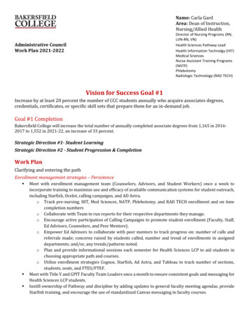
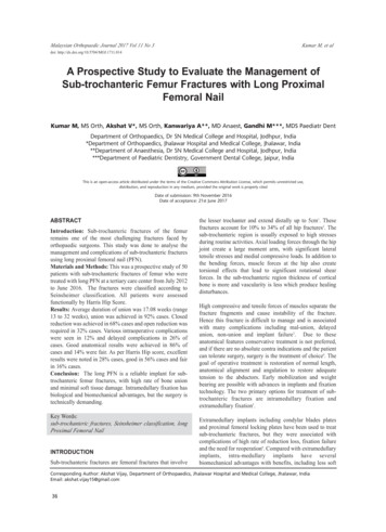
6-204 OA1 11/28/17 5:19 PM Page 39Management of Sub-trochanteric Femur Fractures(a)(b)(c)(d)Fig. 2: AP radiographs (a) Pre-op, (b) Post-op, (c) three month follow-up and (d) six month follow-up.(a)(b)(c)Fig. 3: (a) Z-effect, (b) Fracture of lateral cortex and (c) Implant failure.height. As per Seinshemier classification there were 28 casesof Type II, 12 cases of Type III, 4 cases of Type IV and 6cases of Type V. The most commonly used compressionscrew was size 100mm and the anti-rotation screw was85mm. Average union duration was 17.08 weeks (range 13 to32 weeks), 32 cases showed union at four months, eightcases at five months and six cases at six months duration.Mean duration of surgery was 60.4 minutes (range 35-105).Duration of surgery was longer in the initially operated casesand in managing sub-trochanteric fracture Type IIb. Averageduration of radiation exposure was 115 seconds. Radiationexposure was high in initial cases due to lack of experienceand more in closed reduction cases and for comminutedfracture with difficult reduction. Average amount of bloodloss was 164.6ml (range 70-280ml) measured by mop count(each fully soaked mop containing 50ml blood). There wasgreater blood loss in open reduction cases but withmeticulous dissection and by preventing damage to theperforator we reduced blood loss significantly. Intraoperativeand delayed complications are summed up in Table I and IIand radiographs (Fig. 3 a-c.) Anatomical results wereassessed by presence or absence of shortening, varusdeformities and range of movements in hip and knee joints.Good results were noted in 86% of cases and 14% fair results(Table III). Functional results assessed by Harris HipScoring System gave excellent results in 14 (28%) cases,good in 28 (56%) cases and fair in 8 (16%) cases (Table III).DISCUSSIONSub-trochanteric fractures are usually the result of highenergy trauma and often subjected to significant39
6-204 OA1 11/28/17 5:19 PM Page 40Malaysian Orthopaedic Journal 2017 Vol 11 No 3displacement with difficulty in closed reduction withtraction. The high incidence of delayed union, malunion andnon-union of fractures has left conservative treatmentabolished in modern trauma care10. Extra medullary fixationwith plating has the potential disadvantages of extensivesurgical exposure, severe soft tissue damage and blood loss,thus leading to problems of fracture union and implantfailure. In addition, the eccentric plating is prone to fatiguebreakage due to mechanical load-sharing effect. In aminimally open approach, intramedullary nailing is closelylinked to “biological internal fixation”, in addition to itsmechanical benefits over plate fixation. Intramedullaryfixation allows the surgeon to minimize soft tissuedissection, thereby reducing surgical trauma, blood loss,infection, and wound complications11. Good reduction withminimal dissection, use of appropriate nail length and properpositioning of the nail and screws are necessary to avoidfailure or revision. The abundant muscles around the subtrochanteric region usually cause significant displacement ofthe fractured fragments, leading to great difficulties in closereduction under traction. Sometimes open reduction througha small incision at the fracture site is inevitable.The average union rate reported in the literature was from85-100%. Delayed union and non-union, which are commoncomplications of these fractures, have been reported to be1-10% in various studies4,8,12-15. In our study union rate in sixmonths was 92%. In 4% cases of Type IIb, union wasdelayed due to inadequate reduction of fracture, for whichdynamization was done with fracture union on follow-up inboth cases. Two cases of non-union due to implant breakageat four and six months were treated with exchange nailingand the fractures united three months thereafter.Open reduction was performed in 32% cases out of which24% were Type IIb fractures, 4% cases each of Type IIIb andType V fractures. Difficulty in reduction was probably dueto the integrity of lesser trochanter and muscle pull whichproduce characteristic displacement of flexion, abductionand external rotation of proximal fragment in Types IIb andIIIb and due to severe comminution at fracture site in Type Vfractures. In the study by Jiang et al8, open reduction wasdone in 34% cases and no intraoperative fracture of bone orbreakage of implant were noted. Zhou et al4 open reduced9% of the cases in their study. Iatrogenic fracture of lateralcortex of proximal fragment was seen in 4% cases, plausiblydue to wrong entry point in initially operated cases. It’sincidence decreased with increased experience. In 4% casesfailure to place both proximal screws together was seen,hence proximal locking was left with only neck screw(compression screw) thus decreasing stability of construct.While drilling over guide wire for applying compressionscrew it broke inside the neck in 4% cases; later on it wasremoved and compression screw placed successfully.Review of literature revealed similar intraoperativecomplications8,12,14. In our study superficial wound infection40Kumar M, et alwas seen in 4% cases, in which open reduction wasperformed, and infection resolved with intravenousantibiotics continued for three weeks. Yadav et al14 in theirstudy reported infection in 6.67% cases. Woundcomplication rate in our study was less compared to otherstudies because of the longer administration of antibiotics incases of open reduction.Various mechanical complications associated with PFN werereported by many authors including Z-effect (cut-out ofscrew), reverse Z-effect, implant failure and failure offixation requiring re-operation. Werner et al16 were the firstto introduce the term Z-effect, detected in 7% of their cases.The Z-effect phenomenon was described as a characteristiclateral migration of the inferior screws, varus collapse of thefracture and perforation of the femoral head by superiorscrew during the postoperative weight bearing period. Theyproposed that fixation of the fracture at a cervico-diaphysialangle of 1250 was a predisposing factor for the Z-effect andreverse Z-effect, as well as for cut-out of screws. Strauss etal17 have reproduced the migration of the cephalic screwsfrom the intra medullary nail in the laboratory with the aid ofa polyurethane model and observed that when compressiveforces on the femoral head and bone density were greaterthan those on the femoral neck, inferior screw migratedlaterally.The reverse Z-effect as described by Boldin et al13 involvesthe lateral migration of the superior screw accompanied bythe medial migration of the inferior screw, which requiredearly removal. Simmermacher et al18 in their study hadimplant failure and cut-out of screw in 0.6% cases each. Rateof cut-out of screw and implant failure in literature variesfrom 1-11% and 1-7% respectively. Average rate of Z-effectin various literature was 3-6% and that of reverse Z-effectwas 2-4%4,8,12-15. In the study by Yadav et al14 shortening 1cmand varus deformity were seen in 4% cases. Rate ofreoperation for non-union or implant failure or Z-effect hasbeen reported from 9% to as high as 29%. In our study, Zeffect was seen in 8% cases and no cases of reverse Z-effectand femoral fracture below the tip of PFN were seen. Reoperation due to implant breakage and Z-effect in our studywas in 12% of cases.Excellent results were noted in 14 (28%) cases, good in 26(56%) cases and fair in 8 (16%) cases, as per Harris HipScore in our study. Other studies4,14,15 in which Harris HipScore was used also showed similar outcomes as our study.Favourable results seen in 84% of patients managed by longPFN may be explained, on the basis as stated by Leung et al,and Radford et al11 that intramedullary nailing by allowing aminimally open approach which is closely linked to“biological internal fixation”, in addition to its mechanicalbenefits over plate fixation also allows the surgeon tominimize soft tissue dissection, thereby reducing surgicaltrauma, blood loss, infection and wound complications.
6-204 OA1 11/28/17 5:19 PM Page 41Management of Sub-trochanteric Femur FracturesCONCLUSIONOsteosynthesis with the proximal femoral nail offers theadvantages of high rotational stability of the head-neckfragment, compression at fracture site and isbiomechanically sound as it is an intramedullary device, thusleading to minimal soft tissue damage and high rate of boneunion. Most of the complications of proximal femoralnailing are related to the surgeon and instruments, which canbe reduced by proper patient selection and good preoperativeplanning. Gradual learning and patience are needed to makethis method truly minimally invasive.CONFLICT OF INTERESTThere was no conflict of interest in this study.REFERENCES1.2.3.4.5.6.7.8.9.Bedi A, Toan Le T. Subtrochanteric femur fractures. Orthop Clin North Am. 2004; 35(4): 473-83.Lavelle DG. Fractures and dislocations of the hip. In: Canale ST, James H, editors. Campbell’s Operative Orthopaedics. 11th ed.Philadelphia: Mosby; 2008. 3237-308.Mclaunn TM, Lawler EA. Treatment modalities for subtrochanteric fractures in elderly. Techniques in Orthopaedics.Philadelphia: Lippincott Williams and Wilkins Inc.; 2004. 197-213.Zhou ZB, Chen S, Gao YS, Sun YQ, Zhang CQ, Jiang Y. Subtrochanteric femur fracture treated by intramedullary fixation. ChinJ Traumatol. 2015; 18(6): 336-41.Bucholz RW, Heckman JD, Court-Brown CM, Tornetta P. Rockwood and Green’s Fractures in Adults. 6th ed. Philadelphia:Lippincott Williams and Wilkins Inc.; 2006. 1827-44.Kregor PJ, Obremskey WT, Kreder HJ, Swiontkowski MF; Evidance-Based Orthopaedic Trauma Working Group. Unstablepertrochanteric femoral fractures. J Orthop Trauma. 2005; 19(1): 63-6.Yoon RS, Donegan DJ, Liporace FA. Reducing subtrochanteric femur fractures: tips and tricks, do’s and don’ts. J OrthopTrauma. 2015; 29(4): 28-33.Jiang LS, Shen L, Dai LY. Intramedullary fixation of subtrochanteric fractures with long proximal femoral nail or long gammanail: technical notes and preliminary results. Ann Acad Med Singapore. 2007; 36(10): 821-6.Verley GW, Milner SA. Wound drains in proximal femur fractures surgery: a randomized prospective trial of 177 patients. J RSoc Med. 1995; 88(1): 42-4.10. DeLee JC, Clanton TO, Rockwood CA Jr. Closed treatment of subtrochanteric fractures of the femur in a modified cast-brace. JBone Joint Surg Am. 1981; 63(5): 773-9.11. Redford PJ, Needoff M, Webb JK. A prospective randomized comparison of the dynamic hip screw and the gamma locking nail.J Bone Joint Surg Br. 1993; 75(5): 789-93.12. Tyllianakis M, Panagopoulos A, Papadopoulos A, Papasimos S, Mousafiris K. Treatment of extracapsular hip fractures with theproximal femoral nail (PFN): long term results in 45 patients. Acta Orthop Belg. 2004; 70(5): 444-54.13. Boldin C, Seibert FJ, Fankhauser F, Peicha G, Grechening W, Szyszkowitz R. The proximal femoral nail (PFN)--a minimalinvasive treatment of unstable proximal femoral fractures: a prospective study of 55 patients with a follow-up 15 months. ActaOrthop Scand. 2003: 74(1); 53-8.14. Yadav S, Sinha S, Luther E, Arora NC, Prasad M, Varma R. Comparison of extramedullary and intramedullary devices fortreatment of subtrochanteric femoral fractures at tertiary level center. Chin J Traumatol. 2014; 17(3): 141-5.15. El-Mowafi HM, Eid TA, El-Sayed AS, Zalalo SH. Fixation of subtrochanteric fracture femur using a proximal femoral nail.Menoufia Med J. 2014; 27: 208-14.16. Werner-Tutschku W, Lajtai G, Schmiedhuber G, Lang T, Pirkl C, Orthner E. [Intra- and perioperative complications in thestabilization of per- and subtrochanteric femoral fractures by means of PFN]. Unfallchirurg. 2002; 105(10): 881-5.17. Strauss EJ, Kummer FJ, Koval KJ, Egol KA. The “Z-effect” phenomenon defined: a laboratory study. J Orthop Res. 2007;25(12): 1568-73.18. Simmermacher RKJ, Bosch AM, Van der Werken C. The AO/ASIF-proximal femoral nail (PFN): a new device for the treatmentof unstable proximal femoral fractures. Injury. 1999; 30(5): 327-32.41
Conclusion: The long PFN is a reliable implant for sub-trochanteric femur fractures, with high rate of bone union and minimal soft tissue damage. Intramedullary fixation has biological and biomechanical advantages, but the surgery is technically demanding. Key Words: sub-trochanteric fractures, Seinsheimer classification, long Proximal Femoral Nail