Transcription
DOI: 10.1111/prd.12288REVIEW ARTICLEBiomaterial and biomechanical considerations to prevent risksin implant therapyEstevam A. Bonfante1 Ryo Jimbo2 Lukasz Witek3 Nick Tovar3 Rodrigo Neiva4 Andrea Torroni5 Paulo G. Coelho61Department of Prosthodontics and Periodontology, University of São Paulo Bauru School of Dentistry, Bauru, Brazil2Department of Applied Prosthodontics, Nagasaki University Graduate School of Biomedical Sciences, Nagasaki, Japan3Department of Biomaterials, New York University, New York City, New York, USA4Department of Periodontology, University of Florida at Gainesville, Gainesville, Florida, USA5Attending Oral and Maxillofacial Surgery, Hansjörg Wyss Department of Plastic Surgery, New York University Langone Medical Center, New York City, NewYork, USA6Department of Biomaterials, Hansjörg Wyss Department of Plastic Surgery, Mechanical and Aerospace Engineering, New York University, New York City, NewYork, USACorrespondencePaulo G. Coelho, Department of Biomaterials, Hansjörg Wyss Department of Plastic Surgery, Mechanical and Aerospace Engineering, New York University,New York, NY, USA.Email: pc92@nyu.eduFunding informationFundação de Amparo a Pesquisa do Estado de São Paulo (FAPESP), Grant/Award Number: 2012/19078‐7 and 2016/18818‐8; Conselho Nacional deDesenvolvimento Científico e Tecnológico (CNPq), Grant/Award Number: 304589/2017‐91 I NTRO D U C TI O NThe threaded‐type implants were further tested in edentulouspatients and the clinical outcomes were presented at the TorontoDental implant therapy is today widely accepted as one of the reli‐1Congress in 1982.7 The 15‐year survival presented surprised everyoneable options for restoring missing dentition. The basic concept ofattending, and since then the application of osseointegrated dental im‐implanting metallic devices in bone was first described by Botheplants has become a major alternative to restore full/partial edentulism.et al2 in the 1940s, followed by Leventhal et al3 in 1951, who de‐Certain prerequisites have been proposed as essential factorsscribed titanium as a potentially biocompatible surgical implantablefor successful osseointegration,8 and the development and evolu‐material. Brånemark4 later reported that the metallic microscopetion of the dental implants progressed based on these factors. In the(titanium optic chamber) made of titanium to observe microvascu‐past, research on each individual factor has been performed and alar circulation in living bone could not easily be retrieved after theplethora of evidence has been established. In particular, research onexperiments. The external geometry of the titanium optic chamberimplant surface topography/chemistry has been of major focus, andpossessed threads so that they mechanically engaged into the bone.new surfaces are constantly applied on implants in an attempt toIt was not in their plan that the titanium microscopes integratedaccelerate osseointegration rates.9 As a result, new implant surfacesfirmly into the bone; however, this coincidence inspired the researchare often launched in the market with claims that the implant wouldgroup to apply this concept to dental implants with hopes that theyosseointegrate faster.Recently, the interplay between factors such as implant macroge‐would functionally restore the dentition.Titanium and its alloys are regarded as bioinert or biocompati‐ometry, topography, and surgical protocols has been of interest.10,11ble materials and are stable in the body owing to the spontaneouslyEvidence suggests that enhanced establishment of osseointegrationformed oxide layer.5 Biomaterial research regarding dental implantsmay not be achieved by a single factor.11 For instance, if the implanthas dominantly utilized titanium as a material of choice, and bothsurface possessed state‐of‐the‐art surface features for increasedbasic and clinical research show that commercially pure titaniumosseoconduction, implants may not present better osseointegrationand several other titanium alloys are osteoconductive and promoteunless other factors generate a host bed for the surfaces to interactosseointegration.6with osteogenic cells.11,12Periodontology 2000. rd 2019 John Wiley & Sons A/S.Published by John Wiley & Sons Ltd 139
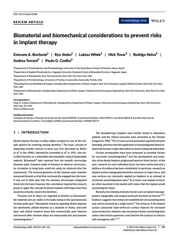
140 BONFANTE et al.This review presents an overview of the current existing evi‐newly formed woven bone and the subsequent evolution toward adence on osseointegrated implants. Factors such as implant microge‐lamellar configuration will eventually render the system with second‐ometry and surface micro‐ and nanotopography will be introduced,ary (biologic) stability that will support the implant device through‐and a critical discussion regarding how they interact with each otherout its lifetime. When evaluated over the long term (5‐10 years)is provided. Of special interest, and less explored in reviews, is howthe interfacial osseointegration pathway results in a compact, ma‐surgical instrumentation, drilling protocols, and drilling methodsture lamellar bone with small marrow spaces.18,19 Figure 1 presentscan be taken into consideration as important factors affecting os‐the main histologic features of the interfacial remodeling healingseointegration. Further, the biomechanical aspect of the titaniumpathway.bulk itself, which had not been discussed much in the past, will bediscussed as a possible factor for periimplant bone loss and, in theworst scenario, implant failure.2 I M PL A NT H A R DWA R E A N DOS S EO I NTEG R ATI O N H E A LI N G PATH WAYS2.2 Healing chamber (intramembranous‐like)osseointegration pathwayDifferently from implant bulk and drilling dimension combina‐tions that lead to high degrees of contact between implant andbone immediately after implantation, healing chamber implantsare devices that present plateaus instead of threads, and areThough a robust body of literature has described implant host re‐tapped into bone osteotomies with the diameter of the implant. 20sponse as a function of time, a large variation in surgical deviceSuch interplay between device and osteotomy dimension allowsdesign, the proportional variation in surgical instrumentation tech‐for minimal primary stability and results in large void spaces be‐niques, and the increasing focus of research on implant surface en‐tween the device and bone, allowing the formation of healinggineering, the attention has eventually shifted to the impact of thechambers.10Although itImmediately after placement, the healing chambers will be filledis acknowledged that implant macrogeometry and the associatedwith a blood clot that evolves toward an osteogenic connective tis‐surgical technique (and thereby implant hardware) are differentsue configuration that will ossify through an intramembranous‐likeparameters, it has been demonstrated that their interplay not onlypathway. 21 This osteogenic connective tissue is highly vascularized atleads to different osseointegration healing pathways but also affectsearly implantation times, allowing osteogenic cell migration through‐how effective micrometer‐ and nanometer‐scale designing shouldout the healing chamber space. 21 Such a vascularization and cell mi‐implant hardware features in bone healing pathways.10,11,13,14ultimately be.The three osseointegration pathways havegration pattern allows for woven bone formation simultaneouslybeen described in detail in previous work,10,11,13,14 and so the maineffects on early‐ and long‐term healing and biomechanical evolutionare summarized.2.1 Interfacial remodelingosseointegration pathwayWhen implant placement results in an intimate contact betweenbone and the device's threaded bulk, consequently rendering highinsertion torque values (tight fit) due to mechanical interlocking,an interfacial remodeling bone healing pathway takes place.10,11This scenario is observed in the majority of marketed implant sys‐tems, where insertion torque levels are chiefly dependent on im‐plant geometry and micrometer‐level surface modifications as wellas osteotomy dimensions, which will eventually regulate the straindistribution in bone. Although debatable, higher primary stabilityhas been associated with the clinical perception of higher rotationalresistance during implant insertion, expressed by insertion torquelevels.15 However, high insertion torque levels result in strain andmicrocrack formation in bone, leading to compression necrosis thattriggers bone remodeling. This scenario has been theoretically sug‐gested,16 and experimentally confirmed,17 by the loss of mechanicalinterlocking between bone and implant due to extensive bone in‐terfacial remodeling after implant placement under stable condition.Through the course of remodeling at the implant‐bone interface,F I G U R E 1 Optical micrograph depicting the main featuresof interfacial remodeling healing pathway at 6 weeks in a caninemodel. The blue line represents the theoretic position of theosteotomy outer diameter closely matching the implant threadsinner diameter resulting in implant high insertion torque atthe expense of interfacial bone compression. The green linedemonstrates the perimeter and extension (green arrows) thecortical shell remodels (native bone osteonic structures, blackarrows), and that woven bone growth occurs primarily throughosteogenic cell migration from the native bone toward the implantsurface that acts as a site for bone growth. The yellow arrowsdepict bone remodeling sites occurring farther away from thebone‐implant interface. Bar, 150 μm
BONFANTE et al.141taking place at the implant surface, osteotomy bone walls, and withininitial stability is proportional to the thread design and the amount ofthe center of the chamber, resulting in rapid device osseointegra‐mismatch between implant outer thread and osteotomy diameters.tion. Shortly after early integration through woven bone formation,Hence, this osseointegration pathway simultaneously presents bonelamellar bone will gradually replace the woven bone, resulting in for‐remodeling where engagement between implant and bone occurred,mation of primary osteonic structures. A series of human retrievalresulting in stability loss that is supposedly compensated by the rapidstudies of plateau root form implants has consistently demonstratedwoven bone formation in the healing chambers formed betweenthat further bone morphologic evolution occurs toward a haversian‐threads. Unlike screw‐type implant systems that result in interfaciallike configuration22-25 that over time increases significantly in me‐remodeling osseointegration and plateau root form implants that os‐chanical properties. 25 Figure 2 presents the main histologic featuresseointegrate through healing chambers, implant systems deliberatelyof the healing chamber healing pathway.designed for this purpose are relatively new in the market.27-31 Thus,long‐term human retrieved samples are not yet available for adequate2.3 Hybrid healing osseointegration pathwayThe hybrid healing osseointegration pathway has been utilized in anassessment of the effect of this healing pathway on long‐term bonemorphologic evolution. Figure 3 presents the main histologic featuresof the interfacial hybrid healing pathway.attempt to obtain devices that are atemporally stable and is a com‐bination of features presented in the interfacial remodeling and heal‐ing chambers osseointegration pathways. Screw‐type implants with3 S U RG I C A L I N S TRU M E NTATI O Nlarge thread pitch and outer to inner thread diameter differences havebeen designed to allow surgical instrumentation sufficiently large forA critical appraisal of the literature will be presented in this sectionthe formation of healing chambers between threads, implant innerto suggest that modest interest and advances have been achieveddiameter, and bone instrumented walls.26-31 For such an implant de‐in the subject of surgical instrumentation since the introduction ofsign configuration, primary stability is obtained by the interactionbetween the outer regions of the threads that engage bone and suchF I G U R E 2 Optical micrograph depicting the main features ofthe intramembranous‐like (healing chamber) healing pathway at6 weeks in a canine model. The blue line represents the theoreticposition of the osteotomy outer diameter closely matching theimplant plateau outer diameter resulting in implant minimalcompression in the bone walls. The green arrows demonstratecortical shell remodeling due to initial compression due to implant‐bone interaction (native bone osteonic structures, black arrows).Woven bone growth occurs from the instrumented native bonesurface toward the healing chamber center, from the implantsurface (white arrows), as well within the healing chamber centralregion. The orange arrows depict bone remodeling sites that at thistime point are starting to replace woven bone by lamellar bone andwill be the site of primary osteonic structures within the chamber.Bar, 150 μmF I G U R E 3 Optical micrograph depicting the main featuresof the interfacial hybrid healing pathway at 6 weeks in a caninemodel. The blue line represents the theoretic position of theosteotomy outer diameter between the implant threads’ innerand outer diameter resulting in implant high insertion torque (atthe expense of bone compression by the thread that results ininterfacial remodeling depicted by green arrows and microcrackingdepicted by red arrows) along with the formation of a healingchamber between the instrumented bone wall and the surfacesbetween implant threads. Worth noting is the extensive wovenbone formation from the native bone surgically instrumentedsurface (retracted from remodeling due to surgical instrumentationtrauma—extension depicted by light blue color arrows) towardthe center of formed healing chamber, from the implant surface(white arrows), as well within the healing chamber central region.The dark blue arrows depict bone remodeling sites that at this timepoint is starting to replace woven bone by lamellar bone and willbe the site of primary osteonic structures within the chamber. Bar,150 μm. The black arrows depict native bone cortical shell osteonicstructures; yellow arrows depict bone remodeling sites occurringfarther away from the bone‐implant interface. Bar, 150 μm
142 BONFANTE et al.under copious saline irrigation, recent preclinical studies have pointedout that substantial work is required to determine the optimal drillingmethod that maximizes osseointegration.For example, a series of previous studies have shown that thesimplified version (pilot drill immediately followed by the final diam‐eter drill for implants of 3.75, 4.2, and 5 mm) of the traditional grad‐ual drilling expansion results in comparable osseointegration relativeto classic protocols.32-37 Recent work has confirmed no statisticaldifference on osseointegration parameters between simplifiedand conventional protocols.32 Alternative to reducing the numberof drills, alteration in drill design to include progressive diameterachievement through the employment of a single progressive drillhas been demonstrated to not decrease early osseointegration.38From a surgical perspective, the total surgical time of the simplifiedprotocol from incision to closure would be significantly shortenedand lead to less postsurgical complications.39 However, the reduc‐tion in the number of drills used in the simplified protocol wouldhave the drawback of reducing the number of attempts to correctlocation or angulation of final implant placement.Drilling speed is an important factor of the surgical techniqueand has an influence on the temperature in the surrounding bone.Excessive drilling speed has been reported to prevent adequate irri‐gation, thus potentially allowing for heat generation that could leadto thermal osteonecrosis.40-42 On the other hand, studies have sug‐gested that low drilling speed can potentially generate more heatwhen compared with high drilling speeds since the surgeon wouldF I G U R E 4 Optical micrograph depicting the main features ofsubtractive drilling and counterclockwise osseodensification ina low‐density sheep hip model at 6 weeks in vivo. A, Uneventfulosseointegration takes place through subtractive drilling, wherenew bone formation occurs through contact osteogenesis fromcells migrating toward the implant surface due to continuitybetween native bone and implant surface, as well as from bonenative tissue. Yellow arrows depict remodeling sites initiallyreplacing woven bone by lamellar bone. B, Counterclockwiseosseodensification drilling resulted in the presence of autograftparticles (bone chips depicted by black arrows) in proximity to theimplant surface. These bone chips did not preclude cell migrationtoward the implant surface and acted as nucleating surfaces thatbridged the implant surface with native bone. At 6 weeks in vivo,extensive bone chip remodeling was observed either in proximitywith the bone chip surface (blue arrows) or directly through bonechip bulk (yellow arrows). Bar, 300 μmpossibly apply not only more vertical compression during drilling, butalso drill for longer periods of time.43,44 Furthermore, it has beensuggested that low drilling speed may potentially increase wobblingand result in overpreparation of the osteotomy site.45,46 Despitethe literature suggesting potential issues with decreasing drillingspeeds, a recent study has pointed that decreasing drilling speeds of1000, 500, and 100 rpm all resulted in successful osseointegration.45Several other studies have pointed out that lower speed drilling didnot decrease osseointegration levels when compared with the mostusually employed higher drilling speeds.It should be noted that the work referenced in this section issystem specific and extreme caution should be taken, since sub‐stantial deviation in drilling recommended dimensions and speedsexists between various implant systems. Nonetheless, these studiespresent variations in subtractive drilling techniques utilizing eachindividual system's drilling methods that may or may not improveimplant therapy. Numerically, preclinical and clinical studies investi‐gating the effects of surgical instrumentation on osseointegrationare at least one order of magnitude lower than the most investigatedsurface treatment effects. In addition, the available literature on thetopic is contradictory, and commonly debated from an anecdotal per‐spective, as dental implant surgical instrumentation has been largelyemployed based on textbook knowledge rather than on informed re‐search platform. While unequivocally high clinical success rates havebeen achieved through the classic protocol of using several subtrac‐tive bone drills of increasing diameter at moderate drilling speedsosseointegration.Whereas the traditional osteotomy using different step drillsis generated by a subtractive method, where bone is drilled away(Figure 4A), a recently developed surgical technique for site drillinghas suggested to enhance implant stability via osseodensificationdrilling (Figure 4B).47,48 The concept of this technique is to executea nonsubtractive multistepped drilling process, which results in thedensification of the osteotomy site wall, creating an environmentthat enhances primary stability due to compaction‐autografting thatgenerates a springback effect that allows bone tissue recoil backinto the implant surface.49 The compaction autografting and the
BONFANTE et al.springback phenomena have been documented in orthopedic litera‐ture,49-52and its effect on increasing implant primary stability is dif‐143It can be speculated that different surgical instrumentationtechniques can create more favorable conditions to promote bothferent than the scenario observed in traditional subtraction drilling,primary stability and osseointegration of implants. The osseodensifi‐where insertion torque levels are mainly dependent on the osteot‐cation technique proved to be effective in improving the biomechan‐omy downsizing dimensions. The densification of the osteotomy wallical properties and secondary stability of implants in translationaland the presence of residual bone chips establishes immediate con‐studies, opening an interesting insight on the role of surgical instru‐tact between the implant system and bone structure, which createsmentation for stability, integration, and an ultimately better out‐stability via physical interlocking, and induces osteoblast nucleationcome of osseointegrated implants. Despite the encouraging result,on the instrumented bone surrounding the implant, thereby acceler‐however, the osseodensification technique remains almost the onlyating bone healing in the proximity of the implant.53-55instrumentation method that has been investigated through a sys‐The classical drilling technique involves a positive rake angle,tematic scientific process, leaving a substantial void in the under‐which is used to extract a small amount of bone as each flute passesstanding of the instrumentation role in the implant integration andto create an osteotomy cleared of bone residue prior to implantsuccess. Further research studies are necessary in order to exploreplacement. In contrast, the osseodensification drilling process uti‐different instrumentation methods, by both bone‐subtractive andlizes a tapered, multi‐fluted bur to create the osteotomy site. Thisbone‐densifying techniques, as well as alternative osteotomy meth‐bur design contains at least four tapered flutes with a negative rakeods (eg, by piezosurgery), in order to identify the ideal integrationangle, which allows for the creation of a layer of compact, densebetween instrumentation protocol, implant geometry, and surfacebone that surrounds the wall of the osteotomy. The densifying buras well as host characteristics (density, morphology, structural, andis capable of controlling the expansion process due to the featuresbiologic features of the alveolar bone) to optimize short‐ and long‐of the cutting chisel and tapered shank, which allows the bur to pro‐term osseointegration.gressively increase in diameter as it drills deeper into the osteotomysite. The expansion process can be operated in both clockwise andcounterclockwise drilling directions and is performed at high drillingspeeds under irrigation.55 The counterclockwise drilling direction ismore efficient at the densification process and is utilized in bone withlow density, whereas the clockwise drilling direction is better suited4 S U R FAC E TR E ATM E NT AT TH EM I C RO ‐ A N D N A N O M E TE R LE N G THS C A LE S : I M P O RTA N C E A S A D H O C S FO ROS S EO I NTEG R ATI O Nfor higher density bone.55 The osseodensification surgical techniquehas been extensively verified in bench top in vitro56 and in vivo ani‐Whereas the field of implant surface engineering has been exten‐mal54,55,57 studies. More recent translational, large animal preclinicalsively explored over the last decades,61,62 especially the modifica‐models have unequivocally shown significant biomechanical im‐tions at the micrometer level, the rationale presented previously inprovements measured through implant primary stability and biome‐this paper will unequivocally demonstrate that surface modificationschanical pullout tests relative to subtractive drilling controls. Thesewill better perform when implant hardware is designed to allow im‐studies have also shown, relative to subtractive drilling controls, sig‐mediate host interaction with the surface.10,11 Also, a correlationnificantly higher osseointegration for implants placed by clockwisebetween the potential benefits and the current contribution of na‐and counterclockwise osseodensification drilling methods.54,55notechnology to implant therapy will be presented.The improvement of quality/quantity of bone surrounding theImplant surface treatment at the micrometer scale has beenimplant to increase primary stability is a theory that has been pre‐the center of focus over the past three decades. This was based onviously explored, but it mainly focused on improving primary sta‐substantial in vitro and in vivo evidence that moderately microtex‐bility after maxillary sinus elevation.58,59 The osteotomy techniquetured surfaces promote osseointegration compared with other sur‐involves the compression of the surrounding bone through an im‐faces.9,36,63-65 As a result, the majority of the commercially existingpaction, where improved primary stability is perceived by cliniciansimplants on the market possess these moderately microtexturedvia increased insertion torque values. Currently, there is insufficientfeatures. Moreover, it has been suggested that the application ofevidence supporting this surgical technique as a superior surgical ap‐nanofeatures on surfaces enhances the osteogenic gene expres‐proach compared with other techniques currently available.60sion,66-68 which in some in vivo studies resulted in enhanced osse‐Surgical instrumentation, in summary, contributes to both theointegration.69 It has been further proven that nanostructures areachievement of initial stability and secondary stability. The degree offurther effective when they exist within hierarchic structures.11,70mechanical interlocking of the implant to the bone can be controlledHowever, in some other studies, the effects of nanofeatures werethrough different surgical techniques, as introduced in this section.not significantly evident, although some tendencies indicate theMoreover, it has been shown that secondary stability (osseointe‐benefits of the nanotexturing.71,72 Numerous reasons for the lessgration) can be promoted through these techniques. Interestingly,effective results could be speculated on; however, one of the majortechniques that were considered to be more invasive than the con‐factors that seems to influence the degree of effectiveness of theventional ones, such as osseodensification, proved to be effective inapplied hierarchic micro‐ and nanotextures is how surgical instru‐enhancing bone healing to the implant.mentation is performed and which type of macrogeometry is applied
144 BONFANTE et al.on the implant.11 Depending on these factors, the surrounding bonemaintenance and failure issues has been explored more often andfollows different healing pathways that affect which cells will firstreported on.7910,11interact with the implant surface.Thus, given the free migrationWhereas it is desirable to discuss the performance of restoredpathway between implant and native bone created by the osteo‐implants based on clinical trials, several challenges are encounteredgenic tissue evolved from the blood clot within healing chambers,that hinder sound comparison between studies. The limitations in‐higher efficiency in hastening bone formation is expected for thisclude the variation in success criteria and/or in selected primaryconfiguration, compared with scenarios of interfacial remodeling,outcome, reduced follow‐up times and patient sample, variations inwhere a cell‐mediated bone resorption is expected prior to boneprosthetic rehabilitation scenarios including implant‐abutment con‐formation.10,29 In hybrid healing, where initial interlocking betweennection design, prostheses type and materials, antagonist, and so on.implant threads and native bone ensure primary stability, the pres‐Therefore, controlled laboratory fatigue testing, while also havingence of healing chambers, hierarchically incorporating micrometer‐its limitations, can be used to simulate a variety of clinical conditionsand nanometer‐level texture for early interaction between implantand serve as a baseline to design proper clinical trials. An emphasissurface and plasma, cells and proteins, will result in rapid bone for‐will be presented on the cumulative damage that inexorably emergesmation; this will maintain implant stability while interfacial remodel‐once a prosthesis is in function, regardless of implant system anding occurs due to compression between implant threads and nativerestoration type.bone. Such a scenario, where concerted bone loss is compensated byrapid bone formation within chamber, may likely result in an implantthat is atemporally stable from a biomechanical standpoint.6 I M PL A NT B U LK M ATE R I A L : TITA N I U MA N D A LLOYS , C E R A M I C S , A N D P O LY M E R S5 I M PL A NT S A N D CU M U L ATI V EDA M AG E D EG R A D I N G TH E I R S TR E N G THMetals, ceramics, or polymers, or a combination, can be used to fab‐ricate the dental implant bulk material. Although titanium has beenpromulgated as a material capable of “growing bone in contact withThe two primary types of complications that may occur with im‐it” since the 1940s, 2 and despite its broadest use amongst implant‐plants are biologic (eg, peri‐implantitis, implant loss) and mechani‐able metallic devices, the quest for end‐stage materials that mimiccal (eg, abutment screw fracture, prostheses veneering materialtooth structures in color and some mechanical properties continues.fracture, implant fracture), with biologic issues generally takingCeramic implants were introduced over four decades ago for ortho‐place earlier than mechanical ones.73 Peri‐implantitis is a major andpedic applications, made of aluminum oxide, with a current robustgrowing problem, as it affects the tissues surrounding dental im‐clinical follow‐up of implanted patients. Owing to quite high failure74Almost all implantrates only for its early versions (up to 13% when fabricated beforesystems are susceptible to biologic complications, and a Swedishplants likely leading to implant loss if untreated.1990s), alumina is currently recommended for less‐demanding load‐clinical trial involving 588 patients revealed the presence of 45%ing scenarios.80 Reports on the use of alumina dental implants wereof peri‐implantitis (bleeding on probing, suppuration and bone lossfrom short term clinical studies,81 but the increased fracture tough‐more than 0.5 mm) and another 14.5% presented sever
terfacial remodeling after implant placement under stable condition. Through the course of remodeling at the implant‐bone interface, newly formed woven bone and the subsequent evolution toward a lamellar configuration will eventually render the system with second ‐ ary (biologic) stability that will support the implant device through‐