Transcription
Articlepubs.acs.org/LangmuirPermeability and Micromechanical Properties of Silk IonomerMicrocapsulesChunhong Ye,†,‡ Irina Drachuk,‡ Rossella Calabrese,§ Hongqi Dai,† David L. Kaplan,§and Vladimir V. Tsukruk‡,*†School of Chemical Engineering, Nanjing Forestry University, Nanjing, Jiangsu 210037, P. R. ChinaSchool of Materials Science and Engineering, Georgia Institute of Technology, Atlanta, Georgia 30332, United States§Department of Biomedical Engineering, Tufts University, 4 Colby Street, Medford, Massachusetts 02155, United States‡ABSTRACT: We studied the pH-responsive behavior of layerby-layer (LbL) microcapsules fabricated from silk fibroinchemically modified with different poly amino acid side chains:cationic (silk-poly L-lysine, SF-PL) or anionic (silk-poly-Lglutamic acid, SF-PG). We observed that stable ultrathin shellmicrocapsules can be assembled with a dramatic increase inswelling, thickness, and microroughness at extremely acidic (pH 2.5) and basic (pH 11.0) conditions without noticeabledisintegration. These changes are accompanied by dramaticchanges in shell permeability with a 2 orders of magnitudeincrease in the diffusion coefficient. Moreover, the silk ionomershells undergo remarkable softening with a drop in Young’s modulus by more than 1 order of magnitude due to the swelling,stretching, and increase in material porosity. The ability to control permeability and mechanical properties over a wide range forthe silk-based microcapsules, with distinguishing stability under harsh environmental conditions, provides an important systemfor controlled loading and release and applications in bioengineering. covalent bonding31 and specific recognition32 can be used asthe driving force for the assembly, which enables the use of awide range of polymers and nanoparticles in the construction ofthe LbL shell. The microcapsule size can be varied from a fewtens of nanometers to hundreds of micrometers, and the shapecan be spherical, tetrahedral or cubic by employing varioustemplates.33 Furthermore, depending on the nature of thepolymer utilized, LbL assemblies can exhibit multiresponsivebehaviors when exposed to different external stimulations, suchas pH, temperature, light, and ionic strength, which enables themanipulation of permeability, morphology and mechanicalproperties.34 41 However, the cytotoxicity of synthetic polymerLbL shells caused by the use of cationic polyelectrolytes is amain limitation for application of these systems in the field ofbiology.42 Microcapsules, with robust shells and stability underharsh environments, such as extreme acidic and basicconditions, high shear force, and biofluids, are rare.Silk fibroin is a natural protein from silkworms and spidersand has been widely utilized in biomaterial composites.43,44 Theversatility of silk is attributed to the outstanding mechanicalproperties (a unique combination of high elastic modulus,elongation to break and toughness),45 near-perfect transparency in the visible range46 and biological properties(biocompatibility and tunable biodegradability), which makeINTRODUCTIONEncapsulation and delivery of active ingredients such as drugs,proteins, flavors or living cells is becoming increasinglyimportant for a wide variety of applications and technologiesin the food industry, for drug delivery, and in biology andbiomedicine.1 13 Intensive research has been directed toaddress the specific requirements of encapsulation: interfacialemulsion polymerization,14 coacervation or gelation ofpolymer,15 fluid extrusion,16 lipid-based liposomes17 andassembly of polymer.18 A versatile method for encapsulationshould provide easily controlled size, permeability, mechanicalstrength, compatibility, and high yield.19,20 Tunable size ofencapsulated media allows flexibility in applications and choicesof encapsulation routines; controlled permeability enablesloading and unloading behavior to be modulated, selectiveencapsulation of macromolecules, and timed release; adjustablemechanical properties can protect entrapped material towithstand varying mechanical loads in different environmentsor enable release by defined shear forces. Finally, biocompatibility of encapsulating media is important for biological andbiomedical applications, such as for cells encapsulation, drugdelivery, and tissue engineering and regeneration.21 25Polyelectrolyte multilayer assemblies fabricated by layer-bylayer (LbL) assembly of different complementary componentsonto the surface of colloid particles, followed by coredissolution, provides a simple and widely adaptable route forencapsulation and release.26 28 A variety of intermolecularinteractions such as ionic pairing,29 hydrogen bonding,30 2012 American Chemical SocietyReceived: June 15, 2012Revised: July 23, 2012Published: July 26, 201212235dx.doi.org/10.1021/la302455y Langmuir 2012, 28, 12235 12244
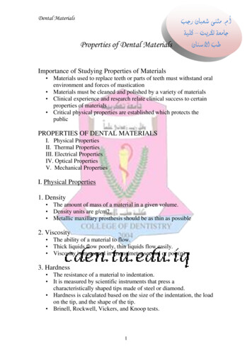
LangmuirArticleFigure 1. Chemical structure of silk-poly(amino acid) ionomers: silk-poly-L-glutamic acid (SF-PG) and silk-poly-L-lysine (SF-PL).Silica spheres with a diameter of 4.0 0.2 μm as 10% dispersions inwater were obtained from Polysciences, Inc. Sodium phosphatedibasic, sodium phosphate monobasic, and hydrofluoric acid (HF 48 51%) were purchased from BDH. Ammonium fluoride (AF) wasobtained from Alfa Aesar. Branched polyethylenimine (PEI) with Mn 10 000, fluorescent isothiocyanate (FITC), and FITC-dextrans withdifferent molecular weights were obtained from Sigma-Aldrich. Thecross-linker, 1-ethyl-3-[3-dimethylaminopropyl] carbodiimide hydrochloride (EDC), was obtained from TCI. All materials were usedwithout further purification. The water used in all experiments wasBarnstead Nanopure water with a resistively above 18.2 MΩ cm.Single-side polished silicon wafers of {100} orientation (UniversityWafer Co) were cut to a typical size of 10 mm 20 mm and cleanedin a piranha solution as described elsewhere.53Fabrication of “PEI-(SF-PG/SF-PL)n” Capsules. Silica sphereswere dispersed in 0.5 mg/mL PEI solution (prepared in 0.1 M NaCl,pH 7) to make a prime layer to stabilize the LbL process. Then silicaspheres were dispersed in 1 mg/mL silk fibroin-poly-L-glutamic acid,SF-PG, prepared in 0.05 M phosphate buffer at pH 5.5, followed bySF-PL deposition as redispersion in 1 mg/mL silk fibroin-poly-L-lysinesolution, SF-PL, prepared in 0.05 M phosphate buffer at pH5.5.Multilayer capsules were obtained by repeating the alternatingdeposition of SF-PG and SF-PL components (Figure 2). Eachsilk-based materials excellent candidates for drug deliverysystems, scaffolds for tissue and biosensor engineering.However, building microconstructions from proteins ischallenging because of their poor stability. Up to now, finalsilk structures (ultrathin films and microcapsules) have beenstabilized by the formation of β-sheets.47 49 However, ourrecent study described a different strategy, based on ion pairingof the poly amino acid functionalized silk ionomers.50 Byutilizing these natural components, stable and robust silk-basedLbL microcapsules were successfully fabricated with highlyreversible pH responsive features and controlled encapsulationand release abilities.51In the present study, we focus on quantitative analysis of thecritical physical (transport and mechanical) properties of thesilk ionomer microcapsules fabricated by combining ionicpairing and covalent cross-linking as has been very recentlyintroduced.51 Both shell permeability and stiffness along withshell morphology have been measured for different fabricationand environmental conditions. These microcapsules demonstrated pH-induced permeability at extreme acidic (pH 2.5)and basic (pH 11.0) conditions with 2 orders of magnitudeincreases in the diffusion coefficient, accompanied by dramaticchanges in morphology but without compromising capsuleintegrity. Furthermore, the silk LbL shell exhibited a higherelastic modulus than traditional hydrogen-bonded microcapsules composed of synthetic polyelectrolytes, and thestiffness is widely tunable by external pH conditions andadditional covalent cross-linking. These silk-based microcapsules with adjustable permeability and outstanding mechanical properties may serve as a new promising platform forbioengineering applications. EXPERIMENTAL SECTIONFigure 2. LbL self-assembly of silk-poly(amino acid) ionomermicrocapsules through ionic pairing followed by covalent cross-linking.Materials. Silk-poly(amino acid)-based ionomers were obtainedusing our previously published method (Figure 1).50 First, the silkfibroin was extracted from Bombyx mori cocoons according to ourestablished procedures.52 The tyrosine and serine residues of the SFbackbone were chemically modified via diazonium coupling andchloroacetic acid reaction, respectively, leading to a silk derivativeenriched in carboxyl groups; subsequently, one of the silk-poly(aminoacid)-based ionomers (SF-PG) was obtained by grafting poly-Lglutamic acid onto silk fibroin (SF) to achieve a high carboxyl content.In contrast, SF-PL represents silk fibroin modified with poly-L-lysineside groups to enrich amine group content.deposition was carried out with slow rotation to avoid the formation ofair bubbles for 15 min, followed by two wash cycles in phosphatebuffer with pH 5.5 by centrifugation at 1000 rpm for 1 min to removethe excess of polyelectrolytes.All capsules were cross-linked with 1-ethyl-3-[3-dimethylaminopropyl] carbodiimide hydrochloride (EDC) according the establishedprocedures.54 The silica spheres with PEI(SF-PG/SF-PL)n multilayerswere added to 5 mg/mL EDC solution (prepared in 0.05 M phosphate12236dx.doi.org/10.1021/la302455y Langmuir 2012, 28, 12235 12244
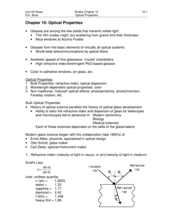
LangmuirArticleFigure 3. Morphology of silk ionomer microcapsule at different pHs in liquid state. (a) AFM topography images of swollen PEI-(SF-PG/SF-PL)5capsules in pH 5.5 and 11.5 (Z-scale: 1,000 nm) and pH 1.5 and 2.0 (Z-scale: 2,000 nm) phosphate buffer solutions. (b) High resolution AFMheight images of swollen PEI-(SF-PG/SF-PL)5 capsules in pH 1.5, 2.0, 2.5, 5.5, 11.0, and 11.5 phosphate buffer solutions (Z-scale: 200 nm).selected flat surfaces without wrinkles from at least threeindividual capsules to ensure representative results.Confocal Laser Scanning Microscopy (CLSM). Confocalimages of capsules were obtained with LSM 510 vis confocalmicroscope equipped with 63 1.4 oil immersion objectivelens (Zeiss). Capsules were visualized by adding FITC solution(1 mg/mL in phosphate buffer at pH 5.5) to the capsulesuspension in Lab-Tek chambers (Electron MicroscopyScience). Excitation/emission wavelengths were 488/515 nm.To investigate capsule permeability, fluorescent recovery afterphotobleaching (FRAP) was investigated by photobleachingFITC-dextran fluorescent molecules inside the microcapsulewith CLSM.62A drop of hollow capsule suspension was added to a Lab-Tekchamber, mixed with 200 μm of 1 mg/mL FITC-labeleddextran solution with different molecular weights (prepared inphosphate buffer at different pHs) and allowed to settle forseveral hours to make sure the fluorescent intensities inside andoutside of the capsule were same. Then a laser beam (488 nm)was focused within a region inside a capsule and pulsed at 100%intensity to photobleach the dye molecules. Each FRAP startedwith 5 prebleached scans, followed by 25 30 iterations ofbleaching to make sure the fluorescent intensity inside thecapsule was reduced by 40 60% while avoiding damage to thecapsule. The fluorescence recovery was monitored by capturingscans of 3 ms exposure at low laser intensity (3% of maximum).The recovery was considered complete when the fluorescenceof the photobleached region leveled off. The FRAP recoverycurves were analyzed using an established procedure.62,63 Therecovery curve of the fluorescence intensity, I, as a function oftime, t, was fit by a functionbuffer at pH 5.5) for 40 min and then washed with phosphate buffer atpH 5.5 to remove excess coupling agent. To dissolve the silica cores,the particles were exposed to 1 M HF/4 M NH4F solution (pH 5.5)overnight, followed by dialysis with slide-A-lyzer dialysis cassettes(100 000 MWCO, Thermal Scientific) against Nanopure water at pH5.5 for 72 h with repeated changes of water (Figure 2).55,56 CHARACTERIZATIONAtomic Force Microscopy (AFM). Surface topography ofthe hollow capsules in dried and swollen states was examinedby AFM.57 The height and phase images were collected with aDimension-3000 AFM (Digital Instruments) in tapping modeusing silicon V-shape cantilevers having spring constants of 46N/m for imaging in air. For the morphology of the swollenstate at different pHs, a scanAsyst-fluid mode on a DimensionIcon (Veeco) was employed using a liquid cell and a tip with aspring constant of 0.7 N/m. Samples were prepared by placinga droplet of hollow capsule suspension on a precleaned siliconwafer and drying in air prior to AFM scanning. During thescanning in liquid, phosphate buffer solution with different pHswas added to the predried capsules. The capsule single wallthickness was determined as half of the height of the collapsedflat regions on dried and swollen capsules using bearing analysisfrom NanoScope software to generate height histograms. Themicroroughness was measured in smooth microcapsule regionsfrom selected 1 μm 1 μm areas without wrinkles.58,59Micromechanical properties of silk ionomer capsules weremeasured by force-volume mode on the Dimension-3000microscope in both dry and swollen states. Force distancecurves were collected by micromapping with 16 16 pointarrays over a 1 μm 1 μm area selected without wrinkles.Silicon nitride tips with spring constants of 6.0 and 0.7 N/mwere used in dry and liquid state, respectively. The springconstants of the cantilevers were determined by thermal tuning.The tip radius was estimated by scanning a reference standardsample with 5 nm gold nanoparticles. Mechanical measurements of capsules in the swollen state were performed with aliquid cell in phosphate buffer adjusted to specified pH values.The collected data were processed with a custom-mademicromechanical analysis (MMA) software according to thewell-established procedure.59 61 A parabolic Snedoon’s modelwas utilized to calculate Young’s modulus by processing theforce distance curves collected in the elastic regime at theI I0(1 e At )(1)where I0 represents the initial fluorescence intensity. Thecoefficient A is related to the diffusion coefficient, D, accordingto equation:A 3P /r 3D/rh(2)Which is valid for diffusion through a spherical wall with radiusr and thickness h. RESULTS AND DISCUSSIONSilk Shell Morphologies. Silk-polyamino acid ionomers,obtained by selective modification side groups of silk protein12237dx.doi.org/10.1021/la302455y Langmuir 2012, 28, 12235 12244
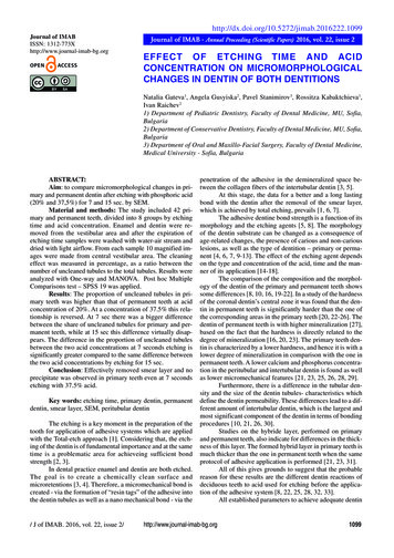
LangmuirArticlebackbone to enrich either cationic (silk-poly L-lysine, SF-PL) oranionic (silk-poly-L-glutamic acid, SF-PG) charges, wereutilized to prepare hollow microcapsules by LbL assembly(Figure1).The capsules were stable over a wide pH range, 1.5 to 12.0,and showed a remarkable degree of reversible swelling/deswelling when exposed to pH conditions below 2.0 andabove 11.0 (Figure 2). As has been demonstrated in ourprevious publication, the diameter of silk ioonomer microcapsule increased for 3.8 0.2 μm at pH 5.5 to 5.3 0.2 μm atpH 1.5 and 5.7 0.2 μm at pH 12.51In order to understand the pH responsive properties of thesilk ionomer microcapsules, the morphology of the capsuleswas monitored upon exposure to different pH conditions(Figure 3a). Upon drying the hollow silk microcapsules on asolid substrate, random wrinkles are formed due to shellcollapse.64,65 Even by exposing the predried capsules to theaqueous solutions, the capsules still kept the wrinkledmorphology. However, upon exposure to the solution with apH above 10.5 and below 3.5 (adjusted by 0.05 M phosphatebuffer), the topography significantly changed. The silk LbLshell swelled for capsules at pH 2.0 when compared to the samebilayer number capsules at pH 5.5. Further reducing thesolution pH to 1.5 resulted in uneven and ruffled morphologiesobserved from AFM. The same phenomenon was obtained atpH above 11.5.The dramatic change of grainy texture of shells was alsorevealed by high resolution AFM images at different pHs(Figure 3b). The surface morphology of silk materialsundergoing partial transformation of secondary structureassociates with the grainy topography at pH 5.5.66 The grainystructure gradually formed as the capsules were exposed to pH2.5, 2.0, and 1.5, respectively. The same change happened uponexposure to basic conditions.The thickness of silk ionomer microcapsules with 5 bilayerswas measured by AFM cross sections in the swollen state atpHs from 1.5 to 11.5 (Figure 4a). The shell thickness increasedgradually from 87 6 nm (pH 5.5) to 107 6 nm (pH 3.5)and 103 8 nm (pH 11.0) and then followed by dramaticswelling to 204 15 nm for pH below 2.0. The capsulesshowed a nonuniform swelling at pH 11.5, where some regionshad high swelling and a reached thickness of 201 35 nm,whereas some regions remained at about the same thickness asat pH 5.5. The values of thickness even in neutral conditionswere significantly higher than typical values for hydrogenbonded capsules within the range from 20 to 35 nm associatedwith the different components and molecule weights.67,68The same trend was observed for changes in shellmircoroughness, as measured in selected smooth (withoutwrinkles) areas of 1 1 μm2. The capsules exhibited an almostconstant roughness (13.5 16.5 nm) over a wide pH range from3.5 to 10.5, much higher than usually observed for electrostaticbased LbL films (around 1 2 nm)69 but common for diffusionenhanced LbL assembly with aggregated and porousmorphologies.70 These features were close in terms ofroughness to those previously reported for hydrogen bondedmicrocapsules employing a PEI-prelayer (in the range of 13 15nm).67Furthermore, the microroughness dramatically increased forpH values below 3.5 and above 10.5, and reached a value of27.5 1 nm at (pH 1.5) and 37.5 3 nm (pH 11.5) (Figure4). Such a remarkable increase in surface microroughness isconsistent with the steady development of domain morphol-Figure 4. pH induced variation of silk ionomer microcapsules physicalfeatures. (a) pH-trigged swelling of PEI-(SF-PG/SF-PL)5 hollowcapsules in pH range from 1.5 to 11.5. (b) Microroughness of PEI(SF-PG/SF-PL)5 hollow capsules as a function of pH.ogies with the dimensions enlarged from 30 40 nm to about300 and 400 nm for pH 11.5 and 1.5, respectively. The grainystructure is a common phenomenon for silk materials,48,71 dueto local aggregation, and the responsive changes of domainsizes are attributed to reduced ionic pair bonding uponexposure to extremely basic and acid pHs.The distinct pH-dependent changes in morphology of silkiomomer capsules can be related to a reduction of ionicbonding in the LbL thin shell at extreme conditions. As known,polyglutamic acid and polylysine utilized here for preparing thepairing of silk ionomers have pKa 3.5 and 9, respectively.72The negative charge of SF-PG gradually decreased due to theprotonation of carboxyl group below its pKa. There is a higherdegree of deprotonation of amino groups on SF-PL and a lowerpositive charge on the SF-PL when the pH is above its pKa. Theresult is consistent with the variation of zeta-potential of hollowcapsules in the pH range from 1.5 to 12.0 which was describedin our previous publication, where the zeta-potential wasnegative in the pH range from 3.5 to 7.5 and then increased topositive values for pHs below 2.5 and more negative above pH7.5.51Only capsules with additional EDC-induced cross-linkingwith amide bonds between carboxylic groups on SF-PG andprimary amine groups on SF-PL exhibited uniform swollenshapes under extremely acidic and basic conditions indicatinglight permanent cross-linking (Figure 2). Indeed, the untreatedmicrocapsules are totally dissolved when exposed to acidic (pH 1.5) or basic environments (pH 11.5). The FTIR spectrafor microcapsules with and without cross-linking did notindicate significant detectable changes thus confirming very lowdegree of cross-linking. In fact, the silk backbone is composedof repeated amino acid units through amide bonds,73 whichresulted in a strong carbonyl peak of amide bonds for silk-based12238dx.doi.org/10.1021/la302455y Langmuir 2012, 28, 12235 12244
LangmuirArticleTable 1. Thickness and Microroughness of PEI-(SF-PG/SF-PL)5 Microcapsules with Different Cross-Linking Times in the Dryand Swollen States (pH 6.0)thickness, nmcross-linked time, min04080160dry48.046.246.744.5 2.11.62.22.4swelling % er, μmmicroroughness, nmswollencapsules even without cross-linking thus masking possiblechanges during cross-linking (Figure 2).54,68,74Significant variation of thickness and microroughness forPEI-(SF-PG/SF-PL)n capsules was observed with differentcross-linked time in dry and swollen states (Table 1). Thedegree of swelling decreased with increasing cross-linking timedue to the higher degree of cross-linking. In addition, thicknessalso decreased from 48.0 2.1 to 44.5 2.2 nm indicatingdensification of the shells upon cross-linking. Comparingovernight cross-linking from the literature,54,75 the cross-linkingtime was set to 40 min to obtain stable capsules at extremelyacidic and basic pHs while minimizing the effect of cross-linkingon capsule properties and avoid excessive stiffening. A grainysurface texture and with significant variation in microroughnessat different pHs of the silk ionomer shells suggests a porousmorphology to facilitate tunable permeability.76Silk Shell Permeability. FRAP analysis of permeability wasconducted by using fluorescein isothiocyanate (FITC)-labeleddextran as a fluorescent probe with confocal microscopy(Figure 5).62,77,78 Focusing the laser beam onto the capsuleinterior and increasing the excitation intensity at 100% ofmaximum resulted in efficient bleaching (Figure 5a, middle).Immediately after bleaching stage and reduction of the laserpower by a factor of 30, the intensity of the capsule interiorgradually increased over time due to the diffusion ofunbleached FITC-dextran surrounding the capsule into theinterior until the intensity inside of the capsule becameconstant (Figure 5a, right). For quantitative analysis, thefluorescence intensity inside the bleached capsule wasintegrated by give intensity values for each time point, whichconstituted the FRAP recovery curves which can be fit with eq1 (Figure 5b,c).Although the number of bilayers significantly affected thecutoff molecular weight for permeation,48,51 the slope of therecovery curve for silk ionomer microcapsules with five bilayerswas only slightly higher than that of seven and nine bilayershells exposed to 4 kDa FITC-dextran at pH 5.5 (Figure 5b).However, much faster recovery was observed at extremely basic(pH 11.5) and acidic (pH 2.0) conditions compared to thesame capsule (5 bilayers) exposed to the same molecular weightFITC-dextran (20 kDa) at pH 5.5 (Figure 5c). This findingindicates that the diffusion rate across shells is changeddramatically by pH variations.The fluorescent intensity recovery curve fitted withtheoretical permeation kinetics equations can provide permeability coefficients, which can be converted into diffusioncoefficients (D) according to eq 2 with use of shell radius andthickness (Figure 6).79 The silk iomoner capsule indicated anexponential build-up thickness with increasing bilyaer number,which resulted in a significantly thicker shell of nine bilayercapsules. However, the diffusion coefficients calculated fromFRAP experiments increased from 7.2 10 11 to 2.3 10 10cm2/s with an increasing number of bilayers (for 4 kDa FITC-dry8.711.511.311.3 swollen1.33.30.31.712.713.617.311.1 2.33.12.71.2swollen3.743.733.803.75 0.060.060.050.06Figure 5. (a) Confocal images of PEI-(SF-PG/SF-PL)5 capsuleexposed to 4 kDa FITC-dextran solution at pH 5.5 during differentstages of FRAP. (b) FRAP recovery curves of capsules with differentnumbers of bilayers for 4 kDa dextran at pH 5.5. (c) PEI-(SF-PG/SFPL)5 capsule FRAP recovering curves for 20 kDa dextran at differentpHs.dextran as a probe) at pH 5.5 indicating the role of porosity(Figure 6B). These values are much higher than those obtainedfor conventional low porosity ionic PAH/PSS shells wherediffusion coefficients were as low as 2.7 10 12 cm2/s for lowmolecular weight dyes for comparable number of bilayers.77On the other hand, the value of the diffusion coefficient wasthe same order of magnitude as that reported for weaklyhydrogen-bonded LbL shells with high porosity.65,80 Wesuggest that the modified silk macromolecular structure, witha hydrophobic backbone and hydrophilic pendant groups, withabundant site for hydrogen bonding contributed to a loose,porous morphology for the silk ionomer shells, resulting in the12239dx.doi.org/10.1021/la302455y Langmuir 2012, 28, 12235 12244
LangmuirArticleFigure 6. Diffusion coefficients of silk ionomer microcapsules. (A) pHinduced diffusion coefficient variation of PEI-(SF-PG./SF-PL)5 hollowcapsules for 20 kDa FITC-dextran. (B) Diffusion coefficient of PEI(SF-PG/SF-PL)n capsules with different bilayer numbers to 4 kDaFITC-dextran at pH 5.5. (C) Diffusion coefficient of PEI-(SF-PG/SFPL)5 capsules with different cross-linking time for 4 kDa dextran at pH5.5.Figure 7. Force distance curves for dried (a) and swollen (b) PEI(SF-PG/SF-PL)5 hollow microcapsules in air and liquid state in anapproaching-retracting cycle.high permeability much close to weakly bonded LbL shells thantraditional ionic-based shells.As expected, a lower diffusion coefficient was found withincreasing labeled dextran molecular weight from 4 to 20 kDa(Figure 6A,B); however, when the same capsules were exposedto the dextran with the same molecular weight (20 kDa) at pH2.0 and 11.5, the diffusion coefficients dramatically increasedfrom 8.5 10 12 to 3.4 10 10 and 1.8 10 10 cm2/s,respectively (Figure 6A). Such an increase by 2 orders ofmagnitude indicates a more porous polymer network associatedwith the shell swelling and stretching at extremely acidic andbasic conditions. Furthermore, the variation of cross-linkingtime had little effect on the diffusion behavior (Figure 6C).Mechanical Properties of Silk Shells. Typical force distance curves, obtained by averaging over multiple points,exhibited dramatically different shapes near the physical contactpoint and remarkable changes of slope in the repulsive contactregime for probing silk capsules in the dry and swollen states(Figure 7). In air, a “jump-in to contact” point was presentedon the approaching curve due to the long-range attractiveforces between the tip and sample, followed by an increasedrepulsive segment, and a high degree of hysteresis on theretracting curve. As known, strong jump-in and pull-off forcesare caused by the tip sample adhesion and capillaryinteractions caused by a few nm-thick layer of water on thecapsule surface at ambient conditions.81 In liquid, the force distance curves revealed lower adhesion due to the diminishedcapillary forces (Figure 7b) and a less sharp contact point ascompared to the curve collected in air, indicating a softrepulsive surface for the swollen silk shell.The loading curves were derived from the force distancedata by analyzing the repulsive section of the approaching curvewith different deformation behavior for the silk shell in differentenvironments (Figure 8).59 The penetration observed from theloading curves is well-defined and less than 10% of thethickness for both dried and swollen capsules, which excludesthe influence of the stiff silicon substrate.82,83 The linearbehavior of the loading curves in Hertzian coordinates for allswollen shells reflects purely elastic deformation under theFigure 8. Loading curves in the Hertzian approximation: (a) PEI-(SFPG/SF-PL)5 shells in air and (b) swollen PEI-(SF-PG/SF-PL)5 shellsat different pH values.loading force.84 86 The loading curve for PEI-(SF-PG/SF-PL)5in the liquid state demonstrated higher penetration whencompared to the dry state even with very light normal loadswhich revealed the softening of swollen silk shells. Furthermore, the varied slope of loading curve obtained at different pHvalues which is directly related to their stiffness, indicates a pHdependent elastic properties (Figure 8b). Penetration increasedby a factor of 5 and 4 as the capsules were exposed to pH 2.512240dx.doi.org/10.1021/la302455y Langmuir 2012, 28, 12235 12244
LangmuirArticleFigure 9. Young’s modulus of PEI-(SF-PG/SF-PL)3 shells in the dried state (a) and Young’s modulus for swollen PEI-(SF-PG/SF-PL)5 shells (b)for different cross-linking times.and 11.5, respectively, indicating more compliant structures inextremely acidic and basic conditions.The Young’s modulus of the LbL silk ionomer shells withdifferent cross-linking was determined from force spectroscopydata to vary from 1.1 to 1.8 GPa in the dry state (Figure 9a).This is the typical range for regular polyelectrolyte LbL film aswell as common flexible polymers below glass transition(usually, from 1 to 5 GPa).87 89 On the other hand, thisvalue is much higher than that measured for hydrogen-bondLbL capsules (600 100 MPa) previously reported by ourgroup for hydrogen-bonded shells67 that can be attributed tothe presence of stronger ionic pairing bonding, the covalentbonds formed due to EDC cross-linking, and the stiffer silkmacromolecule backbone.44As exposed to liquid, the stiffness of the swollen capsules wassignificantly reduced by more than 2 orders of magnitude, tothe range of around 10 MPa, due to significant swelling of thesilk material in liquid, a common trend for highly compliantswollen materials with a high content of liquid.90 The Young’smodulus obtained here for silk ionomer shells is comparablebut higher than that of earlier reported hydrogen-bondedcapsules a with elastic modulus around 4 MPa,68,80 which canbe attributed to the covalent cross-linking and ordered domainorganization of silk backbones.
Silk fibroin is a natural protein from silkworms and spiders and has been widely utilized in biomaterial composites.43,44 The . Single-side polished silicon wafers of {100} orientation (University Wafer Co