Transcription
Contributions of vitamin D response elements and HLApromoters to multiple sclerosis riskDavid Nolan, Alison Castley, Monika Tschochner, et al.Neurology; Published online before print July 11, 2012;DOI 10.1212/WNL.0b013e318263c407This information is current as of July 19, 2012The online version of this article, along with updated information and services, islocated on the World Wide Web 11/WNL.0b013e318263c407Neurology is the official journal of the American Academy of Neurology. Published continuouslysince 1951, it is now a weekly with 48 issues per year. Copyright 2012 by AAN Enterprises, Inc. Allrights reserved. Print ISSN: 0028-3878. Online ISSN: 1526-632X.
Published Ahead of Print on July 11, 2012 as 10.1212/WNL.0b013e318263c407Contributions of vitamin D responseelements and HLA promoters to multiplesclerosis riskDavid Nolan, PhDAlison Castley, BScMonika Tschochner,PhDIan James, PhDWei Qiu, PhDDavid Sayer, PhDFrank T. Christiansen,MDCampbell Witt, PhDFrank Mastaglia, MDWilliam Carroll, MDAllan Kermode, MDCorrespondence & reprintrequests to Dr. e: The identification of a vitamin D–responsive (VDRE) motif within the HLA-DRB1*15:01promoter region provides an attractive explanation for the combined effects of HLA-DR inheritance and vitamin D exposure on multiple sclerosis (MS) risk. We therefore sought to incorporateHLA-DRB1 promoter variation, including the VDRE motif, in an assessment of HLA-DRB1–associated MS risk.Methods: We utilized 32 homozygous HLA cell lines (covering 17 DRB1 alleles) and 53 heterozygote MS samples (20 DRB1 alleles) for HLA-DRB1 promoter sequencing. The influence of HLADRB1 variation on MS risk was then assessed among 466 MS cases and 498 controls.Results: The majority of HLA*DRB1 alleles (including HLA-DRB1*15:01) express the functionalVDRE motif, apart from HLA-DRB1*04, *07, and *09 alleles that comprise the HLA-DR53 serologic group. Allele-specific variation within functional X-box and Y-box motifs was also associatedwith serologically defined HLA-DR haplotypes. Incorporating these results in an analysis ofMS risk, we identified a strong protective effect of HLA-DRB1*04, *07, and *09 (DR53) alleles(p 10 12) and elevated risk associated with DRB1*15 and *16 (DR51) and *08 (DR8) alleles(p 10 18).Conclusions: HLA-DRB1 groups corresponding to serologic HLA-DR profiles as well as promoterpolymorphism haplotypes effectively stratified MS risk over an 11-fold range, suggesting functional relationships between risk-modifying HLA-DRB1 alleles. An independent contribution ofVDRE motif variation to increase MS risk was not discernible, although vitamin D–dependentregulation of HLA-DR expression may still play an important role given that HLA-DRB1*04/*07/*09 (DR53) alleles that express the “nonresponsive” VDRE motif were associated with significantly reduced risk of MS. Neurology 2012;79:538–546GLOSSARYABMDR Australian Bone Marrow Donor Registry; AH ancestral haplotype; HARP heterozygous ambiguity resolvingprimer; MS multiple sclerosis; OR odds ratio; PDDD Perth Demyelinating Disease Database; VDR vitamin D receptor;VDRE vitamin D–responsive element.Supplemental data atwww.neurology.orgThe strong effect of geographic location on multiple sclerosis (MS) risk, described in 19211 andrefined in studies incorporating meteorologic data,2,3 reflects a protective influence of sunlightexposure that is most evident in early life,4 and may even be relevant to in utero development.5,6These benefits are mediated at least in part via vitamin D, derived naturally from sunlightexposure or through supplementation.7 Irrespective of the source, activated vitamin D exerts arange of immune-modulating functions8,9 via vitamin D receptors (VDR) and their targetnuclear vitamin D response elements (VDREs).10In this context, recent data11 have suggested a direct link between vitamin D and the dominant genetic risk factor HLA-DRB1*15:01, present in more than 50% of MS cases.12 ThisFrom the Department of Clinical Immunology (D.N., A.C., F.T.C., C.W.), Royal Perth Hospital, Perth; Institute for Immunology & InfectiousDiseases (D.N., M.T., I.J.), Murdoch University, Western Australia; Department of Neurology (W.Q.), the Third Affiliated Hospital of Sun Yat-SenUniversity, Guangzhou, China; Conexio Genomics (D.S.), Fremantle; School of Pathology and Laboratory Medicine (F.T.C., C.W.), University ofWestern Australia, Nedlands; Department of Neurology (F.M., W.C., A.K.), Sir Charles Gairdner Hospital, Perth; and Centre for Neuromuscularand Neurological Disorders (F.M., W.C., A.K.), Australian Neuromuscular Research Institute, Sir Charles Gairdner Hospital, University of WA,Perth, Western Australia.Study funding: This study was funded in part by the McCusker Charitable Foundation.Go to Neurology.org for full disclosures. Disclosures deemed relevant by the authors, if any, are provided at the end of this article.538Copyright 2012 by AAN Enterprises, Inc.
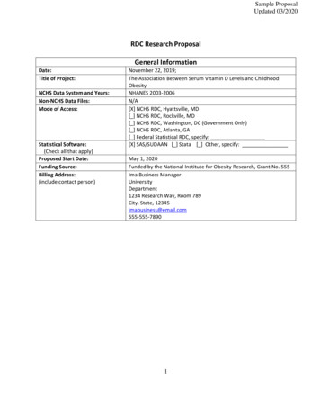
study identified a VDRE motif within theHLA-DRB1*15:01 promoter region that enhances gene expression in response to vitaminD stimulus11 (also observed for HLA-DRB1*03:01 in an unrelated study13), while otherHLA-DRB1 alleles studied were found tocarry an alternative promoter sequence that isnonresponsive to vitamin D. These authors11provided a cogent argument that for HLADRB1*15 bearing individuals, a lack of vitamin D during early life could allowautoreactive T cells to escape thymic deletion,11 providing a plausible common pathway for the effects of sunlight exposure andHLA-DRB1*15:01 carriage on MS susceptibility. We have sought to further delineatethis effect through a comprehensive analysisof HLA-DRB1 promoter sequence variationand allelic diversity in relation to MS diseaserisk.METHODS Research participants. A total of 466 Caucasian participants in the Perth Demyelinating Disease Database(PDDD) with a diagnosis of clinically definite or probable MSaccording to the Poser criteria14 or of MS using the McDonaldcriteria15 were included in the study. Of these, 425 (91.2%) wereclassified as relapsing-remitting MS. The study populationincluded 354 female cases (76%), of whom 94% had relapsingremitting MS, and 112 male cases (81% with relapsingremitting MS). Patients were assessed by the same 2 neurologists(A.G.K. and W.M.C.) at the time of collecting blood samples.The control group consisted of 498 healthy Caucasian individuals (288 [58%] female) from the Australian Bone Marrow DonorRegistry (ABMDR). With regard to participation in other studies of genetic susceptibility to MS, 1 case only from these 466cases was included in the International MS Genetic Consortium,and 90 cases were included in the ANZGene study cohort. Nocontrol subjects were involved in these studies.Standard protocol approvals, registrations, and patientconsents. The study protocol was approved by the Sir CharlesGairdner Hospital Human Research Ethics Committee, and informed consent was obtained from all participants in the PDDDcohort. Approval for use of control samples was provided by theABMDR ethics committee.B-cell lines. To ascertain DRB1 promoter sequences we utilized DNA extracted from cell lines with known ancestral haplotypes (AH). Thirty-two cell lines were selected to obtainsequence data for 17 DRB1 alleles; some represented more thanonce in these cell lines (AH 8.1, 7.1, 52.1, 58.1, 37.1, 46.1,44.2, and 57.1). In addition, 53 samples from the PDDD cohortwere selected based on HLA-DRB1 alleles present—including 8homozygous and 42 heterozygous HLA-DRB1*15:01-positivesamples—in order to obtain sequence data for 20 DRB1 alleles.HLA-DRB1 typing. High-resolution 4-digit HLA-DRB1genotyping was performed in the Department of Clinical Immunology, PathWest, Royal Perth Hospital by DNA sequencing forboth MS cases and control samples, using a previously reportedmethod.16 Automated sequencing was carried out on the ABIPrism 3730 and 3730xl Genetic Analyzers and HLA-DRB1analysis was carried out using ASSIGN V4.0.1.36 (ConexioGenomics). All HLA typing results for MS samples were resolvedto the 4-digit level using heterozygous ambiguity resolving primers (HARPs) where applicable. For control samples, sequencebased typing methods provided 2-digit resolution and NMDPcodes of possible heterozygous HLA-DRB1 combinations (seereference 17 for further information).HLA-DRB1 promoter genotyping. HLA-DRB1 promotergenotyping was also performed in the Department of ClinicalImmunology PathWest, Royal Perth Hospital, using the following methods. DNA was extracted from whole blood using theQIAGEN BioRobot M48 machine using QIAGEN MagattractDNA Blood Mini M48 kit. The PCR was performed in a 30 Lvolume, consisting of 2 L genomic DNA (at approximately 50ng/ L), 10 PCR buffer (15 mM MgCl2, 100 mM Tris-HClpH 8.4, 500 mM KCl, 1 mg/mL of gelatin, 0.2% of NP-40), 1mM of each dNTP (Life Technologies, Rockville, MD), and 2.5units of Taq Platinum polymerase (Life Technologies). The forward primer sequences 5 CAATTAAAGTTTTACATG and5 CAATTAAAGTGTTTTACACG were tagged with the universal M13F sequence, and the reverse primer 5 TGTCCCCAGMCAAAGCCAGT was tagged with the universalM13R sequence. Concentrations of all primers were initiallytested at 20 pmol and adjustments to individual primer concentrations were made if an allele was suboptimally amplified.The DNA were amplified following initial denaturation at96 C for 6 minutes followed by 35 cycles of 96 C for 30 seconds, 70 C for 30 seconds, and 72 C for 2 minutes. A finalextension step was performed at 72 C for 10 minutes. PCR amplification was confirmed following electrophoresis using 1% SEAgarose gel and the PCR amplicons were purified using MIOBIO PCR purification system. Automated sequencing was performed using ABI Big Dye Terminator chemistry, as permanufacturer’s instructions, using universal M13F and M13Rsequencing primers as well as group-specific sequencing primers(5 TCTGACCAGCGACTGAT and 5 TGTCAGAACTGCCATGCA) when required. Samples were purified using theAgencourt CleanSEQ PCR purification system prior to sequencing on ABI Prism 3730 and 3730xl Genetic Analyzers.Finally, the HLA-DRB1 promoter analysis was carried out usingASSIGN ATF 1.0.2.45 (Conexio Genomics).Statistical analysis. Comparisons of carriage frequencies ofalleles between cases and controls were carried out by Fisherexact tests for individual alleles or case-control logistic regressions for multivariable analyses. p Values of 0.05 were considered statistically significant. Analyses were carried out using theTIBCO Spotfire S statistical package (TIBCO Software Inc.,Palo Alto, CA). For analyses of HLA-DR associations with MSrisk, 2-digit resolution HLA results were utilized in order tomaximize data availability among controls.RESULTS HLA-DRB1 allele-specific VDRE sequencevariation. We first sought to examine sequence variationin the HLA-DRB1 promoter region, including the previously identified VDRE motif. As shown in figure 1and table 1, we confirmed the association betweenHLA-DRB1*15:01 and the VDRE motif 211GGGTGGAGGGGTTCA225 associated with vitamin D–responsive HLA-DRB1 gene expression. This VDRENeurology 79August 7, 2012539
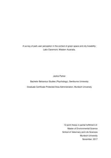
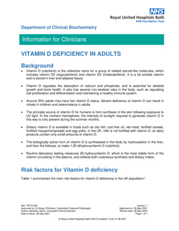
Figure 1Sequence variation in the HLA-DRB1 promoter regionHLA-DRB1 5 UTR reference sequence (HLA-DRB1*15:01) showing variation sites (red)identified by sequence-based typing (see table 1), relative to known regulatory motifs(in blue, as defined in reference 17) and vitamin D–responsive element motif (in green,reference 11).sequence was also found in all HLA-DRB1 alleles examined other than HLA-DRB1*04, *07, and *09 alleleswhere an alternative GGGTGGAGAGGGGT-CA sequence was identified (differences italicized) that introduced an internal GA insertion between positions 216and 217 of the DRB1*15:01 reference sequence shownin figure 1, which is likely to disrupt VDR binding byshifting the adenine from position 7 where it is invariably located within functional VDRE motifs.10 The resulting loss of VDRE function is consistent withprevious findings that the “alternative” (HLADRB1*04/07/09-associated) VDRE is not responsiveto vitamin D stimulation.11 Among the 85 samples inwhich promoter sequencing was undertaken, all conformed to the expected patterns of allele-specific promoter polymorphism identified in table 1, including 53DRB1*15:01-positive samples (3 homozygous cell linesand 50 MS cohort samples: 8 homozygous and 42heterozygous at the DRB1 locus).HLA-DRB1 promoter sequence variation. Investigating the promoter region more generally (table 1), weidentified sequence identity throughout the entirepromoter region between HLA-DRB1*15:01 andthe closely related DRB1*15:02 and DRB1*16:01alleles that make up the DR51 haplotype group,18as well as HLA-DRB1*01 alleles (4 DRB1*01:01,1 DRB1*01:02, and 3 DRB1*01:03-positive samples) that share a distant ancestral history withHLA-DRB1*15:0119 but which display only limited sequence similarity and epitope bindingpreferences.20,21 It is also notable that the HLADRB1*15:01, DRB1*15:02, and DRB1*16:01 alleles within the HLA-DR51 haplotype group sharean association with HLA-DRB5, while loss of thisadditional HLA-DRB locus has been identified as a540Neurology 79August 7, 2012feature of the evolutionary divergence of HLADRB1*01 from the ancestral HLA-DR51 group.19All 53 HLA-DRB1*15:01-positive samples showedsequence identity across the promoter region (figure 1).Looking beyond the VDRE motif region, weidentified HLA allele-associated polymorphismwithin regulatory X-box and Y-box motifs (see figure1) that have been previously shown to influenceconstitutive and cytokine-stimulated HLA-DR expression, respectively.18,22 With regard to the MSassociated HLA-DRB1*15:01 allele, its X-boxsequence has been associated with high constitutivegene expression, 4- to 5-fold higher than observedwith DR53-associated HLA-DRB1*04, *07, and *09alleles, in which the canonical motif is disrupted bythe A-to-G transition at position 106 (figure 1 andtable 1).22 Similarly, polymorphic residues at positions 139 (G) within the Y-box domain and 164 (C)within the CCAAT domain of HLA-DRB1*15:01have been associated with 2-fold increase in DRB1expression in response to cytokine stimuli (e.g.,interferon- and tumor necrosis factor- 18), whilethe HLA-DR53-associated sequence (139C, 164T)was again associated with nonresponsiveness.18Taken together, these findings provide evidencefor shared promoter sequence variation within theHLA-DR51 group (DRB1*15:01, 15:02, 16:01) andHLA-DRB*01:01, characterized functionally byhigh constitutive as well as inducible gene expressionin response to vitamin D/VDR as well as cytokinemediated stimuli. This functional VDRE motif isalso shared by HLA-DRB1 alleles within the HLADR52 group (table 1) as well as DRB1*10:01 andDRB1*08, although these alleles lack the cytokineresponsiveness conferred by the DR51-specific Y-boxand CCAAT motifs. In contrast, the HLA-DR53group of alleles (DRB1*04, *07, and *09) share promoter polymorphisms that confer both lower constitutive gene expression as well as nonresponsiveness toboth vitamin D– and cytokine-mediated stimuli.HLA-DRB1 alleles and VDRE variants as MS riskfactors. Having established associations between pro-moter sequence variation and HLA-DRB1 alleles, wesought to investigate the potential relevance of theseobservations to genetic risk of MS among CaucasianMS cases (n 466) and controls (n 498). Table 2describes the rates of HLA DRB1 allele carriage andhomozygosity frequencies among cases and controls,with odds ratios (ORs) for HLA allele carriageamong MS cases and controls represented in figure 2.As expected, HLA-DRB1*15 was overrepresentedamong cases (n 256, 54.9%) compared with controls (n 135, 27.1%), providing an OR of 3.27( p 0.001). The closely related HLA-DRB1*16 allele, although low in prevalence (17 cases, 3.6%; 8
Neurology 79August 7, 5555DRB3, 4, 5DRB3, 4,5 125CCCCCCCCCCCCCCCCCCCCCCCCCG139Changes in SNP sites when compared to reference ss otherwise specified, all results are derived from homozygous cell lines. DRB1*12:01 and *14:04 results were obtained from DRB1*15:01 heterozygous samples. Other results from heterozygote samples:DRB1*01:02 and *01:03 share sequence identity with DRB1*01:01 (variant 1); DRB1*011:04 shares sequence identity with DRB1*11:01 (variant 3); DRB1*04:03, *04:04, and *04:06 share sequence identity withDRB1*04:01 (variant lleleHLA-DRB1 promoter sequence variation identified in this studya7.1HLAhaplotypeTable 1
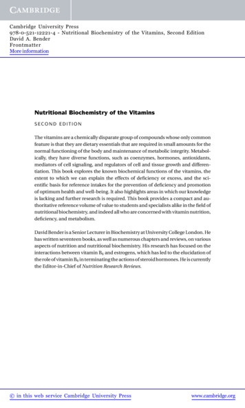
Table 2HLA allele carriage (phenotype) andhomozygosity frequency in casesand controlsAllele 66)Controls(498)No.No. %No.%51.0DRB1*03 110 23.6 122 24.5 153.2 102.0DRB1*04 113 24.2 186 37.351.1 224.4DRB1*0784 18.0 157 2.600.000.0DRB1*1071.5102.000.000.0No.DRB1*01%%83 17.8 106 21.310.2DRB1*1153 11.455 1110.210.2DRB1*12121510.210.2DRB1*1375 16.185 17.140.961.2DRB1*14172200.000.0DRB1*15 256 54.9 135 27.1 398.4 142.8DRB1*160.00.0172.63.63.683.04.41.600controls, 1.6%), was also associated with increasedMS risk (OR 2.32, p 0.066), particularly amongHLA-DRB1*15-negative individuals (OR 3.62, p 0.005). Similarly, carriage of HLA-DRB1*08 was as-Figure 2sociated with MS risk in the overall dataset (29 cases,6.2%; 16 controls, 3.2%: OR 2.00, p 0.032) witha more pronounced effect after exclusion of HLADRB1*15 (OR 2.90, p 0.006). No other HLADRB1 alleles were associated with increased MS risk,either within a restricted dataset of relapsingremitting disease (table e-1 and figure e-1 on theNeurology Web site at www.neurology.org) or inthe presence or absence of HLA-DRB1*15 (figuree-2A and e-2B).Reduced MS risk was associated with carriage ofthe prevalent alleles HLA-DRB1*04 (113 cases,24.2%; 186 controls, 37.3%: OR 0.54, p 0.0001)and HLA-DRB1*07 (84 cases, 18.0%; 157 controls,31.5%: OR 0.48, p 0.0001). A trend toward reducedrisk was also found for the low-prevalence HLADRB1*09 allele (5 cases, 1.1%; 13 controls, 2.6%: OR0.40, p 0.096). Notably, HLA-DRB1*04 remainedprotective among HLA-DRB1*15 heterozygous individuals (OR 0.55, p 0.023), while the protectiveeffect of HLA-DRB1*07 was observed in the HLADRB1*15-negative group (OR 0.42, p 0.0001),but not among HLA-DRB1*15 carriers (OR 1.48,p 0.29).Overall, the effects of HLA-DRB1 allele carriageon MS risk show consistent correlations with theoriginal HLA-DR serologic groups defined prior tothe availability of sequence-based HLA typing tech-Relative risk of multiple sclerosis according to HLA carriage: Odds ratios and 95%confidence intervalsHLA serologic groups are indicated by color and defined in the left-hand column. Alleles that share promoter sequenceidentity with HLA-DRB1*15:01 are indicated with bold italics.542Neurology 79August 7, 2012
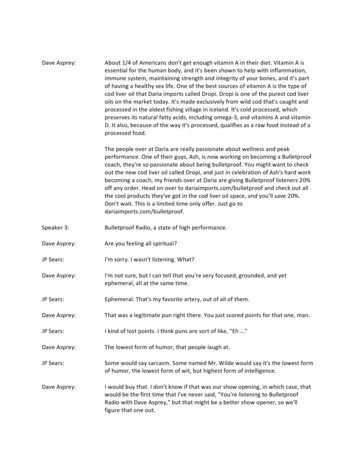
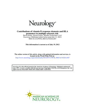
Figure 3Combined effects of HLA-DR profiles on multiple sclerosis riskStratification of multiple sclerosis risk according to combined HLA allele profiles, including prevalence rates in cases (n 466) and controls (n 498): odds ratios and 95% confidence intervals.niques (figure 2), which reflect both the peptidebinding preferences of these HLA-DRB1 allelegroups19 –21 as well as their haplotypic associationswith additional HLA-DRB loci (i.e., HLA-DRB5[DR51], DRB3 [DR52], and DRB4 [DR53]). Thus,HLA-DRB1 alleles from the DR53 group appear tobe protective, while those from the DR51 and DR8group are associated with excess risk. The remainingDR52 and DR1-associated HLA-DRB1 alleles didnot significantly influence MS risk in this population, and it is notable in particular that carriage ofHLA-DRB1*01 was not associated with increasedMS risk in the overall group (OR 0.80, p 0.19) orin the absence of HLA-DRB1*15 (OR 0.95, p 0.84) despite sharing complete promoter sequenceidentity with HLA-DRB1*15 and DRB1*16 alleles.Thus, there was no evidence that promoter sequencevariation— either within the VDRE motif or acrossthe promoter region more generally—acted as adominant independent risk factor for MS in thisstudy.Considering these findings in terms of the inheritance of serologic HLA-DR profiles, there was a clearlinear effect of HLA-DR53 allele copy number ( pvalue for trend 1.9 10 12) as well as HLA-DR51/DR8 allele copy number ( p 10 18). Utilizing these data to stratify genetic risk (figure 3), theoverall effect of these compound genotypes appearsto be consistent with a dominant influence of “highrisk” HLA-DR51 and HLA-DR8 alleles (present in61.8% of cases and 30.9% of controls), and a recessive protective effect of HLA-DR53 alleles (21.0% ofcases and 49.4% of controls), although independentallele dosage models cannot be excluded.DISCUSSION The identification of a vitamin D response element in the promoter region of the HLADRB1 gene,11 and the role of allele-specific sequencevariation within this VDRE motif in determining responsiveness to vitamin D stimulus,11,13 has providedan attractive mechanism by which the 2 dominantrisk factors for MS—HLA-DRB1 allele carriage andlow environmental vitamin D exposure—may be directly linked. In this study we have further exploredthe role of promoter polymorphism in relation toHLA-DRB1 alleles represented in Caucasian populations, demonstrating that the HLA-DRB1*15:01associated VDRE motif is widely represented amongCaucasian HLA-DRB1 alleles with the exception ofHLA-DRB1*04, DRB1*07, and DRB1*09 alleles.Neurology 79August 7, 2012543
Furthermore, we identified promoter sequence variation at other regulatory sites within the DRB1 promoter region that have been previously shown toinfluence both constitutive and cytokine-stimulatedexpression levels.18,22 In this regard, the MS riskassociated HLA-DRB1*15:01 allele is characterizedby relatively high constitutive expression as well asresponsiveness to both vitamin D/VDR as well ascytokine stimulation, combined traits that are sharedby several DRB1 alleles that show sequence identitythroughout the promoter region (DRB1*15:02, 16:01,and 01:01). Conversely, those DRB1 alleles that lack avitamin D–responsive VDRE motif (DRB1*04,DRB1*07, and DRB1*09) are also characterizedby promoter sequence variation within X-box andY-box domains that confer low constitutive geneexpression and nonresponsiveness to cytokine stimuli.18,22In light of these observations, it is interesting thatrisk of MS in a large case-control study involvingComment:The HLA region in multiple sclerosisNolan et al.1 investigate human leukocyte antigen (HLA)-DRB1 promotersequence variation, including a previously characterized vitamin D responseelement (VDRE),2 addressing the challenge of a mechanistic explanation for theassociation of HLA with multiple sclerosis (MS) and the suggested link withvitamin D.2The HLA-DRB1 gene is highly polymorphic, with many different forms orhaplotypesthat are evolutionarily related. The latest MS genome-wide association studyidentified DRB1*1501, DRB1*03:01, and the group of DRB1*13:03 and DRB1*08:01 haplotypes as increasing risk of MS.3 Nolan et al. reveal differences in thesequence of functional promoter elements among haplotypes, ranging fromhaplotypes with high constitutive expression levels that can be further induced byvitamin D and cytokines (DRB1*15, *16 and *01 haplotypes), to those with lowconstitutive gene expression and nonresponsiveness to vitamin D and cytokines(DRB1*04, *07, and *09 haplotypes).1 Combining promoter sequence differenceswith high-resolution association studies, not all cytokine and vitamin D responsivehaplotypes are associated with MS. For example, the high-risk MS haplotype HLADRB1*15:01 (in the Nolan et al. study) is identical in promoter sequence, includingthe VDRE, to related haplotypes that are not associated with MS, such asDRB1*15:02.1,3 This study hence suggests that none of the HLA-DRB1 promotercharacteristics can on its own account for the association of HLA with MS.Whereas the current knowledge of MS genetics provides further evidence for boththe HLA and the vitamin D metabolism pathways,3 the study by Nolan et al.1 doesnot support response to vitamin D as a major explanation for the association ofHLA with MS. Further studies coupling detailed knowledge of sequence variationto imputation and conditional analyses in large datasets are needed to gainadditional insights.1. Nolan D, Castley A, Tschochner M, et al. Contributions of vitamin D responseelements and HLA promoters to multiple sclerosis risk. Neurology 2012;79:XXX–XXX.2. Ramagopalan SV, Maugeri NJ, Handunnetthi L, et al. Expression of the multiplesclerosis-associated MHC class II allele HLA-DRB1*1501 is regulated by vitaminD. PLoS Genet 2009;5:e1000369.3. The International Multiple Sclerosis Genetics Consortium, The Welcome TrustCase Control Consortium 2. Genetic risk and a primary role for cell-mediatedimmune mechanisms in multiple sclerosis. Nature 2011;476:214 –219.An Goris, PhDA. Goris receives/has received funding from the Research Foundation Flanders,Research Fund KU Leuven, Belgian Charcot Foundation, Belgian NeurologicalSociety, and Wetenschappelijk Onderzoek Multiple Sclerose. Go to Neurology.orgfor full disclosures.544Neurology 79August 7, 2012Caucasian participants from Western Australia couldbe effectively stratified according to groups of HLADRB1 alleles that reflect these promoter haplotypesin part, but which are also defined by their evolutionary ancestry,19,21 antigen-binding preferences,20 andlinkage disequilibrium with additional HLA-DRBloci (i.e., HLA-DRB3/4/5).23 In this respect it is alsonotable that the serologic HLA-DR groups originallydefined before the advent of high-resolutionsequence-based typing methods appear to provide avery useful classification system for assessing MS riskin this study. Thus, the HLA-DR53 group of alleles(DRB1*04, DRB1*07, and DRB1*09, each in linkage disequilibrium with HLA-DRB4) demonstrateda consistent association with reduced MS risk, whileincreased MS risk was associated with the HLADR51 group (DRB1*15:01, DRB1*16:01, associated with HLA-DRB5 carriage) as well as DR8(DRB1*08, which has no additional HLA-DRB lociwithin the extended HLA-DR haplotype). Both protective and risk effects were dependent on allele doseand were highly significant, contributing to a geneticprofile for this study population that could effectively stratify low- and high-risk groups, providing an11-fold range in disease susceptibility across thestudy population (ORs 0.28 –3.06, figure 3). Theseresults need to be interpreted in light of the limitations of the study, both in terms of the influence ofsample size on statistical power as well as the use oflow-resolution (2-digit) DRB1 typing results in thisanalysis, so that we were unable to assess any differential effects of closely related alleles (e.g.,DRB1*15:01 vs 15:02) on MS risk. Nevertheless, itis interesting that similar results were obtained froman analysis of HLA-DR serologic profiles in a Finnish study.24With regard to the potential influence of VDREpolymorphism on MS risk, we can conclude that thetrait of vitamin D–responsive HLA-DRB1 gene expression is not specific to the high-risk DRB1*15:01allele, and indeed that the functional VDRE motif iscommon among a broad range of Caucasian HLADRB1 genotypes, including DRB1 alleles that haveno discernible influence on MS risk. This finding,however, does not exclude the possibility that earlylife vitamin D exposure may interact with HLADRB1 inheritance as a risk factor for the disease, orindeed that interactions between the HLA-DRB1promoter region and environmental- or infectionderived stimuli such as vitamin D and inflammatorycytokines may play an important role in modulatingdisease risk through epigenetic mechanisms.25 Wedid not assess sunlight exposure as a potential variable in the analysis of MS risk in this cohort, butanticipate that such a study would be more usefully
undertaken in a more geographically diverse population given that study participants were largely derived from metropolitan Perth, which is located at alatitude (32 ) where early-life UV exposure is likelyto be sufficient to protect against excess MS risk.2,3These findings suggest that the functional relationships between HLA-DR alleles, both in terms oftheir promoter sequence characteristics18,22 as well astheir peptide-binding characteristics,19 –21,26 can beusefully explored to identify HLA-DRB1 allelespecific and shared genetic traits that contribute toMS risk. We therefore submit that the results of thisstudy provide a platform for further analyses in largerstudy populations with sufficient statistical power tocomprehensively examine the influence of HLADRB1 promoter sequence variation, allele distribution, and serologic profiles on MS risk. In this regard,the collaborative efforts of the International MultipleSclerosis Genetic Consortium have now provided anunprecedented opportunity to conduct such analysesbased on existing data,27 with the add
The online version of this article, along with updated information and services, is . TIBCO Spotfire S statistical package (TIBCO Software Inc., Palo Alto, CA). For analyses of HLA-DR associations with MS risk, 2-digit resolution HLA results were utilized in order to . ples) that share a distant ancestral history with HLA-DRB1*15:0119 (1 .