Transcription
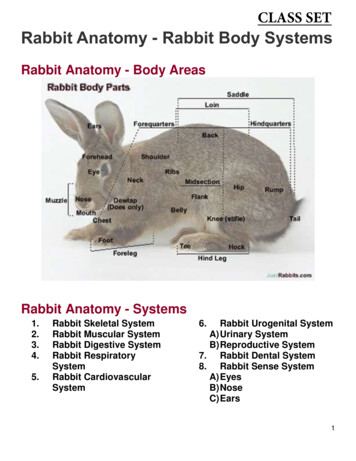
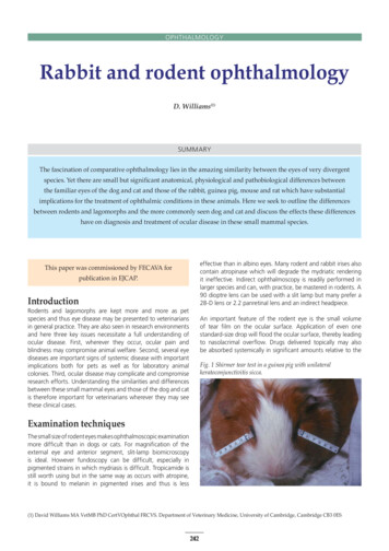
OPHTHALMOLOGYRabbit and rodent ophthalmologyD. Williams(1)SUMMARYThe fascination of comparative ophthalmology lies in the amazing similarity between the eyes of very divergentspecies. Yet there are small but significant anatomical, physiological and pathobiological differences betweenthe familiar eyes of the dog and cat and those of the rabbit, guinea pig, mouse and rat which have substantialimplications for the treatment of ophthalmic conditions in these animals. Here we seek to outline the differencesbetween rodents and lagomorphs and the more commonly seen dog and cat and discuss the effects these differenceshave on diagnosis and treatment of ocular disease in these small mammal species.effective than in albino eyes. Many rodent and rabbit irises alsocontain atropinase which will degrade the mydriatic renderingit ineffective. Indirect ophthalmoscopy is readily performed inlarger species and can, with practice, be mastered in rodents. A90 dioptre lens can be used with a slit lamp but many prefer a28-D lens or 2.2 panretinal lens and an indirect headpiece.This paper was commissioned by FECAVA forpublication in EJCAP.IntroductionRodents and lagomorphs are kept more and more as petspecies and thus eye disease may be presented to veterinariansin general practice. They are also seen in research environmentsand here three key issues necessitate a full understanding ofocular disease. First, wherever they occur, ocular pain andblindness may compromise animal welfare. Second, several eyediseases are important signs of systemic disease with importantimplications both for pets as well as for laboratory animalcolonies. Third, ocular disease may complicate and compromiseresearch efforts. Understanding the similarities and differencesbetween these small mammal eyes and those of the dog and catis therefore important for veterinarians wherever they may seethese clinical cases.An important feature of the rodent eye is the small volumeof tear film on the ocular surface. Application of even onestandard-size drop will flood the ocular surface, thereby leadingto nasolacrimal overflow. Drugs delivered topically may alsobe absorbed systemically in significant amounts relative to theFig. 1 Shirmer tear test in a guinea pig with unilateralkeratoconjunctivitis sicca.Examination techniquesThe small size of rodent eyes makes ophthalmoscopic examinationmore difficult than in dogs or cats. For magnification of theexternal eye and anterior segment, slit-lamp biomicroscopyis ideal. However fundoscopy can be difficult, especially inpigmented strains in which mydriasis is difficult. Tropicamide isstill worth using but in the same way as occurs with atropine,it is bound to melanin in pigmented irises and thus is less(1) David Williams MA VetMB PhD CertVOphthal FRCVS. Department of Veterinary Medicine, University of Cambridge, Cambridge CB3 0ES242
EJCAP - Vol. 17 - Issue 3 December 2007significantly among rodent and lagomorph species [5]. We donot know what contribution to the tear film they make and whythey differ so markedly between species. Yet from a pathologicalperspective these glands may prolapse, in the same way that thenictitans gland does in the dog. Similarly the orbital vascularplexus, present in rodents and lagomorphs, differs substantiallybetween species and understanding its anatomy is important inorbital surgery and enucleation [6].The small size of the globe in many species also complicatesmethods for measuring intraocular pressure. The Tonopen hasbeen favorably evaluated in rabbits [7] and the small eyes of rats[8] but the footplate is too small for mice. The new reboundtonometer (TonoVet) (Fig. 3) is small enough to provide accuratemeasurements of intraocular pressure in even the smallest ratand mouse eyes as shown experimentally [9-10] yet its accuracyand repeatability have yet to be reported in the clinical settingfor any rodent species. The paucity of reliable evidence in thetwo basic measures of tear production and intraocular pressurein small mammals just goes to show how much basic work therestill is to undertake in this whole area.Fig. 2 The phenol red thread test used in a hamster.size of the animal. This has important implications in both thetreatment of ocular disease and potential side effects, as thedrug may be acting through circulating blood levels as well asby direct ocular penetration.When interpreting ocular findings in experimental species orpet animals derived from laboratory strains the prevalence ofinherited disease must be seen as a background against whichother ocular disease is noted. This is particularly importantamong inbred strains, in which recessive genes may occur in agiven strain not being used to study that specific trait.Ancilliary ophthalmic tests include determination of tearproduction and measurement of intraocular pressure. TheSchirmer tear test is applicable to rabbits and guinea pigs (Fig. 1)but the test strip is too large for rats and mice. Breed differencesare significant, with Netherland dwarf rabbits having an unusuallyhigh Schirmer tear test reading of 12.0 2.5 mm/min comparedwith an average of 5.3 2.9 mm/min in other breeds, and a rangefrom 0 to 15 mm/min in 142 normal eyes [1]. Evaluation of thetear film in smaller rodents is difficult, if not impossible, with theSchirmer tear test strip. Here the Phenol Red Thread Test may beuseful (Fig. 2). This test has been used to measure tear volumerather than production in the mouse [2] while another study [3]compared the PRTT with the Schirmer tear test in rabbits findingmean wetting of the Schirmer test strip of 4.9 2.9mm/min andmean PRTT wetting of 20.9 3.7mm/15 s. A recent paper on tearevaluation in the guinea pig gave a mean STT of 0.6 1.83 mmwetting/min and a mean PRTT-value of 16 4.7 mm wetting/15s) [4]. The glands contributing to the precorneal tear film differThe rat and mouseMuch has been written on the ocular diseases of rats, summarizedin a recent review paper [11]. A superb overview of the mouseeye has been provided by Smith et al [12]. Further information isavailable through those references.Conjunctivitis is common in rodents and may often relateto mycoplasmal respiratory disease, though other agentscan also be involved [13-15]. Young animals are often moreseverely affected [16]. Environmental changes rather thanFig. 3 The Rebound Tonometer is invaluable for measuringintraocular pressure in the small eyes of rodents.Fig. 4 Chromodacryorhoea in a rat.243
Rabbit and rodent ophthalmology - D. WilliamsFig. 5 Murine microphthalmos with engorged episcleral and iridalvessels. Courtesy Dr. Peter Lee.Fig. 6 Corneal dystrophy in a CD1 mouse. Courtesy Dr. Peter Lee.primary infectious agents may be the most important factors inconjunctival inflammatory disease: ventilation currents in rodentcages may produce airborne suspensions of fine bedding mattergiving rise to a severe keratoconjunctivitis [17]. Investigating aproblem such as conjunctivitis in such a group of rodents thusrequires a full and thorough history and clinical examination ofindividual animals as well as of the group as a whole.A common problem in laboratory rodents is microphthalmoswith a valuable overview provided by Smith et al [25]. A numberof transgenic rodents are inadvertently affected by this conditionwith clinical features [26] including microcornea, engorgedepiscleral vessels, and abnormal ocular vasculature (Fig. 5) andwelfare implications given visual dysfunction and the propensityto develop defective tear drainage together with microbialcontamination of the deep conjunctival sac [27].Perhaps the most severe adnexal disease in laboratory rats issialodacryoadenitis (SDA) virus infection [18]. This coronavirusinfection causes ocular irritation with conjunctivitis andperiorbital swelling, followed by sneezing and cervical swelling[19]. The condition is usually self-limiting within one to twoweeks, whereas resolution of secondary signs may take longer.The immune status of the animals is a critical factor in diseaseseverity [20]; introduction of a naive group of rats into asubclinically affected animal house may provoke clinical onsetof severe disease in these animals. Serologic testing has shownthe agent to be present in 45% of rat colonies within the UnitedKingdom, though the incidence of overt disease is significantlylower [21].Corneal opacification is relatively common in rodents.Spontaneously occurring dystrophic lesions manifest as ellipticalparacentral corneal opacities, characterized by a deposition ofbasophilic material in the subepithelial stroma (Fig. 6). Someworkers consider them heritable [29] while others suggest theFig 7 Post-anaesthetic exposure keratopathy in a rat.Many laboratory rodents exhibit red crusting around their eyesin cases of ocular irritation, upper respiratory tract infection,and stress (Fig. 4). Porphyrin pigmented tears are producedin normal amounts by the Harderian glands in several rodentspecies [22] but particularly in the rat but also some otherrodent species, excess tear production with characteristic reddeposits on the periorbital fur, nose, and paws after wiping theeyes, so-called chromodacryorhhoea, is seen [23]. Diseases suchas mycoplasmosis and sialodacryoadentitis (SDA), nutritionaldeficiencies, and other physiologic stresses are factors that maycause chromodacryorrhea. Appropriate remedial action shouldbe taken to remove the stressors involved be they infectious,environmental or managemental [24].244
EJCAP - Vol. 17 - Issue 3 December 2007Fig. 8 Glaucoma with buphthalmos in a rat.Fig. 9 Dyscoria in a young mouse, probably colobomatous inorigin.lesions are sequelae to excess ammonia in the cage bedding[30]. Inter-strain variation in rats suggests that there is probablyan interaction between genetic factors and environmentalinfluences, in lesion pathogenesis. Exposure keratopathy inrodents causes corneal ulceration in the interpalpebral bandduring prolonged anesthesia (Fig. 7) when the lids are wideopen unless they are prophylactically taped shut during surgeryor protected with a lubricant. The problem is more associatedwith xylazine and ketamine than with other anaesthetics [32].Abnormalities of the fundus may be congenital lesions, inheritedretinal dystrophies, inflammatory lesions, degenerations anddetachments [41]. Most congenital lesions of the rodent fundusare either abnormalities of the retinal and hyaloid vasculatureas noted above, or colobomatous defects of the retina oroptic disc. In young rats persistence of the hyaloid vasculatureis common and while these vessels regress over the next fewmonths considerable vitreal hemorrhage may occur during thisperiod (Fig. 11).Glaucoma has also been noted in rodents (Fig. 8) [33]. Thefailure of aqueous drainage here, however, is often not caused byiridocorneal angle abnormality, as in many inherited glaucomas inthe dog, but rather results from persistent pupillary membranescausing pupil-block glaucoma or from peripheral anteriorsynechiae in uveitis preventing drainage. Lesions of the anterioruvea in rodents are mostly either congenital or associated withinflammatory disease. The former include adhesions between thecornea and lens and the persistence of the pupillary vasculature.Synechiae from iritis may cause opacities in either the cornea orlens [34], Blood in the anterior segment is, however, more likelyto be associated with persistent embryonic lens vasculaturewhile blood in the vitreous often relates to persistent hyaloidsvessels. Dyscoria and other abnormalities in pupil shape amongyoung rodents and especially mice (Fig. 9) are considered bysome to be colobomatous lesions, but others suggest thateccentric pupils are associated with iritis [35].Inherited retinal dystrophies and degenerations occur amongexperimental animals bred specifically for study of theseconditions. The widespread nature of these dystrophic genesthroughout rat and, particularly, mouse strains, however, meansthat such blindness may occur in a supposedly normal groupof rodents. The rd gene, for instance, is seen in C57BL/6J,CBA, C3H, and various outbred albino mouse strains withFig. 10 Congenital nuclear cataract in a mouse.In the rat and mouse, cataracts may occur spontaneously,congenitally (Fig. 10) [36] and in aging animals [37]. In ratswith retinal degeneration cataracts may occur secondarily,because of the release of various metabolic byproducts [38].In mice, cataracts may arise from infection with a helicalspirochete termed the suckling mouse cataract agent [39] butmore commonly in pet and laboratory strains cataracts will beinherited [40].245
Rabbit and rodent ophthalmology - D. Williamsretinal degeneration and blindness [42]. Laboratory rodentsare also sensitive to the toxic effects of light on the retina withdevelopment of lesions causing blindness [43].The rabbitFor many years the rabbit has been widely used in ophthalmicresearch, both in drug and chemical testing using the now rightlyinfamous Draize test [44] and in basic anatomic, physiologic andpharmacologic work. More recently the rabbit has moved frombeing a mere childrens’ pet to becoming, at least in the UK, thethird most commonly encountered pet in small animal practicewith a large number of house rabbits kept by adults as valuedcompanion animals. Associated with this increase in veterinaryattention, much has been learnt about the healthy and diseasedrabbit eye, as detailed here.The rabbit eye has several anatomical peculiarities thatdifferentiate it from that of dogs and cats. An importantfeature is the retrobulbar venous plexus, vital to note duringenucleation. Performing this surgery transconjunctivally ratherthan transpalpebrally and remaining as close as possible tothe globe during dissection, obviates this problem in the vastmajority of cases. An unusual manifestation of disease relatedto the retrobulbar venous plexus is exophthalmos occurringduring stress when venous drainage is compromised by a spaceoccupying thoracic or cervical mass [45]. Another anatomicaldifference concerns the nasolacrimal duct. There is a singlenasolacrimal punctum in the rabbit and a duct which has aconvoluted passage through the lacrimal and frontal bones,passing close to the molar and incisor tooth roots [46] and thusis likely to be affected by dental disease. Malocclusion of themolar arcades in particular results in retropulsion of the toothinto the weakened maxillary bone, with subsequent nasolacrimalocclusion. Incisor malocclusion can, perhaps more commonly,produce this same result.Fig. 11 Persistent hyaloids vasculature with haemorrhage in a rat.Conjunctivitis and dacryocystitis are thus common and potentiallyproblematic conditions in domestic rabbits; distinguishingbetween the two is important [47]. Purulent ocular dischargewith conjunctival hyperemia often relates not just to conjunctivitisbut also to nasolacrimal duct infections (Fig. 12). The diagnosis ofinfective conjunctivitis and dacryocystitis should be approachedon the basis of understanding the normal bacterial flora of theconjunctival sac. Pasteurella sp. is considered by many to be themost common bacterial pathogen in the rabbit, but it is importantnot to forget Staphylococcus aureus. In a survey of staphylococcaldisease in rabbits, more than 60% had nasal exudate withconjunctivitis [48] and in another report of conjunctival florain rabbits with conjunctivitis and dacryocystitis, Pasteurella wasnot the most commonly isolated species: bacteria were isolatedfrom 78% of swabs with Staphylococcal species found in 42%of isolates while Pasteurella species were only detected in 12%[48]. Culture of nasolacrimal flushes from affected rabbits [46]showed a wide range of organisms, including Neisseria sp.,Moraxella sp. and Bordetella sp among others but Pasteurellamultocida was not detected in animals affected by epiphorarather than by dacryocystitis. The same diversity of organismswas found in nasolacrimal flushes from unaffected rabbits.Fig. 12 Dacryocystitis in the rabbit.Fig. 13 Dacryocystorhinogram of rabbit with dilated cysticnasolacrimal duct. Courtesy of Francis Harcourt-Brown.246
EJCAP - Vol. 17 - Issue 3 December 2007Fig. 14 Cannulation of the rabbit nasolacrimal duct.Fig. 15 Florid conjunctivitis with dacryocystitis.Dacryocystorhinography can show substantial lakes of dischargein dilated portions of the duct (Fig. 13), demonstrating whythe discharge can be profuse and continuous. Treatment ofdacryocystitis in the rabbit is by cannulation of the singlenasolacrimal punctum and flushing of the duct (Fig. 14). Ifcannulation from the ocular punctum is difficult, cannulation ofthe duct opening at the nasal meatus is possible, but the smalldiameter of the duct at its nasal end renders this proceduredifficult. The proximal end of the nasolacrimal duct can alsobe difficult to visualize in a rabbit in which dacryocystitis hasextended to produce a florid conjunctivitis (Fig. 15). Pressingon the lower eyelid will often manifest the duct as a pair oflighter pink lips “pouting” through the darker red, inflamedconjunctiva. In most cases, cannulation and flushing of the ductwith a drug such as orofloxacin or gentamicin will resolve theproblem but this will require repetition potentially over severaldays to weeks. When this does not have the desired effect, theduct can be cannulated in a more permanent manner with finemonofilament nylon. There are problems and potential hazardswith this technique, however, especially given the tortuosity ofthe rabbit nasolacrimal duct. Nevertheless, sometimes there isno other option in stubborn cases of dacryocystitis.Blepharitis in the rabbit may be associated with Treponemacuniculi, the agent of rabbit syphilis. The diagnosis here is madeon the basis of identifying the spirochaete on conjunctivalscrape cytology. Treatment is three injections of penicillin G at40,000 IU/kg given at 7-day intervals. Conjunctival disease inthe rabbit can be caused by viral as well as bacterial agents.The myxoma virus causes inflammatory and edematous lesionsof the lids and conjunctiva as well as of the mouth, anus, andgenitals (Fig. 16). In the acute form, death may supervene beforeany obvious ocular signs occur but conjunctival hyperemia maybe the only sign before death. In the more common subacuteor chronic form, conjunctival hyperemia progresses to chemosis,with a copious ocular exudate. The profound white ocularexudate in the disease may be caused by Pasteurella multocidadacryoadenitis in all cases, because the pathogenesis of thisdisease involves profound immunosuppression and, often,subsequent multifocal infection with Pasteurella sp. [49].Fig. 17 Perioxular myxomas in a vaccinated rabbit withmyxomatosis.Fig. 16 Myxomatosis in a rabbit with blepharitis and discharge.247
Rabbit and rodent ophthalmology - D. WilliamsFig. 18a Retobulbar abscessationin a rabbit.Fig. 18b Magnetic imaging study revealing the extent ofabscessation.Vaccinated rabbits will not succumb to this severe manifestationbut may develop the myxomas from which the virus derivesits name (Fig. 17). Thus, the ocular signs of myxomatosis are acomplex mixture of virally induced ocular signs and secondaryinfections because of a reduced immune response.An unusual abnormality in rabbits is aberrant overgrowthof conjunctiva (Fig. 19). This condition is poorly documentedin the literature and may be termed precorneal membranousocclusion, conjunctival centripetalisation or pseudopterygium[50]. The latter term is inaccurate as pterygium in man is aninflammatory conjunctivalisation of the cornea itself while inrabbits the conjunctiva has a free margin and is not attached tothe underlying cornea except at the limbus. It appears as a thinannulus extending a few millimtres form the limbus or may cover aconsiderable portion of the cornea. Surgical removal results onlyin reformation of the aberrant tissue, whereas suturing the foldback onto the sclera or using topical cyclosporine postsurgicallyare more effective method of preventing recurrence. The causeof this condition is unknown.Pasteurella is also often associated with retrobulbar abscessationin rabbits, often linked to a tooth root abscess (Fig. 18). Orbitalexenteration is the only option in such cases, with the additionaluse of antibiotic-impregnated methacrylate beads being useful insome circumstances just as they are used in dentistry for infectedtooth roots and orthopaedics to treatment osteomyelitis. In alltoo many cases eventual recurrence of the abscess occurs withbreakdown of the enculeation woundsite, euthanasia being thepreferred option on welfare grounds.Entropion, which is relatively commonly seen in rabbits, is acondition rarely of sufficient interest to warrant reporting in theliterature, but the few reports that have appeared confirm thislesion can be severe and is only corrected by surgery.Corneal epithelial dystrophy in the rabbit similar to that seen inepithelial basement membrane dystrophy in the boxer dog orReis-Buckler’s or mapdot fingerprint epithelial dystrophy amonghumans [51]. As in the dog, treatment by debridement withor without grid keratotomy but followed by protection of theFig. 19 Conjunctival overgrowth in a rabbit.Fig. 20 Glaucoma in a bu/bu New Zealand White Rabbit.248
EJCAP - Vol. 17 - Issue 3 December 2007Fig. 22 Encephalitozoan culiculi phacoclastic uveitis.is a recessive trait that is also semilethal, with heterozygotesgiving birth to small litters of unthrifty offspring.Fig. 21 Staphylococcal endophthalmitis in a rabbit.Uveitic changes in the rabbit eye may be associated with infectiousdisease such as Pasteurella or Staphylococcal panophthalmitis(Fig. 21) but are more likely to be related to lens induced uveitislinked to capsular rupture caused by intralenticular infection bythe protozoan Encephalitozoon cuniculi [53]. While the former ischarcaterised by a yellow-white exudate filling the eye, the latterhas a more defined and often vascularised white mass in the irisoften associated with cataract formation (Fig. 22). Treatmentis lens removal, predominantly by phacoemulsification, withconcurrent topical anti-inflammatory medication or by medicaltreatment with the antiparasiticides fenbendazole or albendazoleas well as topical anti-inflammatory medication.The finding of cataract in a rabbit should prompt the evaluationofserum titres of antibody to E. cuniculi, since the parasite isresponsible for a large, but as yet unknown, proportion of rabbitlens opacities. Congenital cataracts have been documented inrabbits with nuclear lenticular opacities with persistent pupillarymembranes in some affected animals but age-related cataractsseem much less prevalent than in the guinea pig (see below).The fundus of the rabbit is merangiotic in nature with a band ofblood vessels and mylelinated nerve fibres traversing the retinain a horizontal plane from the optic disc (Fig. 23). Optic disccupping is seen in glaucoma in the rabbit although it can bedifficult to differentiate from the deep physiological cups whichcan be part of normal variation in this species. No spontaneousdiseases of the rabbit fundus have been reported but for oneretinal degeneration in a strain of laboratory rabbits.Fig. 23 Merangiotic fundus of the rabbit.corneal surface with a contact lens can resolve the persistentulceration seen in this condition.Hereditary glaucoma in the New Zealand white rabbit has beenwell-researched since the early 1960s but can also be seen in otherbreeds of rabbit (Fig. 20) [52]. Neonatal bu/bu homozygoteshave normal intraocular pressure (15–23 mm Hg), but after 1 to3 months of age, the pressure rises to between 25 and 50 mmHg. Histopathologic features of these glaucomatous eyes involvegoniodysgenesis of the pectinate ligaments and trabecularmeshwork. Eyes enlarge (become buphthalmic, hence the bugene terminology) with cloudy corneas, but whereas vision islost at this stage, the eyes do not appear to be painful, probablybecause of the gradual increase in size accompanying the raisedpressure . Over a period of several months, pressure reduces,probably associated with ciliary body degeneration renderingmedical treatment for this condition unnecessary. The bu geneThe guinea pigThere is very little work published on ocular disease in thisspecies although they are used widely in research and regularlykept as pets. Our as yet unpublished findings suggest that 45%of pet guinea pigs in a survey of over a thousand animals hadsome degree of ocular pathology, mostly involving incompletelens opacification. Congenital defects ranging from thoseas severe as clinical anophthalmos to mild posterior polarsubcapsular cataract are seen in a sizeable proportion of guineapigs, particularly those of Roan x Roan matings. Ocular surfacedefects in young animals may be caused by trichiasis in Texel249
Rabbit and rodent ophthalmology - D. WilliamsFig. 24 Heterotopic bone formation in a guinea pig.Fig. 25 Diabetic cataract and subconjunctival fatdeposit in a guinea pig.Other rodents: the chinchilla,degu and hamsteranimals where the coat is composed of short bristly hairs whichcan easily abrade the eye in the first few days of life. Vaseline canbe used to direct periocular hairs away from the ocular surfacebut even so corneal ulceration and edema can still occur.The scientific literature yields little regarding diseases of thechinchilla eye. Peiffer and Johnston (236) report the examinationof 14 aged chinchillas revealing a shallow orbit, a rudimentarynictitating membrane, a large cornea, a densely pigmented iriswith a vertical slit pupil, and an anangiotic fundus with variablevascularization of the optic disc. Mean intraocular pressure was18.5 5.8 mmHg while glaucoma with lens luxation has beennoted. Bilateral posterior cortical cataracts and asteroid hyalosiswere observed in 2 animals. Dental disease is common in petanimals and can lead to epiphora [60]. Exophthalmos, while itmay be related to molar retropulsion, has also been reportedwith parasitic invasion of the orbit [61].Conjunctivitis among guinea pigs has been associated withchlamydial organisms for over forty years [54]. Some animalshave only slight reddening of the eyelid margins, whereasothers have thick, purulent exudate. Other infectious causes ofconjunctivitis in guinea pigs include listeriosis and salmonellosis.Infectious agents are not the only cause of conjunctival lesionsin this species; because they are incapable of forming their ownvitamin C, guinea pigs are at considerable risk of scurvy, one ofthe early signs of which is conjunctivitisExcess lipid deposition in the inferior conjunctiva occurs in obeseanimals while smaller pink masses in the medial canthus areprobably analogous to prolapsed nictitating membrane in thedog. Calcium deposition is reported in the ciliary body of guineapigs (Fig. 24) and termed heterotopic bone formation [55]. Inone recent report, a link was suggested between secondaryglaucoma and osseous choristoma [56] but our findings suggestthat intraocular pressure is generally lower in affected animalsrather than higher. Regarding the cause of this bone formation,ciliary body concentrates plasma ascorbic acid into the aqueoushumor which may be important as ascorbic acid is known topromote bone formation in the presence of a rich blood supply,such as occurs in the ciliary body.Degu’s have unremarkable eyes expect for their propensity todevelop diabetes with secondary cataract [62]. The high level ofaldose reductase in their lens may have a role to play in the rapiddevelopment of lens opacity in these situations.Few reports exist of ophthalmic abnormalities in hamsters, butindividual cases of conjunctivitis, keratoconjunctivitis sicca, eyelidmelanoma with pulmonary metastasis [63] and retinal dysplasia[64] are seen. Such single reports suggest that more assiduousevaluation of ocular disease in this species would reveal yet morepathology. Over-enthusiastic handling of hamsters by scruffingthe neck will lead to globe prolapse which can be treated bytarsoraphy but with a generally poor prognosis for full eyefunction subsequently.As noted above cataracts are commonly seen in guinea pigs– 18% of outbred animals in our study were affected whileinherited cataracts have been reported in the N13 strain ofguinea pigs [57]. Diabetic cataarcts occur commonly in thespecies rapidly progressing to maturity (Fig. 25).ConclusionThe similarity of the eyes of these rodents and rabbits yet alsothe differences in anatomy, pathology, treatment and prognosisrender laboratory mammal ophthalmology a continuallyfascinating and challenging area. Much still remains to bediscovered with new diagnoses and improved treatments tobe determined and evaluated. It is hoped that this review willprovide a platform for such further study.The guinea pig has an anangiotic retina but no reports of fundusabnormalities have been reported in the literature although arecent report documents a spontaneous disorder of rod functionas determined by electroretinography in a group of animals as aresult of consanguineous mating [58].250
EJCAP - Vol. 17 - Issue 3 December 2007References[22] SAKAI (T.) - The mammalian Harderian gland: morphology,biochemistry, function and phylogeny. Arch Histol Jpn 1981,44:299–333.[23] EIDA (K.), KITUTANI (M.) - Harderian gland. IV. Porphyrin formationfrom delta aminolaevulin in the Harderian gland of rats. ChemPharm Bull 1969, 17:927–931.[24] HARKNESS (J.E.), RIDGWAY (M.D.) - Chromodacryorrhoea inlaboratory rats (Rattus norvegicus): etiologic considerations. LabAnim Sci 1980, 30:841–844.[25] SMITH (R.S.), RODERICK (T.H.), SUNDBERG (J.P.) - Microphthalmiaand associated abnormalities in inbred black mice. Lab Anim Sci1994, 44:551–560.[26] LEE (P.) - Ophthalmic findings in laboratory animals. Anim Eye Res1989, 8:1–12.[27] SUNDBERG (J.P.), BROWN (K.S.), BATES (R.) - Suppurativeconjunctivitis and ulcerative blepharitis in 129/J mice. Lab AnimSci 1991, 41:516–518.[28] SHIBUYA (K.), TAJIMA (M
Rabbit and rodent ophthalmology D. Williams(1) Fig. 1 Shirmer tear test in a guinea pig with unilateral keratoconjunctivitis sicca. 243 EJCAP - Vol. 17 - Issue 3 December 2007 . Serologic testing has shown the agent to be present in 45% of rat colonies within the United Kingdom, though the incidence of overt disease is signifi cantly lower [21].