Transcription
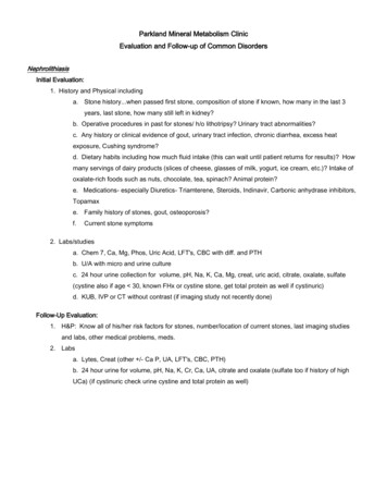
Parkland Mineral Metabolism ClinicEvaluation and Follow-up of Common DisordersNephrolithiasisInitial Evaluation:1. History and Physical includinga. Stone history.when passed first stone, composition of stone if known, how many in the last 3years, last stone, how many still left in kidney?b. Operative procedures in past for stones/ h/o lithotripsy? Urinary tract abnormalities?c. Any history or clinical evidence of gout, urinary tract infection, chronic diarrhea, excess heatexposure, Cushing syndrome?d. Dietary habits including how much fluid intake (this can wait until patient returns for results)? Howmany servings of dairy products (slices of cheese, glasses of milk, yogurt, ice cream, etc.)? Intake ofoxalate-rich foods such as nuts, chocolate, tea, spinach? Animal protein?e. Medications- especially Diuretics- Triamterene, Steroids, Indinavir, Carbonic anhydrase inhibitors,Topamaxe. Family history of stones, gout, osteoporosis?f.Current stone symptoms2. Labs/studiesa. Chem 7, Ca, Mg, Phos, Uric Acid, LFT's, CBC with diff. and PTHb. U/A with micro and urine culturec. 24 hour urine collection for volume, pH, Na, K, Ca, Mg, creat, uric acid, citrate, oxalate, sulfate(cystine also if age 30, known FHx or cystine stone, get total protein as well if cystinuric)d. KUB, IVP or CT without contrast (if imaging study not recently done)Follow-Up Evaluation:1. H&P: Know all of his/her risk factors for stones, number/location of current stones, last imaging studiesand labs, other medical problems, meds.2. Labsa. Lytes, Creat (other /- Ca P, UA, LFT's, CBC, PTH)b. 24 hour urine for volume, pH, Na, K, Cr, Ca, UA, citrate and oxalate (sulfate too if history of highUCa) (if cystinuric check urine cystine and total protein as well)
OsteoporosisInitial Evaluation:1. History and Physical includinga.Bone fracture history if any, including h/o trauma and general assessmentof fall risk. Symptoms of bone pain? Kyphosis/scoliosis on exam? Blue sclerae?b.Other medical conditions including childhood illness, immobilization, menopause, chronic illness.c.Medications present or past, especially use of steroids, hormone replacement, anticonvulsants,previous treatment for osteoporosis.d. Cigarettes, alcohol use? Physical activitye. History of diarrhea (# BM/day)f.Intake of dietary calcium and supplemental Ca/vitamin Dg. Family history stones, osteoporosis, cancerh. Physical exam to rule out secondary risk factors2. Labs/studiesa. Chem 7, Ca, Mg, Phos, LFT's, CBC with diff., TSH, PTH ( /- ESR and SPEP)b. 24 hour urine collection for volume, Na, Ca, Creat, N-telopeptides ( /- cortisol and UPEP)c. Strongly consider AP and Lateral of T/L/S-spine ( /- AP pelvis, sites of pain)d. BMD (nuclear medicine) DEXA spine and hip (phone 25120)Follow-Up Evaluation:1. H&P: Know all the risk factors for osteoporosis, number/location of fractures, last BMD/x-rays, key medicalproblems and meds2. Labsa. Lytes, Creat, Ca ( /- PTH, AP, TP, albumin)b. 24 hour urine Na, Ca, Creat. ( /- N-telopeptide)c. BMD q year (initially and then at increasing intervals when stable)
Primary Hyperparathyroidism1. History and Physical (initial and follow-up)a.history or symptoms of stones, abdominal pain, heartburnb. history of fractures, bone painc.d.exposure to neck radiation (get hyperparathyroidism 40 years later)medication history, steroids, lithium, thiazides, calcium, vitamin D, vitamin A?e. family history hypercalcemia, kidney stones, pituitary tumor, thyroid tumor, ulcer disease,abdominaltumor, pheochromocytoma, severe hypertension, sudden death?f.physical exam—R/O hypertension, Cushingoid/Marfanoid, band keratopathy, neck mass2. Labs/Studiesa. Lytes, Creat, Ca, P, Mg, Alk Phos, Albumin, PTH q 6 months (initial and F/U)b. 24 hour urine for Na, Ca, Mg, Creat, N-telopeptide initial and selected follow-up-R/O pheo if hypertensivec. BMD q year (make sure to include the radius)Paget's DiseaseInitial Evaluation:1. History and Physical focusing on sites involved and related symptoms (bone pain, change in bone shapeespecially legs, skull and sites of pain or known involvement (document where involved), hearing loss?).Note any deformity or warmth in sites of involvement.2. Labsa. Lytes, Creat, Ca, P, UA, PTH, CBC, LFT's (?PSA)b. 24 Hour urine for Na, Ca, Creat, N-telopeptidec. X-rays and/or bone scanFollow-Up Evaluation:1. History (as above)2. Labsa. Creat, Ca, P, Alk Phos q visit (every 3-6 months)b. 24 hour urine for Na, Ca, Creat, /- N-telopeptide q 3-6 months (may not need if AP correlates well)c. /- BMD
GENERAL COMMENTS ABOUT PREVENTING KIDNEY STONESCenter for Mineral Metabolism and Clinical ResearchUT Southwestern Medical Center at DallasPhone: 214-590-5676Kidney stones are formed when the urine is overly concentrated particularly when there is lack of inhibitorsagainst stone formation. We analyze twenty-four hour urine samples to observe what defects are present, so thatappropriate treatment may be started. Below are listed our usual recommendations based on the results of anevaluation. Many of these treatments have been shown to reduce kidney stone recurrence in published studies.Urine Volume (or TV)All stone formers should increase fluid intake so that daily urine output is greater than ½ a gallon (about 2liters) of urine per day. One study found that this reduced stone recurrence rate by about 60%. Usually, about a quartof fluid is lost by the body daily due to sweating, breathing, and going to the bathroom. So, ¾ a gallon (about 3 liters) offluid intake is probably needed to result in enough urine volume. With increased sweating (due to warm environment,exercise or fever) or other external fluid losses (such as vomiting or diarrhea), even more fluid is needed to replacebody losses. An easy way to tell that you are drinking enough fluid is by examining the color of the urine.If the urine is dark, drink more.If the urine is almost as clear as water, you’ re doing well.Once the fluid is absorbed into the body, the kidneys quickly filter it. So, it’ s important to space out the fluidintake to avoid exposing the kidneys to high concentrations of stone-forming salts. Your goal should be to: drink 16ounces with each of your three daily meals, 8 oz. between meals, 8 oz. at bedtime, and 4-8 oz. during the middle ofthe night. The late night fluid is probably the most important in avoiding kidney stones, because the morning urine isusually the most concentrated. If you are not used to having 10 glasses of fluids/day, try to get on the schedule above.First, divide your current number of glasses/day into this schedule. Until you reach your goal intake, add 4 oz. every 510 days at a time slot you haven’ t yet filled.If your urine volume is still not high enough on the schedule above, it is best to increase your fluid intakeslowly by drinking one more ounce (1 sip) with every glass every 5-10 days until the goal urinary volume is reached.This gradual increase in fluid intake will help your body adjust so you won’ t feel bloated. High fluid intake has beenproven to reduce kidney stone recurrence in a controlled study, and of course, it is the cheapest treatment anddoesn’ t cause serious side effects.Urinary Calcium (or Ca)If urine calcium is high and the bone density is normal, we cautiously limit dietary calcium. At the moment,recommended dietary calcium intake is controversial, but it seems unwise to completely exclude dairy intake. Highcalcium intake is known to raise urine calcium. On the other hand, you may have seen news about high calcium intakeprotecting against stones in those who have not previously had stones. In fact, there is evidence that the higher theurinary calcium in stone-formers, the higher the risk for kidney stones. Unfortunately, the long-term effect of increasing
calcium intake alone has not yet been tested formally in stone-formers, so we do not know for sure if the higher dairyintake protects against stone formation or increases the risk.Most of dietary calcium is in dairy products (skim products have slightly more calcium), so we limit them to oneserving per day (8 oz. milk or breakfast yogurt or calcium fortified orange juice; 1 ½ thin slices of cheese; or about 12oz. of ice cream, frozen yogurt or cottage cheese) in patients with high urinary calcium. This restriction is graduallyrelaxed each follow-up visit if possible base on urinary calcium. Salt, demonstrated by urinary sodium (or Na), and acidload (from animal protein), estimated by urinary sulfate or (SO 4), may increase urinary calcium and reduce theeffectiveness of dietary and/or medication therapy. For these reasons, salt should be avoided and animal proteinintake should be limited (goal urine values of Na 150 meq/d; SO4 25). A good website for dietary options on a lowsalt diet is www.saltfreelife.com. I believe it costs 5.00 to become a member.We often test urine calcium after an overnight fast and after a 1gram oral calcium load. If the 2 hour fastingurinary calcium is increased ( 0.11mg/mg creatinine), we are suspicious of too much loss of calcium from the bone orkidney. If the 4-hour postload urinary calcium is increased ( 0.20mg/mg creatinine), we are suspicious of excessiveintestinal calcium absorption. In contrast, if the 4 hour postload urinary calcium is 0.10mg/mg creatinine, we areworried about in adequate calcium absorption. This latter finding may require follow-up by your primary care physicianto make sure that there is no underlying intestinal disease.If the urine calcium remains high, a thiazide diuretic or Indapamide have been shown to decrease the risk offurther kidney stones and to protect against bone loss. Do not take any diuretic containing triamterene, which mayactually cause kidney stones. It is important to avoid potassium loss usually caused by thiazides, so potassium citrateis usually added. This potassium supplement is preferred over potassium chloride because studies show thatpotassium citrate also prevents kidney stones. Urinary calcium should be monitored while on treatment because thedrug often loses effectiveness (usually after 2 years of treatment). If this happens, effectiveness is restored bychanging medication (for example: hydrochlorothiazide to Indapamide). The bone density should be followedperiodically to make sure that mild dietary calcium restriction or the predisposition to high urinary calcium does notresult in loss of bone mass. Whenever urine calcium is found to be very high ( 300mg/day) or very low ( 100mg/day), it is important to be evaluated by a physician to rule out underlying disease such as primaryhyperparathyroidism (high urinary calcium) or vitamin D insufficiency (low urinary calcium).Urinary uric acid (or UA)If urinary uric acid is very high, there is increased risk of calcium and uric acid stones. Animal protein from thediet (beef, chicken, fish, etc) is metabolized into uric acid. When we measure a high sulfate (SO 4) levels in the urine, ittells us that your animal protein intake is high. So, the first treatment option is reducing animal protein intake to about 8oz. per day. Allopurinol, which lowers uric acid production and decreases the risk of kidney stones, is added ifnecessary. If the urine is acidic (pH 5.5), there is additional risk of uric acid and calcium stones, so potassium citrateis added to raise pH to the 6 to 6.5 range. Potassium citrate has been shown to reduce recurrent stones in this group.
Urinary oxalate (or Ox)If urinary oxalate is high, dietary oxalate restriction is very important. Food with high oxalate content includenuts (and peanut butter), green leafy vegetables (such as spinach), brewed tea, chocolate, and rhubarb. Vitamin Cshould not be taken in excess ( 500mg/day) since it can be metabolized to oxalate. It is important to have evaluationby your doctor if your urine oxalate is high because it may represent other important underlying conditions (diarrhea,primary metabolic disorder and vitamin B6 deficiency). Calcium binds oxalate in the intestine and thereby may lowerurine oxalate by 10 to 20%. Calcium administration will only help if given during the meal and if urinary calciumremains well-controlled. Sometimes, vitamin B6 decreases urine oxalate very effectively.Our basin approach is to limit 2 to 3 foods common in your diet that are the most likely cause of elevatedurinary oxalate (we have a more detailed oxalate content handout). If repeat urinary oxalate is high, further dietaryrestriction is added and medical treatment with vitamin B6 (50 to 100mg twice daily) or calcium is considered. Thistreatment is done not only for those with high urinary oxalate, but also for those with high normal urinary oxalatecombined with high or high normal urinary calcium.Urinary citrate (or Cit)Citrate is a known inhibitor of calcium stones, so low urinary citrate is a risk factor for kidney stones. Theaverage urinary citrate is about 640mg/day. The lower limit of normal urinary citrate is 320mg/day. First line treatmentis to avoid environmental causes of low citrate such as diarrhea, urinary tract infection, excessive exercise, and highintake of animal protein or salt.Although dietary measures may raise citrate (lemonade, citrus drinks, high intake of fruits/vegetables, lowintake of animal protein), additional medication will be necessary for most people. We use potassium citrate, which hasbeen shown to reduce recurrent kidney stones. Urocit-K is the only slow-release tablet form of potassium citrateavailable. A wax remnant (the carrier) in the stool should be expected and is not a sign of poor absorption on themedication. There are several liquid preparations but due to their immediate release, they must be taken morefrequently and they have more side effects. In subjects with chronic diarrhea, the liquid preparations are preferred dueto rapid intestinal transit. Treatment with potassium citrate has been shown to reduce kidney stone recurrence. Otheravailable medications that will raise medication include sodium citrate, potassium bicarbonate, and sodiumbicarbonate. We prefer to avoid sodium-based therapies due to the hypothetical concerns that they will raise urinarycalcium and reduce the citrate raising effects.Urinary pHIf the urine is acidic (pH 5.5), there is increased risk of calcium or uric acid stones. Potassium citratecorrects this problem by raising the pH towards normal. Low urinary pH is common in those who suffer from gout. Lowurinary pH is the easiest stone risk factor to treat. If the measurement of urine pH, on the other hand, is very high ( 7.0), there is increased risk of calcium phosphate stones and urinary tract infection should be ruled out.So, our goal pH is about 6 to 6.5 to avoid either extreme. This can be accomplished by diet or medicaltreatment. High animal protein intake (acid load) and strenuous exercise (only during exercise) may lower urine pHwhile fruit and vegetable intake may raise it. Potassium citrate, which provides alkali load, is used to raise urinary pH.At the present, we do not give medication to lower pH 7, but potassium citrate and other alkali should be avoided inthis setting.
Urinary Magnesium (or Mg)Magnesium, when given with food, is believed to inhibit stone formation by binding oxalate. Initial controlledstudies with magnesium have shown benefit; however, later, controlled studies showed no benefit over placebo (sugarpill). The latter studies were not done on magnesium deficient patients, so it is still possible that magnesiumsupplementation would be useful in that setting to avoid kidney stones. In about 6% of our patients, the only detectablephysiologic defect is urinary magnesium less than 60mg/day. In these patients, we would consider treatment withmagnesium supplements such as magnesium oxide or gluconate (often 2 pills three or four times a day are needed) ormilk of magnesium (2.5ml two to three times per day). The problem with magnesium supplements is that they oftencause diarrhea, which would increase stone risk by at least four ways-lower urinary volume, pH and citrate and higherurinary oxalate.Infection stonesSometimes, kidney stones result directly from urinary tract infection. Certain bacterial organisms create veryalkaline urine (pH often 8.0) and produce ammonia, a combination that predisposes to infection stones such asstruvite (magnesium ammonium phosphate) and carbonate apatite. Infection may also complicate existing calciumstones. When kidney stones are caused by recurrent urinary tract infections, surgery and antibiotics are almost alwaysnecessary. Lithostat, a medication that lowers the urine pH by inhibiting production of ammonia, has been shown to beeffective additional medical therapy to help reduce recurrent stone formation in these patients. Since it has possibletoxic side effects, it should be used carefully.Other testsParathyroid hormone (PTH) is an important hormone that maintains our blood calcium level. Sometimes, anabnormal gland or glands make too much of this hormone and too much calcium is taken out of the bone, absorbedfrom the intestine and excreted into the urine. If your PTH is 65pg/ml, you may have an overactive parathyroid glandthat is causing your kidney stones.You may have had a bone mineral density test since the bone mass if often low in stone-formers. Bonemineral densitometry is the most sensitive tool we have to estimate bone mass and predict future risk of fracture. Thetwo types of bone (trabecular and cortical) are distributed differently at specific skeletal areas, so we measure thedensity in three different sites. In the lumbar spine, mostly trabecular or spongy bone is found. At this site, bone masspreferentially declines after the loss of male or female sex hormones or with glucocorticoid treatment. Arthritis in thespine and calcification of the aorta (very common in individuals older than 60 years) falsely elevates this measurement.The long bones (legs and arms) have primarily compact or cortical bone, which is primarily lost with conditions ofexcess parathyroid hormone. We use the distal radius of the arm as an indicator of cortical bone. The femoral neck ofthe hip has a mixture of both types of bone. This is an important site to measure because it is the best predictor for themost serious complication of low bone mass, hip fracture.
Osteoporosis Evaluation and TreatmentI.Scope of Problem – 40% of women and 13 % of men suffer osteoporotic fracture/yr; 1.5 million osteoporoticfractures/yr (700,000 vertebral, 300,000 hip) at a cost of 10-20 billion; fracture may result in death, disabilityand loss of independence (180,000 nursing home admissions/year)II.Definition – loss of bone mass and disturbance of skeletal architecture that predisposes to fracture.Remaining bone is normally mineralized.III.BackgroundA. Bone Composition and Types:1. Composition – mineral (hydroxyapatite crystals – Ca10PO4(OH)2) and protein ( 90% collagen), 70-80%of the body’ s bicarbonate2. Type of bonea. Cortical – 80% of skeleton, 20% of surface area, mechanical function, 4% turnover/yearb. Trabecular – 80% of surface area, metabolic function (99% calcium, 70-80% HCO3), 20%turnover/yearB. Bone Remodeling Unit – organelle composed of osteoclasts and osteoblasts responsible for remodelingthe bone. Multiple units are active at any one time. An individual unit is active for a few months. Osteoclastsstart the process by removing the old or damaged bone. Then, osteoblasts lay down and organize new bone.C. Bone acquisition pattern- rapid bone acquisition occurs in puberty and peak bone mass is reached. Then,bone mass is stable for 2 to 3 decades. In women, rapid loss occurs with menopause lasting 3 to5 years. Inboth sexes, gradual loss (0.5-1%/year) occurs after age 40 or 50.IV.Make the diagnosis with Bone Mineral Density (BMD)A. Indications- postmenopausal (especially age 65), osteopenia by x-ray, key risk factors (personal history oflow trauma fracture, family history of fracture, hyperparathyroidism, steroids), monitoring on treatment.B. Types of densitometers1. DEXA (dual energy x-ray absorptiometry)- gold standard2. QCT (quantitative computed tomography)- less precise, better for spine in elderly3. Ultrasound- cheaper, no radiationC. Interpretation1. T-score- compared to peak bone mass; by W.H.O. criteria, normal within 1 standard deviation,osteopenia- loss of 1 to 2.5 S.D., osteoporosis- loss of 2.5 S.D. Every loss of 1 S.D. doubles the risk offracture. This is relative risk, so the risk may still be low in young patients.2. Z-Score-matched for age and sex; loss 2 S.D. suggests secondary bone loss; yet, those with secondaryrisk factors may certainly have Z-score loss of smaller magnitude.3. Artifact- carefully match positioning, arthritic changes, scoliosis, fracture
4. Site- spine- trabecular (loss with hypogonadism, steroids); radius- cortical (loss with Ca deficiency orhyperparathyroidism); femoral neck- mixture of cortical and trabecular bone. This is now the preferred site tomeasure since it correlates best with risk of hip fracture (-1 S.D. RR 2.6)5. Frequency- Generally, follow patients every 2 or more years (acceptable to insurance companies). If newpatient with low BMD or monitoring treatment, recheck in 1 year. In a very high risk patient (especially withnew glucocorticoid treatment). Consider rechecking in 6 months. Increase intervals between BMD whenstable.V.Establish risk factors and rule out secondary causes of osteoporosis if density is low (pneumonic: The Medic)by history, physical and labs (V). Rule out osteomalacia.A. Tumor – multiple myeloma, mastocytosis, mestastasesB. Hereditary – family hx of fracture (hip), hip geometry, race, size, osteogenesis imperfecta, homocystinuria,Marfan’ s disease, Ehlers Danlos, Gaucher’ sC. Endocrine – menopause, male hypogonadism (mumps), timing of puberty, hyperthyroidism,hyperparathyroidism, Cushing’ s syndrome.D. Medications – steroids, heparin, anticonvulsants (Phenobarbital, dilantin), aromatase inhibitors, PPIsE. Environmental – diet (calcium, salt, protein), vitamin D (sunlight), cigarettes, alcoholF. Diarrhea – (malabsorption)G. Immobilization – bedrest, sedentary lifestyleH. Chronic disease – heart, kidney, liver, rheumatic disease, infection, severe illness during childhood,prevalent fracturesVI.Work-upA. Blood – SMA, CBC, TSH, PTH ( ESR, 25-D, SPEP)B. Urine – Na, Ca, Cr ( UPEP, cortisol, bone turnover markers)C. X-rays – spine (interpret density, asymptomatic fractures) and painful areasD. Other – consider biopsy of the bone or skin, stool fat, etc., in selected casesVII. TreatmentA. Calcium – 1200-1500mg/d (including diet) if 65 years or untreated postmenopausal; otherwise1000mg/d; protects against bone loss and fracture (particularly when given with vitamin D). Dietary calcium ismainly from dairy (300 mg per 8 oz. milk or yogurt, 1 ½ oz. cheese, 12-16 oz. cottage cheese, frozen yogurtor ice cream). Risks: constipation, bloating. Consider adding magnesium to combat constipation side effects.B. Vitamin D – 600 to 800IU/day; Pharmacologic doses if UCa 50-100mg/d. Risks – hypercalcemia.C. Thiazide/Lozol – if UCa 300 mg/d; protects against bone lossD. Estrogen - density over 3 years by 5-10% at spine, 2-5% at femoral neck and 0-2% at arm. fracture30-60%. Other benefits: improved cholesterol ( LDL, HDL). Other potential benefits: improvement invasomotor function, in atrophy of the mucosa of vagina and outer 1/3 of urethra, prevention of tooth loss andwrinkling. Risks: Women’ s health initiative: (oral Premarin/Provera) found higher risk of heart attack,stroke, dementia, thromboembolism and breast cancer with decreased colon cancer and fracture: (Premarin)higher risk stroke and lower risk fracture. Other risks: vaginal bleeding, mastalgia, breast cancer, uterine
cancer (negated by progestins), hypertriglyceridemia, gallstones. Indication: hot flashes and prevention ofosteoporosis in postmenopausal women (indication will likely be more limited soon; dose 0.3 to 0.625 mg/dconjugated; 50 to 100 mcg transdermal).E. Raloxifene - density at spine and hip by 1-2% over 2 years. fracture risk at spine by 30%. Otherbenefits - LDL, may reduce breast cancer risk; Risks: thromboembolism. Indication: treatment/prevention at60mg/day.F. Alendronate - density over 3 years by 6-10% at spine, 3-6% at femoral neck and 0-1% at arm. fractureby 50% spine, hip and 30-50% peripherally. Other benefits – apoptosis of cancer cells?; Risks: esophagitis,hypocalcemia, accumulation in renal failure ( 35cc/min), slight increase in atypical femoral shaft fractures withlong-term use. Indication – treatment 10 mg/d or 70 mg/wk;prevention 5 mg/d or 35 mg/wk.G Risedronate – bisphosphonate class like alendronate. density over 3 years by 5.4% at spine, 2-3% atfemoral neck and 0-1% at arm. fracture by 40-50% spine and hip and 30-40% peripherally. Risks: same asalendronate. Indication – treatment/prevention 5mg/d or 35mg/wk or 150 mg/month.H. Other Bisphosphonates – ibandronate has been approved for osteoporosis in daily or monthly oral dose(similar risks and benefit but hip fracture efficacy not yet demonstrated). Intermittent intravenous therapy withzoledronic acid (Reclast) improves bone mineral density and reduces fracture risk. 5 mg IV once a year.I. Calcitonin - density at the spine by 0-2%. No apparent protection at the hip or arm. risk of spinefracture by 33%. Other benefits – central analgesia, no severe risks; Risks: (nasal spray) – nasal ulcer,coryza, nausea. Indications – treatment 200IU puff daily (alternate nostrils).J. Teriparatide (PTH 1-34) - density over 18 months by 9.7% at spine, 2.8% at femoral neck and 2.1 atradial shaft. vertebral fracture by 65% and nonvertebral fracture by 53%. Risks: osteogenic sarcoma?,orthostatic hypotension, leg cramps, hypercalcemia. Indication – treatment of “ severe” osteoporosis.20mcg/d sq (approved for 24 months in specific patients).K. Denosumab. Monoclonal antibody against RANKL. Given as 60 mg SQ injection every 6 months. Powerfulanti-resorptive agent with proven fracture efficacy. Reversible mechanism of action.L. Exercise/Hip protectors – exercise mainly prevents bone loss. Walkin 30 minutes 3x/week is effective.Active exercise may raise bone mass up to 3%. More importantly, by improving strength and balance,exercise may prevent fall and fracture. Hip protectors may reduce hip fracture 50% (studies are mixed).Evaluate balance and consider evaluation by physiatrist and visit by occupational therapist to reduce fracturerisk.VIII. Six risk factors for fractureA. Bone DensityDouble the risk for every loss of 1 S.D. from peakB. AgeDouble the risk for every 5 to 10 years over 50C. FractureSynergistic with bone densityD. Tendency to FallPoor balance, visual perception, weakness, flexibility; drugsE. Bone TurnoverSynergistic with bone densityF. Family hx of hip fractureDouble the risk of hip fracture
IX. Suggested ReadingsA. Rubin C, Grand Rounds, UT Southwestern, June 18, 1998.B. Hui SL, et al. Age and bone mass as predictors of fracture in a prospective study. J Clin Invest 1988; 81:1804C. Dawson-Hughes, B. Effect of calcium and vitamin D supplementation on bone density in men and women65 years of age or older. N Engl J Med 1997; 337: 670D. Lufkin EG, et al. Treatment of postmenopausal osteoporosis with transdermal estrogen. Ann Intern Med1992; 117: 1.E. Ettinger B, et al. Reduction of vertebral fracture risk in postmenopausal women with osteoporosis treatedwith raloxifene: results from a 3 year randomized clinical trial. JAMA 1999; 282: 637.F. Chesnutt CH, et al. A randomized trial of nasal spray salmon Calcitonin in postmenopausal women withestablished osteoporosis: The prevent recurrence of osteoporotic fractures study. Am J Med 2000; 109: 26776.G. Black, DM et al. Randomized trial of effect of alendronate on risk of fracture of women with existingvertebral fractures. Lancet 1996; 348: 1535.H. Cummings SR, et al. Effect of alendronate on risk of fracture in women with low bone density but withoutvertebral fracture. JAMA 1998; 280: 2077.I. Harris ST, et al. Effects of risedronate treatment on vertebral and non vertebral fractures in women withpostmenopausal osteoporosis: a randomized controlled trial. JAMA 1999; 282: 1344.J. Kannis P, et al. Prevention of hip fracture in elderly people with use of a hip protector. N Engl J Med 2000;343: 1506.K. Risks and benefits of estrogen plus progestin in postmenopausal women: Principle results from thewomen’ s health initiative randomized control trial. JAMA 2002; 288: 321L. Neer RM, et al. Effect of parathyroid hormone (1-34) on fractures and bone mineral density inpostmenopausal women with osteoporosis. N Engl J Med 2001; 344; 1434.
Key Risks benefits of Anti-Osteoporotic Medications and Hip ProtectorsMineral ClinicIf you are started on any new medication, you should contact your physician for new persistent symptoms.1) Bisphosphonates: Alendronate (Fosamax)/Risedronate (Actonel) – both protect against fracture of the hip, spineand peripheral bones. They are as effective as estrogen for bone gain. Most bone gain occurs within 3 years (usually5-10% at the spine and 2-5% at the femoral neck). Thereafter, bone mass is stable (may gain 4% over the next 7years). They reduce the risk of fracture at the spine by 50%. They likely reduce fracture at the hip and arms by 3050%. Both drugs are approved for the prevention and treatment of postmenopausal osteoporosis. They are alsoapproved for treatment of steroid induced osteoporosis (risedronate for prevention also). Alendronate is approved forosteoporosis in men. Key side effects include heartburn, pain in the chest or abdomen, trouble swallowing, and bonepain. Both of them may be given daily or once/week. Your must take medication as noted on our bisphosphonatehandout. A new drug, Ibandronate (Boniva), is given by mouth only once/month. It has similar effect on bone gain andhas bee
Parkland Mineral Metabolism Clinic Evaluation and Follow-up of Common Disorders Nephrolithiasis Initial Evaluation: 1. History and Physical including a. Stone history.when passed first stone, composition of stone if known, how many in the last 3 years, last stone, how many still left in kidney? b.


