Transcription
Graduate School for Cellular and Biomedical SciencesUniversity of BernFunctional impact of neuronal nitric oxidesynthase (nNOS) on angiogenesis and themolecular characterization of its isoformexpression pattern in skeletal musclePhD Thesis submitted byFelicitas Anna Maria Lauerfrom GermanyThesis advisorProf. Dr. Hans HoppelerInstitute of AnatomyMedical Faculty of the University of Bern
Copyright NoticeThis document is licensed under the Creative Commons Attribution-Non-CommercialNo derivative works 2.5 -nc-nd/2.5/ch/You are free:to copy, distribute, display, and perform the workUnder the following conditions:Attribution. You must give the original author credit.Non-Commercial. You may not use this work for commercial purposes.No derivative works. You may not alter, transform, or build upon this work.For any reuse or distribution, you must take clear to others the license terms of thiswork.Any of these conditions can be waived if you get permission from the copyrightholder.Nothing in this license impairs or restricts the author’s moral rights according toSwiss law.The detailed license agreement can be found 5/ch/legalcode.de
Accepted by the Medical Faculty, the Faculty of Science and theVetsuisse Faculty of the University of Bern at the request of theGraduate School for Cellular and Biomedical SciencesBern,Dean of the Medical FacultyBern,Dean of the Faculty of ScienceBern,Dean of the Vetsuisse Faculty Bern
Table of ContentsAbstract . 5Abbreviations. 81 Introduction.101.1 Skeletal muscle .101.2 Nitric oxide synthase .131.2.1 Genes . 131.2.2 Nitric oxide . 141.2.3 Neuronal nitric oxide synthase . 161.3 Angiogenesis .201.4 Exercise.222 Methods and Results.252.1 Project I .252.2 Project II .342.2.1 Methods . 352.2.2 Results . 372.3 Project III .452.3.1 Methods . 462.3.2 Results . 503 Discussion and Outlook.55References .64Acknowledgments .75Curriculum Vitae .76Declaration of Originality .79
AbstractAbstractIn rodents and humans skeletal muscle neuronal nitric oxide synthase (nNOS) ispresent at the sarcolemma of skeletal muscle fibres. nNOS produces highconcentrations of the signalling molecule nitric oxide (NO), which could play animportant role in the molecular integration of muscle metabolism and themicrovascular supply in skeletal muscle.The association of nNOS expression with the capillary supply in human skeletalmuscle fibres was assessed in response to endurance exercise (Project I).Vastus lateralis (VL) muscle biopsies from ten sedentary male subjects before andafter moderate training were investigated. nNOS is mainly expressed as an alphaisoform with the additional mu exon and to a lesser extent without the mu exon asanalysed by RT-PCR. Only five of the ten subjects exhibited significant (P 0.05)elevations in the C/F ratio ( 25%) and capillary density ( 21%), indicative ofangiogenesis. Therefore, the subjects were divided into angiogenesis responder (AR)and non-angiogenesis responder (NR) groups. The mRNA levels of nNOS tended tobe elevated after training ( 34%; P 0.05) in the VL biopsies of all subjects analysedby real-time PCR. However, when the AR group was analysed separately, the nNOSmRNA levels were significantly up-regulated ( 128%; P 0.05) in response toexercise. Moreover, these mRNA levels correlated positively (r 0.08; P 0.01) withexercise-induced differences in the C/F ratio. The quantification of the nNOS-specificimmunoblots showed that the nNOS protein expression was significantly increased( 82%; P 0.05), but only in the VL biopsies of the subjects of the AR group.These data might represent an additional evidence for the functional impact of upregulated nNOS on exercise-induced angiogenesis in skeletal muscles.
AbstractVarious isoforms of nNOS have been characterized at protein level, but the primarystructure of these variants has not yet been elucidated. Therefore, the expressionand structure of nNOS isoforms in skeletal muscles of mice were characterized inmore detail (Project II).The nNOS protein expression was analysed in skeletal muscles of C57Bl/6 mice byimmunoblotting. Two bands at 165 and 160 kDa represented the nNOS alphaisoform, with and without the additional mu exon. The nNOS beta isoform with andwithout exon mu was detected at 145 and 140 kDa. Immunofluorescencehistochemistry localized the nNOS alpha and beta isoforms at the sarcolemma ofskeletal muscle fibres. Performing RT-PCR in skeletal muscle of C57Bl/6 mice, threecDNA variants (1500 bp, 650 bp and 300 bp) were detected using the primer pairspecific for exon 1c and exon 5. The remaining nNOS sequence was covered usingseven different primer pairs. One cDNA variant was amplified between exons 2-15and exons 17-28. Using the primer pair specific for exon 14 and exon 18, a doubleband was amplified which represented isoforms with and without the alternativespliced mu exon (102 bp). Nucleotide sequencing revealed that in C57Bl/6 mice thenNOS alpha isoform (1500 bp) had the following exon sequence: 1c-pi-rho-sigmatau-4 to 29. The shorter nNOS beta isoform (650 bp) consisted of: 1c-pi-rho-4 to 29.Each of the isoforms existed with and without exon mu. Unexpectedly, two cDNAvariants (450 bp and 300 bp) were detected using the primer pair specific for exon 1cand exon 5 in skeletal muscles of nNOS-knockout mice. The expression pattern ofthe remaining nNOS sequence was similar to the pattern in C57Bl/6 mice with theexception that no cDNA band was detected using the primer pair specific for exon 2(containing the initiation codon) and exon 6. In nNOS-knockout mice, none of thecDNA variants were translated.Taken together, in mice skeletal muscle, four nNOS cDNA variants were translated:the nNOS alpha isoform, with and without the mu exon, and the nNOS beta isoform,with and without the mu exon.
AbstractThe functional impact of the nNOS alpha isoform on angiogenesis should be furthercharacterized in mice skeletal muscle by gene electrotransfer (Project III).Therefore, the full-coding nucleotide sequence (4676 bp) of nNOS was isolated,cloned into the pIRES2-ZsGreen1 vector and over-expressed in hind limb musclesfrom mice by gene electrotransfer. The pIRES2-ZsGreen1-nNOS plasmid and thecontrol pIRES2-ZsGreen1 plasmid were injected into the tibialis anterior (TA)muscles. After the gene electrotransfer, the mice were separated into one sedentaryand one exercise group. The successful gene electrotransfer of both plasmids wasconfirmed by detection of the ZsGreen1 fluorescence in muscle fibres. However, noclear nNOS over-expression was detected in muscle fibres transfected with thepIRES2-ZsGreen1-nNOS plasmid by immunofluorescence. The real-time PCRanalysis showed that the nNOS mRNA expression was not significantly up-regulatedin the TA muscles with the pIRES2-ZsGreen1-nNOS plasmid compared to the TAmuscles with the control plasmid of the sedentary as well as exercise group.Therefore, further characterization of the function of the nNOS alpha isoform with thisapproach was not possible so far.
AbbreviationsAbbreviations3’ UTR3’ Untranslated region5’ UTR5’ Untranslated regionAMPKAdenosine monophosphate-activated protein kinaseAng-1/-2Angiopoetin-1/-2ARAngiogenesis responderBH4TetrahydrobiopterinBSABovine serum -dependent protein kinasecDNAComplementary DNAcGMPCyclic guanosine monophosphateDMDDuchenne muscular dystrophyDNADeoxyribonucleic acidEDLExtensor digitorum longusEDRFEndothelium-derived relaxing factoreNOSEndothelial nitric oxide synthaseERR-alphaEstrogen-related receptor alphaESTExpressed sequence tagFADFlavin adenine dinucleotideFGFFibroblast growth factorFMNFlavin mononucleotideGLUT4Glucose transporter 4HIF-1alpha Hypoxia inducible factor-1alphaHSPHeat shock proteiniNOSInducible nitric oxide synthaseLGMDLimb-girdle muscular dystrophiesL-NAMEL-N-arginine methyl ated protein kinaseMCSFAMean cross-sectional fibre area88
AbbreviationsMHCMyosin heavy chainMLCMyosin light chainMMPMatrix metalloproteinasemRNAMessenger RNANADP/HNicotinamide adenine dinucleotide phosphateNMDAN-methyl-D-aspartatenNOSNeuronal nitric oxide synthaseNONitric oxideNOSNitric oxide synthaseNRNon-angiogenesis responderNRF-1/-2Nuclear respiratory factor-1/-2PBSPhosphate buffered salinePDGF-BPlatelet-derived growth factor BPGC-1alpha Peroxisome proliferator-activated gamma coactivator 1 alphaPINProtein inhibitor of nNOSPPARPeroxisome proliferator-activated receptorRNARibonucleic acidROSReactive oxygen speciesRT-PCRReverse transcription polymerase chain reactionSEMStandard error of the meanSMTCS-methyl-L-thiocitrullineTATibialis anteriorTSP-1Trombospondin-1VEGFVascular endothelial growth factorVEGFR-2Vascular endothelial growth factor receptor 2VLVastus lateralisVO2maxMaximal oxygen uptake99
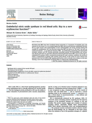
Introduction1Introduction1.1 Skeletal muscleSkeletal muscle is one of the major muscle types besides cardiac and smoothmuscle and is also known as striated muscle under control of the central nervoussystem. A fibrous fascia, also called the epimysium, surrounds the whole muscle andfurther divides it into several fasciculi. Each fasciculus is surrounded by theperimysium and contains bundles of muscle fibres which build a specific mosaic inevery mammalian skeletal muscle. Each muscle fibre is surrounded by moreconnective tissue called endomysium. The endomysium consists of collagenfilaments and proteoglycans in which capillaries and nerve fibres are embedded.Each single muscle fibre is surrounded by a membrane called the sarcolemma andconsists of myofibrils which are surrounded by the sarcoplasm (figure 1) (Billeter etal. 1994; Baechle et al. 2000; Schünke et al. 2005).Figure 1: Detailed structure of a skeletal muscle (Baechle et al. 2000).1010
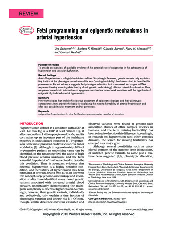
IntroductionSkeletal muscles have an abundant supply of blood vessels and nerves which isrelated to the primary function of skeletal muscles, mechanical contraction.Contraction of various muscle fibres is controlled by a single motor-neuron.Depolarization of the membrane during contraction leads to an action potential whichinnervates a group of fibres at the same time (Monti et al. 2001).Skeletal muscle fibres can be attributed to different metabolic and contractilephenotypes, which differ in their oxidation and glycosylation potential, as well as theirvelocities of contraction. Myosin, the motor of contraction, exists in different isoformsin skeletal muscle fibres. Although isoforms of both myosin heavy chain (MHC) andmyosin light chain (MLC) portions of the molecules exist, research is mainly focusedon the expression of MHC isoforms (Canepari et al. 2010). Different skeletal musclefibres can be identified by their MHC composition (Schiaffino et al. 1996). In humansthree different fibre types are determined, while in rodents four fibre types are known.In both species, type 1 slow-twitch, oxidative fibres are slow in force generation andhave an oxidative profile whereas type 2A fibres are fast in force generation andhave a similar oxidative profile as type 1 fibres. In humans type 2X fibres are fast inforce generation and have a glycolytic profile. In contrast, rodent type 2X fibres arefast, oxidative, glycolytic and type 2B fibres are glycolytic and have an even fastertwitch profile (Schiaffino 2010).The functional heterogeneity of skeletal muscles can be explained by the existenceof mixed fibre types having different contractile and energetic properties (Canepari etal. 2010). The differentiation of skeletal muscles into fast and slow also correspondsto a morphological difference. Fast muscles appear white and slow muscles red. Theredness is due to high amounts of myoglobin and a high capillary density (Gödeckeet al. 1999; Scott et al. 2001). The fibre type composition of different skeletal musclesdepends on their function. Muscles used for long, endurance work consist mainly ofslow, type 1 fibres (e.g. soleus) in contrast to muscles used for short and intensework which consist of fast, type 2 fibres (e.g. vastus lateralis) (figure 2). Othermuscles contain a rather balanced fibre type distribution (e.g. gastrocnemius). Forthis reason long distance runners have rather slow twitch fibres whereas sprintershave more fast twitch fibres (Saltin et al. 1977; Hoppeler 1983).1111
IntroductionFigure 2: Percentage of slow twitch fibres in different human skeletal muscles (Saltin et al.1977).Changes in the environment like temperature, nutritional conditions and exercise canlead to specific adaptations in skeletal muscle tissue. These structural and functionalmodifications are reversible and maintained as long as the stimulus exists (Hoppeleret al. 2002). Physical activity can result in nuclear and mitochondrial improvedenduranceperformance or myofibre hypertrophy and force development (Hood et al. 2006). Themodifications can be regulated by several signalling pathways which are present inthe skeletal muscle. These signalling pathways include the Ras / mitogen-activatedprotein kinase (MAPK), the calcium / calmodulin-dependent protein kinase (CaMK),the peroxisome proliferator-activated gamma coactivator 1 alpha (PGC-1alpha) andthe vascular endothelial growth factor (VEGF) (Zierath et al. 2004; Arany et al. 2008).Further details of the signalling pathways are provided in sections 1.3 and 1.4.1212
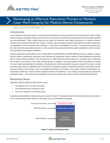
Introduction1.2 Nitric oxide synthase1.2.1 GenesThree different isoforms of the nitric oxide synthase (NOS) are known, named aftercells or tissues in which they were originally found and purified (figure 3). Theneuronal NOS (NOS1, nNOS) was purified by cDNA cloning of brain tissue, theinducible NOS (NOS2, iNOS) from activated macrophages and the endothelial NOS(NOS3, eNOS) from endothelial cells (Schmidt et al. 1992; Stamler et al. 2001).The nNOS is prominent in neurons, epithelial cells of the lung, in macula densa cellsof the kidney and in skeletal muscle fibres. As signalling molecule, nNOS-derived NOis an endogenous mediator in neural transmission. In skeletal muscle, nNOS hasdifferent functions as a paracrine and autocrine signalling molecule (Schmidt et al.1992; Kobzik et al. 1994; Ortiz et al. 2003). The eNOS is highly expressed in theendothelial tissue of vessels where it increases cGMP levels leading to relaxation ofsmooth muscle cells and thus influencing blood pressure (Li et al. 2002; Kojda et al.2005). The iNOS is rapidly upregulated in all cell types following immunologicalstimuli and serves a nonspecific immune function (Mungrue et al. 2003; Li et al.2011).The activity of nNOS, iNOS and eNOS is regulated at transcriptional, translationaland posttranslational levels. Typically, iNOS is upregulated by cytokines whereasnNOS expression is increased by muscle activity and aging process. eNOS isspecifically upregulated by chronic exercise and in blood vessels by shear stress. Allisoforms are transcriptionally regulated by hypoxia (Stamler et al. 2001). The activityof nNOS and eNOS is regulated by calcium-dependent calmodulin binding whereasthe activity of iNOS is described as calcium-independent but presumes a tightinteraction with calmodulin (Brenman et al. 1995; Mungrue et al. 2003).1313
IntroductionMoreover, all NOS isoforms catalyse the reaction of arginine, NADPH and oxygen tonitric oxide (NO), citrulline and NADP. NO and many NO-related molecules (e.g. Snitrosothiols, peroxynitrite) are generated from the catalytic conversion of L-arginineto L-citrulline. It seems that the availability of cofactors (BH4, FAD, FMN, haem) aswell as subcellular localisation can influence the enzymatic product (Alderton et al.2001; Stamler et al. 2001; Mungrue et al. 2003).Figure 3: Domain structure of nNOS, eNOS and iNOS with the corresponding binding sitesfor the cofactors and -substrates (Alderton et al. 2001).1.2.2 Nitric oxideNitric oxide is a diffusible gaseous radical with impact on many physiological andpathological processes. NO has a short half-life which is influenced by the interactionwith various proteins (e.g. haemoglobin, myoglobin). As NO is electrically neutral, itdiffuses across the plasma membrane and thus has intracellular as well asextracellular effects (Stamler et al. 2001). Until the work of Ignarro et al. (1988) andFurchgott et al. (1989), NO was mainly linked to cell toxicity. But these groups couldshow that endogenously produced NO is a biological mediator, produced bymammalian cells, and identical to the endothelium-derived relaxing factor (EDRF).1414
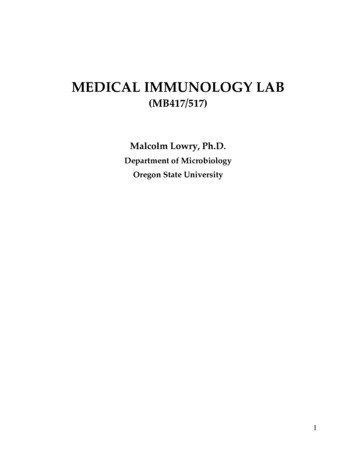
IntroductionNO as well as EDRF are produced and released by the endothelium and causesmooth muscle cell relaxation by activating the guanylate cyclase (Ignarro et al.1988; Furchgott et al. 1989; Alderton et al. 2001).Physiologically NO plays a role in many processes like regulation of vascular tone,insulin secretion, angiogenesis, mitochondrial oxygen metabolism and developmentof the nervous system. Furthermore, pathologically it plays a role in immuneresponse, septic shock and host defence against pathogens and tumours(Grozdanovic 2001). NO is involved in mitochondrial biogenesis at several levels. Itdirectly regulates the supply of oxygen to mitochondria via binding and release fromhaemoglobin. Moreover, NO indirectly regulates the supply of respiratory substratesto mitochondria via blood flow regulation. Mitochondrial activity has a critical role invarious processes in skeletal muscles like fibre type switching and muscleregeneration. NO can induce the change in energy metabolism in these processes(Clementi et al. 2005).Figure 4: Schematic overview of the main target sites of NO in skeletal muscle. ROS:reactive oxygen species; XO: xanthine oxidase; OR: oxidoreductase; GC: guanylate cyclase(Grozdanovic 2001).1515
Introduction1.2.3 Neuronal nitric oxide synthaseStructureThe nNOS gene is more than 4500 bp long and consists of 29 exons. The initiationcodon is located in exon 2 and the termination codon in exon 29. The nNOS proteinis expressed as a dimer with an oxidase domain at the N-terminal and a reductasedomain at the C-terminal end. Several binding domains for cofactors and -substrates(e.g. L-arginine, haem, BH4, FAD, FMN and NADPH) are distributed along thesequence (Nathan et al. 1994; Stamler et al. 2001; Newton et al. 2003).Most nNOS isoforms contain a characteristic PDZ/GLGF motif of 100 amino acidsat the N-terminal end which is linked to the dystrophin glycoprotein complex locatedat the sarcolemma. This connection is due to the direct binding of nNOS toα-syntrophin via PDZ-PDZ interactions (Kobzik et al. 1994; Brenman et al. 1995;Brenman et al. 1996). A further binding of the β-hairpin finger loop, located betweenresidues 101-130 of nNOS, to the peptide binding groove in the PDZ domain of αsyntrophin is involved in the localisation of nNOS to the sarcolemma (Abdelmoity etal. 2000). If dystrophin is absent in the muscle, like in human Duchenne musculardystrophy (DMD) patients and in its’ animal model mdx mice, nNOS is dislocatedfrom the sarcolemma (Brenman et al. 1995).In skeletal muscle, different nNOS isoforms are known (figure 5). The nNOS alphaisoform consists of all 29 exons and produces a 160 kDa protein. The alpha isoformuniquely manifests a 237 amino acid long stretch, which is encoded by exon 2. Thisexon contains the characteristic PDZ domain that assures sarcolemmal anchorage.The nNOS beta isoform expresses a truncated N-terminus with a deletion of 236amino acids, which is initiated at the boundary between exon 1 and exon 3. Thisdeletion is owing to a shift in the frame-reading that is caused by an alternativesplicing event. The beta isoform lacks the PDZ domain which leads to adelocalization from the sarcolemma but retains full enzymatic activity. It produces a136 kDa protein. The nNOS gamma isoform is similar to the nNOS beta isoform butwith a deletion of the first 335 amino acids of the nNOS alpha isoform. As exon 2 withthe initiation codon and the PDZ domain is missing, the initiation codon is dislocatedin exon 5. The nNOS gamma isoform produces a 125 kDa protein. The nNOS mu1616
Introductionisoform, especially known in skeletal muscle, has an alternatively spliced 102 bp (34amino acid residues, 5 kDa) long stretch between exon 16 and 17 (Brenman et al.1996; Silvagno et al. 1996; Brenman et al. 1997; Alderton et al. 2001; Stamler et al.2001).Figure 5: Schematic diagram of the nNOS splice variants: nNOS alpha (160 kDa), nNOSbeta (136 kDa), nNOS gamma (125 kDa) and nNOS mu (165 kDa) (Alderton et al. 2001).Due to alternative splicing and the alternative usage of multiple promoters, nineindependent exon 1 mRNA variants are detectable and designated as exons 1a, 1b,1c, 1d, 1e, 1f, 1g, 1h and 1i in different tissues (Hall et al. 1994; Xie et al. 1995;Brenman et al. 1996; Lee et al. 1997). As these exon 1 mRNA variants are 5’upstream of the coding sequence, they do not encode in different protein variants butaffect the translational efficiency of the nNOS gene. Multiple exon 1 variants might besimultaneously expressed in one cell type e.g. exon 1a, 1b and 1c are highlydemonstrated in skeletal muscle (Wang et al. 1999; Newton et al. 2003).nNOS-knockout mice were generated with a targeted disruption of exon 2 whichcontains the translation initiation codon (Huang et al. 1993). Consequently, the PDZdomain, which is important for the association of the protein to the sarcolemma, isalso deleted with the segment. The nNOS-knockout mice do not show anyreproductive or survival defects (Huang et al. 1993; Huang 1999). An additional1717
IntroductionnNOS-knockout strain was generated by targeted deletion of the haeme-bindingregion present within exon 6 (Gyurko et al. 2002). These mice completely lack allnNOS activity in comparison to the nNOS-knockout mice of Huang et al. (1993)which still exhibit low levels of nNOS activity (Mungrue et al. 2003).LocalisationThe nNOS is found in several tissues of different species including humans, rats,mice, chicken and hamsters. Nevertheless, it was shown, that nNOS expression ishigher in skeletal muscle tissue than in brain tissue where it was first isolated. It isdetected in several different skeletal muscles including gastrocnemius, tibialisanterior (TA), quadriceps, soleus and extensor digitorum longus (EDL) (Nakane et al.1993; Stamler et al. 2001).The expression of nNOS in the different skeletal muscle fibre types is speciesspecific. An equal distribution of nNOS expression was shown in human musclesenriched in slow-twitch, type 1 fibres (soleus) and fast-twitch, type 2 fibres (EDL,gastrocnemius) (Frandsen et al. 1996; Grozdanovic et al. 1996). Whereas in mice, ahigher nNOS activity was only detected in muscles enriched in fast-twitch, type 2skeletal muscle fibres (Kobzik et al. 1994; Kusner et al. 1996; Kapur et al. 1997;Planitzer et al. 2001).The nNOS localization was shown along the sarcolemma of skeletal muscle fibres(Kobzik et al. 1994; Frandsen et al. 1996). At the inner surface of the sarcolemma aswell as at subsarcolemmal areas near mitochondria, an association of nNOS, α1syntrophin and dystrophin was detected (Wakayama et al. 1997). Moreover, inskeletal muscle homogenates, the nNOS activity resides mainly in the particulatefraction (Brenman et al. 1995; Chao et al. 1996; Kameya et al. 1999).The activation of nNOS due to extracellular signals increases the Ca2 concentrationand facilitates the interaction with calmodulin. Caveolin-3 inhibits the activity of nNOSand may also inhibit the activities of iNOS and eNOS in skeletal muscle (Venema etal. 1997). At neuromuscular junctions, nNOS is enriched and colocalizes with the Nmethyl-D-aspartate (NMDA) type glutamate receptor (Brenman et al. 1996;Grozdanovic et al. 1997). Denervated muscle fibres have a decreased expression ofnNOS with preserved expression of dystrophin but upon reinnervation the enzyme1818
Introductionlevel is restored (Tews et al. 1997).Urazaev et al. (1995) reported that in ratdiaphragm NOS activity is stimulated through NMDA-mediated influx of calcium andregulates the resting potential (Urazaev et al. 1995).FunctionSkeletal muscle continuously produces NO and reactive oxygen species (ROS)which are involved in various complex processes. As one of the main NO producer,nNOS has various functions as autocrine and paracrine signalling molecule inskeletal muscle (reviewed in (Stamler et al. 2001)). nNOS-derived NO lowers thecontraction force, hinders the depletion of energy stores and supports the uptake ofglucose. Furthermore, nNOS generated NO controls the cGMP-dependent relaxationof vascular smooth muscle cells, thereby contributing to vasodilation (Lau et al. 2000;Stamler et al. 2001; Da Silva-Azevedo et al. 2009). The submaximal contraction forcein mammalian skeletal muscle was increased by several NOS inhibitors (e.g. L-Narginine methyl ester (L-NAME) or L-N-monomethylarginine (L-NMMA)) andhaemoglobin (Kobzik et al. 1994). Thus, NO has an effect on skeletal muscle forceproduction by shifting the force-frequency curve rightward. Contraction force is alsoincreased if cGMP signalling is decreased. This indicates that the NO / cGMPsignalling inhibits contraction (Kobzik et al. 1994; Huang 1999; Reid 2001). Inpatients with DMD and in mdx mice, the nNOS is dislocated from the sarcolemmaand the expression is down-regulated. The loss of nNOS leads to muscle damagedue to degeneration of muscle fibres and increased fatigability. These observationslead to the assumption that nNOS plays a key role in the pathogenesis of this muscleillness (Brenman et al. 1995; Tidball et al. 2007; Lai et al. 2009).Furthermore, NO participates in scavenging of ROS (e.g. superoxide) as this reactionis faster than the dismutation of ROS (Nathan et al. 1994). The scavenging of ROSprevents the damage of proteins and lipids inside the skeletal muscle fibres, inparticular by the reaction of superoxide to peroxynitrite (Smith et al. 2006; Jackson2009). ROS increases calcium transients and thereby force in contracting muscle. Asantioxidant, NO prevents the effect of ROS and decreases the contraction forceindirectly (Reid 2001; Jackson et al. 2007). Indeed, the ROS metabolism has been1919
Introductionshown to be up-regulated in the skeletal muscles of nNOS-knockout mice (Da SilvaAzevedo et al. 2009).1.3 AngiogenesisAll muscle fibres in skeletal muscles are surrounded by a characteristic number ofcapillaries that provide the tissue with oxygen and nutrition. The microvascular bedcan be enlarged beginning with the growth of capillaries, with larger arterioles beingformed from the capillary network (Hudlicka et al. 1992). The process underlying thegrowth of the microvascular system is called angiogenesis. The capillary supply canbe increased due to different signals like exercise (Hudlicka 1998). Two differentmechanisms of angiogenesis are known: sprouting and intussusception. Sproutingangiogenesis, the most common type, forms new vessels whereas intussusceptionangiogenesis splits one existing vessel into two new ones (Djonov et al. 2003). Inboth mechanisms, signalling molecules like VEGF, fibroblast growth factor (FGF),and others are activated (Hudlicka et al. 2009).VEGF-A and the VEGF receptor-2 (VEGFR-2) as well as the angiopoetins,angiopoetin-1 (Ang-1) and angiopoetin-2 (Ang-2), are essential factors in theangiogenic process in skeletal muscle (Gustafsson et al. 2007). Ang-1 and Ang-2 actas antagonists as the capillary stability or instability is promoted by the ratio of theirconcentrations (Gavin 2009). Beside the positive effect of VEGF on angiogenes
Therefore, the full-coding nucleotide sequence (4676 bp) of nNOS was isolated, cloned into the pIRES2-ZsGreen1 vector and over-expressed in hind limb muscles from mice by gene electrotransfer. The pIRES2-ZsGreen1-nNOS plasmid and the control pIRES2-ZsGreen1 plasmid were injected into the tibialis anterior (TA) muscles.