Transcription
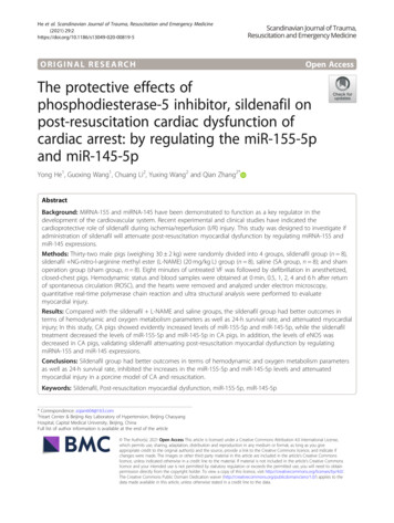
He et al. Scandinavian Journal of Trauma, Resuscitation and Emergency Medicine(2021) INAL RESEARCHOpen AccessThe protective effects ofphosphodiesterase-5 inhibitor, sildenafil onpost-resuscitation cardiac dysfunction ofcardiac arrest: by regulating the miR-155-5pand miR-145-5pYong He1, Guoxing Wang1, Chuang Li2, Yuxing Wang2 and Qian Zhang2*AbstractBackground: MiRNA-155 and miRNA-145 have been demonstrated to function as a key regulator in thedevelopment of the cardiovascular system. Recent experimental and clinical studies have indicated thecardioprotective role of sildenafil during ischemia/reperfusion (I/R) injury. This study was designed to investigate ifadministration of sildenafil will attenuate post-resuscitation myocardial dysfunction by regulating miRNA-155 andmiR-145 expressions.Methods: Thirty-two male pigs (weighing 30 2 kg) were randomly divided into 4 groups, sildenafil group (n 8),sildenafil NG-nitro-l-arginine methyl ester (L-NAME) (20 mg/kg L) group (n 8), saline (SA group, n 8); and shamoperation group (sham group, n 8). Eight minutes of untreated VF was followed by defibrillation in anesthetized,closed-chest pigs. Hemodynamic status and blood samples were obtained at 0 min, 0.5, 1, 2, 4 and 6 h after returnof spontaneous circulation (ROSC), and the hearts were removed and analyzed under electron microscopy,quantitative real-time polymerase chain reaction and ultra structural analysis were performed to evaluatemyocardial injury.Results: Compared with the sildenafil L-NAME and saline groups, the sildenafil group had better outcomes interms of hemodynamic and oxygen metabolism parameters as well as 24-h survival rate, and attenuated myocardialinjury; In this study, CA pigs showed evidently increased levels of miR-155-5p and miR-145-5p, while the sildenafiltreatment decreased the levels of miR-155-5p and miR-145-5p in CA pigs. In addition, the levels of eNOS wasdecreased in CA pigs, validating sildenafil attenuating post-resuscitation myocardial dysfunction by regulatingmiRNA-155 and miR-145 expressions.Conclusions: Sildenafil group had better outcomes in terms of hemodynamic and oxygen metabolism parametersas well as 24-h survival rate, inhibited the increases in the miR-155-5p and miR-145-5p levels and attenuatedmyocardial injury in a porcine model of CA and resuscitation.Keywords: Sildenafil, Post-resuscitation myocardial dysfunction, miR-155-5p, miR-145-5p* Correspondence: zqian604@163.com2Heart Center & Beijing Key Laboratory of Hypertension, Beijing ChaoyangHospital, Capital Medical University, Beijing, ChinaFull list of author information is available at the end of the article The Author(s). 2021 Open Access This article is licensed under a Creative Commons Attribution 4.0 International License,which permits use, sharing, adaptation, distribution and reproduction in any medium or format, as long as you giveappropriate credit to the original author(s) and the source, provide a link to the Creative Commons licence, and indicate ifchanges were made. The images or other third party material in this article are included in the article's Creative Commonslicence, unless indicated otherwise in a credit line to the material. If material is not included in the article's Creative Commonslicence and your intended use is not permitted by statutory regulation or exceeds the permitted use, you will need to obtainpermission directly from the copyright holder. To view a copy of this licence, visit http://creativecommons.org/licenses/by/4.0/.The Creative Commons Public Domain Dedication waiver ) applies to thedata made available in this article, unless otherwise stated in a credit line to the data.
He et al. Scandinavian Journal of Trauma, Resuscitation and Emergency MedicineBackgroundMorbidity and mortality from cardiac arrest (CA) remains unacceptably high, yet effective treatments for CAhave proven to be elusive [1]. Global ischemia and reperfusion injury induced by cardiopulmonary resuscitation(CPR) causes so-called post-resuscitation syndrome [2].Postresuscitation myocardiac dysfunction, an importantcomponent of the postcardiac arrest syndrome, is causedby ischemia/reperfusion (I/R) injury and includes primary manifestations such as arrhythmias, myocyte apoptosis, and contractile dysfunction [3]. Furthermore, postresuscitation myocardial dysfunction is considered theleading cause of death within 72 h after successful CPR[4]. Therefore, studies of new medications that aim toimprove post-resuscitation myocardial dysfunction areof great urgency and importance.MicroRNAs (miRNAs/miRs) are small non-codingRNAs that are able to negatively regulate gene expression via binding to the 3′-untranslated region (UTR) oftarget mRNAs [5]. Previous studies have indicated thatmiRNAs participate in numerous cellular and molecularevents, and the roles served by miRNAs in the pathogenesis of several diseases have been reported [5, 6]. MiR155 and miR-145 have been demonstrated to function asa key regulator in the development of the cardiovascularsystem [7, 8]. Additionally, nitric oxide (NO) is producedby NO synthase (NOS), an enzyme present in largequantities in the endothelium, in which the expressionof NOS is controlled by flow-induced shear stress [9,10]. After I/R, endothelial dysfunction and inhibition ofNOS with reduced NO availability are commonly observed, due to oxidative stress [10]. It was reported thatthe impairment in miRNA functions during normoxiacould upregulate the expression of eNOS, thus implicating miRNAs in the general epigenetic mechanisms involving the posttranscriptional modification of eNOSexpression [11]. Preliminary data suggested that miR155 and miR-145 were shown to directly bind to NOSmessenger RNA (mRNA) during normoxia [7, 8].Sildenafil, is a selective inhibitor of the isoform 5 ofthe enzyme phosphodiesterase (PDE5), which is responsible for the breakdown of 39, 59-cyclic guanosinemonophosphate (cGMP) in smooth muscle cells [12]. Asthe intracellular level of cGMP is controlled by theactivity of PDE5, it is expected that pharmacological inhibition of PDE5 by sildenafil might improve cardioprotection in the myocardium [12]. Our previous animalexperiments showed that sildenail improved postresuscitation perfusion of the heart and improved cardiac function by enhancing the activation of eNOS production and acts on myocardial ischaemia-associatedmiRNAs [12, 13]. Additionally, it is well established thatmiR-155 and miR-145 are involved in the processes of I/R injury via regulating the expression of eNOS and the(2021) 29:2Page 2 of 10production of NO [7, 8]. Based on this background, thepresent study was designed to test the hypothesis thatsildenafil attenuating post-resuscitation myocardial dysfunction by participating in the regulation of miRNA155 and miRNA-145 expression.Materials and methodsEthics statementThis study was carried out in strict accordance with theguideline for animal care and use established by theCapital Medical University Animal Care and Use Committee. The study’s experimental protocol was approvedby the Committee on the Ethics of Animal Experimentsof Capital Medical University (Permit Number: 2019-D014). Animals used in this study were handled in compliance with the Guiding Principles for the Care and Useof Animals expressed in the Declaration of Helsinki [14].All animals were maintained in a specific pathogen-freeenvironment in our facility, and were fed with standardchow and had free access to water. All surgery was performed under anesthesia and analgesia, and all effortswere made to minimize suffering.Animal preparationThirty-two male domestic pigs aged 11 to 13 monthswith an average weight of 30 2 kg were used in eachpart of this study [15]. The strain of those pigs is mixedbreed. Those animals were supplied by a single sourcebreeder (Experimental Animal Center of Capital MedicalUniversity, Beijing, China). The piglets were randomlyassigned into 4 groups, sildenafil group (n 8), sildenafil NG-nitro-l-arginine methyl ester (L-NAME) (20 mg/kgL) group (n 8), saline (SA group, n 8); and sham operation group (sham group, n 8). Sildenafil was obtained from a 25-mg Viagra (Pfizer Australia) tablet thatwas dissolved in 50 ml saline, filtered and stored at 4 C.In sildenafil group, this solution was given once intraperitoneally in the dose of 0.5 mg/kg 30 min prior to VF[16]. In sildenafil L-NAME group, sildenafil (0.5 mg/kg)and L-NAME (20 mg/kg), pretreatment was administered once intravenously at 30 min before VF [17]. Thedrugs were delivered in a randomized manner by thesealed envelope method, as we previously described [12].The vehicle (0.9% NaCl) was administered in the samemanner and volume. After premedication with 0.5 mg/kg intramuscular midazolam, the animal was anesthetized by ear vein injection of propofol (1.0 mg/kg) andmaintained in a surgical plane of anesthesia with intravenous infusion of sodium pentobarbital (8 mg/kg/h).All animals were intubated by a cuffed 6.5-mm endotracheal tube and ventilated by a volume-controlled ventilator (Servo 900C; Siemens, Munich, Germany) using atidal volume of 8 mL/kg and a respiratory frequency of12 breaths/min with room air. End-tidal CO2 was
He et al. Scandinavian Journal of Trauma, Resuscitation and Emergency Medicinemeasured by an inline infrared cacographic (CO2SMOplus monitor; Respirometric Inc., Murrysville). Respiratory frequency was adjusted to maintain end-tidal CO2between 35 and 40 mmHg before CA was induced.Room temperature was adjusted to 26 C, and bodytemperature was maintained at 37 C under an infraredlamp, and all efforts were made to minimize suffering.Fluid losses were compensated by an infusion of 30 mL/kg acetated Ringer’s solution during the first hour ofpreparation, followed by a continuous infusion of 2.5%glucose-electrolytes solution 8 mL/kg/h and acetatedRinger’s solution 20 mL/kg/h. All investigators performing CPR and interpreting the outcome assessments wereblinded to the medication.An angiographic catheter was inserted from the femoral artery into the aortic arch for collecting blood samples and for measuring aortic pressure. A Swan-Ganzcatheter (7 Fr; Edwards Life Sciences, Irvine, CA) wasadvanced from the right femoral vein and flow-directedinto the pulmonary artery for measurement of rightatrial pressure, mean pulmonary arterial pressure(MPAP) and cardiac output (CO). The electrocardiogram and all hemodynamic parameters were monitoredwith a patient monitoring system (M1165; HewlettPackard, Palo Alto, CA). Animals with self-adhesive defibrillation electrodes located on the chest wall. Pigs inthe sham group that were not subjected to CA wereused as controls. Arterial blood gas values were measured regularly using an ABL 520 Blood Gas Analyzer(Radiometer, Bronshoj, Denmark) at six time points: atbaseline, 30 min, and 1, 2, 4, 6 h after ROSC. Mean aortic pressure (MAP) was monitored via the right femoralarterial catheter. The amounts of infused fluid and urineoutput were also monitored during the experiment. Coronary perfusion pressure (CPP) was calculated as the difference between decompression diastolic aortic andtime-coincident right atrial pressure measured at theend of each minute of precordial compression. DuringCPR, CPP was calculated as the difference between themean aortic and mean right atrial pressures during diastole (spontaneously beating) or decompression (CPR).CPR compression force, rate, and depth were controlledand continuously recorded during all experiments to assure that all groups received identical CPR quality.Experimental protocolAfter establishment of vascular catheters, the animalswere allowed to equilibrate for 30 min to achieve a stableresting level. Baseline measurements and arterial bloodgases were obtained. Mechanical ventilation was established as described above. The temporary pacemakerconductor was inserted into the right ventricle throughthe right sheathing canal and connected to an electricalstimulator (GY-600A; Kaifeng Huanan Equipment Co,(2021) 29:2Page 3 of 10Ltd., Kaifeng, China) programmed in the S1S2 mode(300/200 ms), 40 V, 8:1 proportion, and 10 ms steplength to provide a continuous electrical stimulus untilVF [18]. VF was defined as a waveform of VF emergingon the monitor and a rapid decline in MAP toward zero.After successful induction of VF, mechanical ventilationwas discontinued. Mechanical ventilation was discontinued after the onset of VF. After 8 min of untreated VF,CPR was performed. Manual chest compressions wereimmediately initiated at a rate of 100 compressions perminute for 2 min and ventilation conducted using a bagrespirator attached to an endotracheal tube with roomair. CPR was performed by the same CPR technicianfrom our laboratory, who compressed the porcine chestto approximately one-third of the anteroposterior diameter. The quality of chest compressions was controlledby a Heart Start MRx Monitor/Defibrillator with Q-CPR(Philips Medical Systems, Best, Holland) [19]. Thecompression-to-ventilation ratio was 30:2. After 2 minof CPR, a single 120 J biphasic electrical shock wasattempted with a Smart Biphasic defibrillator (PhilipsMedical Systems,Andover, MA). If the first defibrillation was unsuccessful, epinephrine (20 μg/kg) wasgiven intravenously followed by 2 mins of CPR, andrepeated every 2 min if ROSC was not achieved. The150 J shocks were used for the second and all subsequent attempts. The study was blinded as to themedication used, and only the principal investigator,who did not take part in any resuscitation effort,knew the assignment of each animal. Furthermore,the investigators involved in data recording, dataentry, and data analysis were also blinded to the allocation. If spontaneous circulation was still notachieved, CPR was continued for a further 2 min, anddefibrillation was attempted once more.ROSC was defined as 10 consecutive minutes of maintenance of systolic blood pressure at 50 mmHg. If spontaneous circulation was not restored within 30 min, weregarded the animal as dead [20]. All the animals received normal saline (10 mL/kg/h) intraoperatively to replenish fluid losses. After successful resuscitation, theanimals were mechanically ventilated with 100% inspiredoxygen for the first 30 min, 50% for the second 30 minand 21% thereafter. With the exception of one jugularvein sheath that was used for fluid administration, allother vascular sheaths and endotracheal tube were removed after a 6 h intensive care period. The animalswere allowed to recover from anesthesia, and were thenplaced in observation cages and monitored for a further18 h. After a period of 24 h, post-resuscitation measurements were completed. All catheters were removed andwounds were surgically sutured. The animals were theneuthanatized with 10 mL of 10 mol/L potassium chlorideintravenously following a bolus of 100 mg of propofol
He et al. Scandinavian Journal of Trauma, Resuscitation and Emergency Medicine(2021) 29:2Page 4 of 10intravenously. Myocardial specimens were harvested andsnap frozen in liquid nitrogen and stored at 80 C.2 ΔΔCt method. All samples were run in triplicatefrom three independent experiments.MeasurementsHemodynamic and oxygen metabolism parameterscollectionUltra structural analysisECG was continuously monitored. The hemodynamicparameters including heart rate (HR), CO, MAP, andMPAP were measured continuously, and we recordedthe values at baseline, and at 30 min, 1, 2, 4, 6 h afterROSC. At the end of each time point, 4 C saline wasinjected into the right atrium through the Swan-Ganzcatheter to determine CO by the transpulmonary thermodilution method as described previously [12]. MAP wasdetermined by the electronic integration of the aorticblood pressure waveform. The amounts of infused fluidand urine output were also monitored during the experiment. Serum lactate level, and arterial blood gas valuesof which temperatures were corrected to 37 C weremeasured regularly using an ABL 520 Blood GasAnalyzer (Radiometer, Bronshoj, Denmark). CPP wascalculated as the difference between decompression diastolic aortic and time-coincident right atrial pressuremeasured at the end of each minute of precordial compression. CPP during VF was defined as the differencebetween the mean aortic and mean right atrial pressures.Oxygen metabolism parameters, including oxygen delivery (DO2) and oxygen consumption (VO2), werecalculated.SurvivalThe survival rate was determined based on the animalsthat survived the experimental protocol starting atROSC until 24 h after ROSC. Animals that died duringsurgical recovery were excluded.Micro-RNA isolation and expressionTotal RNA samples were extracted using Trizol (Invitrogen, USA) from cultural myocytes. MiR-155-5p, miR145-5p and eNOS level were quantified by the mirVanaqRT-PCR (quantitative real-time PCR) miRNA Detection Kit (Ambion, USA) in conjunction with real-timePCR with SYBR Green I (Applied Biosystems, USA)[21]. The following primers were used for PCR detection: miR-155-5p [5′-GCGCGTTAATGCTAATTGTGA-3′(forward); 5′-AGTGCAGGGTCCGAGGTATT3′ (reverse)]; miR-145-5p [5′- CGGTCCAGTTTTCCCAGGAA 3′ (forward); 5′ AGTGCAGGGTCCGAGGTATT 3′ (reverse)], U6 was used as an internal control. eNOS [5′-TCC CAG ACC CCA TAA CAA CAG3′ (sense) and 5′-TGA GGG TGC AGCGAA CTT TA3′ (antisense)]. The relative expression of miR-155-5p,miR-145-5p and eNOS mRNA was calculated using theThe remaining tissue was preserved in 10% formaldehyde and 4% paraformaldehyde to observe pathologicand ultra structural changes of the myocardium undertransmission electron microscope (TEM) (H-7650; Hitachi, Tokyo, Japan). The pathologic data were assessed byreviewers blinded to the experimental groups.Statistical analysesContinuous variables were presented as mean standarddeviation (SD) when data were normally distributed oras a median (25th, 75th percentiles) when data were notnormally distributed. Student t test was used for comparisons between every two groups. Differences at different time points were compared with repeated-measuresanalysis of variance (ANOVA) with Bonferroni correction for post hoc comparison. The Kruskal-Wallis testwas used to compare continuous variables in multiplegroups. For these comparisons, the Bonferroni correction was applied to control for the multiple testing. Survival analysis was performed using the method of Kaplanand Meier, and comparisons between groups were madeusing the log-rank test. A value of p 0.05 was considered as statistically significant. All analyses were conducted using the SPSS 17.0 software (SPSS Inc., ChicagoIII) and GraphPad PRISM version 6 (GraphPad SoftwareInc., San Diego, CA).ResultsBaseline statusBaseline hemodynamic measurements and oxygen metabolism measurements are shown in (Table 1). None of the variables (body weight, HR, MAP, MPAP, CO, lactateconcentration, DO2, VO2 and extraction of oxygen (ERO2)differed significantly among the four groups (p 0.05).Resuscitation outcomes and survivalResuscitation outcomes are shown in Table 2. None ofthe 24 animals restored spontaneous circulation afterinitial defibrillation attempts. By comparison, the cumulative defibrillation energy was significantly lower in sildenafil group than in sildenafil L-NAME group andSA group (p 0.05). ROSC was achieved in eight pigletsin the both sildenafil group and in the sildenafil LNAME group, and in seven of eight piglets in the SAgroup. In the SA group, six piglets died at 14mins, 70mins, 196 mins, 256 mins, 322 mins and 416 mins afterROSC. In the sildenafil L-NAME group, four pigletsdied at 10 mins, 75 mins, 180 mins and 210 mins afterROSC. While, in the sildenafil group, only one pigletdied at 48 min after ROSC. A significant difference in
He et al. Scandinavian Journal of Trauma, Resuscitation and Emergency Medicine(2021) 29:2Page 5 of 10Table 1 Baseline characteristicsSHAM group (n 8)SA group (n 8)Sildenafil group (n 8)Sildenafil L-NANE group (n 8)Weight (kg)29.13 2.1630.63 0.9230.38 0.9230.44 0.89HR99.00 7.44101.38 8.30100.50 10.04101.52 9.84MAP, mmHg103.12 5.19101.88 5.2299.00 5.81102.41 5.21MPAP, mmHg23.42 4.3224.13 5.2424.34 4.5623.72 4.68CO, L/min2.86 0.222.99 0.202.99 0.192.89 0.22DO2, ml/min444 39445 34450 28439 31VO2, ml/min112 12112 9115 12113 10ERO2, %24.17 2.3425.44 2.7025.46 1.4925.36 1.76Lac, mmol/L1.21 0.491.31 0.721.23 0.4011.24 0.42Values are mean SD or number (n)SHAM sham, SA saline, HR heart rate, CO cardiac output; MAP mean aortic pressure, MPAP mean pulmonary arterial pressure, DO2 oxygen delivery, VO2 oxygenconsumption, ERO2 extraction of oxygen, Lac lactatesurvival to the end of the 24-h experiment between sildenafil, sildenafil L-NAME and SA groups were demonstrated using the Kaplan-Meier survival curve and thelog-rank test (p 0.05) (Fig. 1).Hemodynamics and left ventricular functionIndeed, HR, MAP and MPAP were significantly increased during the first 4 h after ROSC compared tobaseline in the three resuscitation groups (p 0.01 vs.Baseline, Table 2). However, sildenafil pretreatmentinhibited this increase of HR, MAP and MPAP at fourtime-points (0 min, 30 min, 2 h and 4 h after ROSC),while the values of CPP and CO were decreased duringthe initial 6 h after ROSC when compared to baselinevalues (p 0.01, respectively) and the CO and CPPvalues were significantly higher in the sildenafil groupthan those in the sildenafil L-NAME group at 1, 2 hand 4 h after ROSC (p 0.03, p 0.02, p 0.04, respectively (Table 3).Arterial blood gasesThere were no significant differences in blood gas valuesat baseline among three resuscitation groups. The serumlevels of PH, HCO3 , PaO2, PaCO2 and lactate were significantly worse than those at baseline in three resuscitation groups. PH, HCO3 , PaCO2 and lactate weresignificantly lower in the sildenafil group than those inthe sildenafil L-NAME group at 0 min, 30 min and 1 hafter ROSC (p 0.04, p 0.02, p 0.02, p 0.03 respectively) and the PaO2 was higher in the sildenafil groupthan those in the sildenafil L-NAME group at 0 min,30 min, 1 h and 2 h after ROSC (p 0.03, p 0.04, p 0.02, p 0.03, respectively, Table 4).Effect of sildenafil on expression of miR-155-5p and miR145-5pReal-time PCR analysis revealed that following CAand post-resuscitation treatment pigs showed evidently increased levels of miR-155-5p (Fig. 2a) andmiR-145-5p (Fig. 2b) compared with sham operatedpigs, whereas that there were the significantly downregulated expression of miR-155 and miR-145 following sildenafil treatment compared with the sildenafil L-NAME group and SA group ((both p 0.01). Similarly, compared with that in the shamgroup, the relative expression of eNOS mRNA (Fig. 2c)were suppressed in the following CA and postresuscitation groups, while the expression of eNOSmRNA was recovered in the sildenafil treatment group.The results suggested that sildenafil may serve a protectiverole in post-resuscitation myocardial dysfunction by participating in the regulation of miRNA-155 and miRNA145 expression.Table 2 Cardiopulmonary resuscitation outcomesSA group (n 8)Sildenafil group (n 8)Sildenafil L-NAME group (n 8)Number of defibrillatory shocks4.31 1.622.91 0.833.72 0.71Cumulative defibrillation energy (J)145.01 33.4195.02 33.41*#115.23 23.14*Duration of CPR before ROSC (min)6.12 2.214.71 1.325.43 1.586-h survival37*6*24-h survival1#7*Values are mean SD or number (n)SA saline, ROSC restoration of spontaneous circulation, CPR cardiopulmonary resuscitation*p 0.05 vs. SA group, #p 0.05 vs. Sildenafil L-NAME group4*
(2021) 29:2He et al. Scandinavian Journal of Trauma, Resuscitation and Emergency MedicinePage 6 of 10improved faster in the sildenafil than in the sildenafil L-NAME group and in the SA group (Fig. 3e-h). Additionally, myocardial damage were further significantlyalleviated in the sildenafil group when compared withthe sildenafil L-NAME group, animals treated with sildenafil exhibited little intracellular damage in the myocardium: partial nuclear chromatin condensation,reduced crest fracture and moderate edema occurred inthe mitochondria and sarcoplasmic reticula (Fig. 3g, h).Fig. 1 Kaplan-Meier survival curve. We found a significant differencein survival between the sildenafil group, sildenafil L-NAME groupand the saline group (P 0.001 by logrank test)Ultra structural changes in cardiomyocytesUnder TEM, normal mitochondria structures were displayed in the sham group (Fig. 3a, b). The myocardialfiber and intercalated disk were obviously disordered,broken, even dissolved in the SA group 24 h after ROSC;most of the mitochondria were severely broken, evenexhibiting vascular with vague, arranged irregularly, ordisrupted cristae (Fig. 3c, d). However, all of them wereDiscussionThe major findings of this study were as follows: (1)Compared with the sildenafil L-NAME and salinegroups, the sildenafil group had better outcomes interms of hemodynamic and oxygen metabolism parameters as well as 24-h survival rate, and attenuated myocardial injury in a porcine model of CA and resuscitation.(2) In this study, CA pigs showed evidently increasedlevels of miR-155-5p and miR-145-5p, while the sildenafil treatment decreased the levels of miR-155-5p andmiR-145-5p in CA pigs. In addition, the levels of eNOSwas decreased in CA pigs, validating sildenafil attenuating post-resuscitation myocardial dysfunction by regulating miRNA-155 and miR-145 expressions.The severity of postresuscitation myocardial dysfunction is the major cause of early death, and no effectivetreatment method has been established [3]. Actively protective treatment to myocardial function is urgent toTable 3 Haemodynamics during cardiopulmonary resuscitation and ROSCGroupSA groupSildenafil groupParameter Baseline**ROSC 30 min**ROSC 1 hROSC 2 h**ROSC 4 hROSC 6 h***HR101.3 8.30 132.2 11.3129.4 10.4120.3 8.9118.9 9.3113.5 17.4101.4 10.4CO2.99 0.200.91 0.07**0.88 0.06**0.89 0.06**0.91 0.05**1.04 0.05**1.15 0.04**MAP91.88 3.22 125.2 7.3**122.5 6.4**115.4 6.1**109.8 5.7**107.6 5.1**101.5 4.8***CPP44.2 4.1222.3 2.5MPAP24.1 2.249.7 6.3**HRCOMAPCPPSildenafil L-NANEgroupROSC 0 min102.1 7.30 123.2 9.3**Δ#2.99 0.20**0.99 0.0689.88 3.22 117.2 7.143.1 3.34**Δ#**Δ27.6 2.8**Δ#MPAP24.6 2.740.3 5.6HR103.2 7.22 128.4 9.1**CO2.99 0.20**MAP88.18 3.22 121.2 6.1**Δ0.98 0.07**Δ**23.4 2.647.3 5.7****Δ#118.4 8.4**1.12 0.03**Δ#115.5 5.9**Δ28.2 2.9**Δ#38.4 4.1125.6 7.3*Δ****25.6 2.743.8 4.7****115.3 7.9**Δ#1.23 0.04**111.8 5.6**32.4 3.1**Δ#37.8 4.1118.5 7.2*39.7 4.2***Δ#110.9 6.3**Δ#1.34 0.08**Δ#103.6 4.735.4 3.6*Δ#33.6 3.5*Δ*33.8 3.7*32.5 3.835.8 3.7*107.5 5.435.1 3.1*Δ#**Δ#1.42 0.07105.4 4.41.36 0.08***Δ#94.5 3.4Δ#39.4 4.3ΔΔ29.8 3.128.7 2.2Δ99.8 4.336.5 3.9112.6 5.7Δ104.7 6.4**115.9 6.6*Δ0.87 0.050.92 0.060.98 0.041.17 0.05**1.21 0.06**120.6 7.4**117.4 6.4**113.8 6.7**110.7 4.6**99.7 4.6*****29.7 3.6******CPP44.2 3.5224.8 2.725.7 2.827.7 2.630.6 3.2MPAP24.6 2.745.2 5.4**Δ46.2 5.3**44.7 4.3**38.4 4.7***32.7 3.635.7 3.4*36.9 3.5*34.3 3.6*Values are mean SD (n)*p 0.05 vs. baseline, **p 0.01 vs. baseline, Δ P 0.05 vs. SA group. # p 0.05 vs. Sildenafil L-NAME group. (Student t test was used for comparisons betweenevery two groups. Partial pressures are given in mmHg. HCO3: bicarbonate in mM; Differences at different time points were compared with repeated-measuresanalysis of variance (ANOVA) with Bon-ferroni correction for post hoc comparison between multiple experimental groups)
(2021) 29:2He et al. Scandinavian Journal of Trauma, Resuscitation and Emergency MedicinePage 7 of 10Table 4 Arterial blood gassesGroupParameterBaselineSA groupPH7.41 0.11 7.14 0.05**PaO2 (mm Hg)89.1 9.3PaCO2 (mm Hg) 39.3 3.2HCO3(mmol/L)LAC (mmol/L)Sildenafil groupPHPaO2 (mm Hg)27.2 4.72.6 0.5ROSC 0 min**ROSC 1 hROSC 2 hROSC 4 hROSC 6 h7.18 0.07**7.20 0.06**7.27 0.09*7.30 0.08*7.32 0.09*******80.7 7.5*55.4 5.160.1 6.270.2 7.148.3 5.1**46.7 5.4**45.4 4.1**43.8 4.7*41.6 4.241.5 4.8****20.4 1.6**21.6 2.722.7 2.6**24.5 3.1*25.2 3.7******19.3 1.211.2 3.37.42 0.13 7.27 0.07**Δ#9.3 2.7**8.8 1.9**Δ#7.29 0.04**Δ#*6.9 2.2*Δ#7.30 0.07**Δ#79.5 7.7*44.5 4.7**Δ#4.7 1.1*5.8 1.3*7.32 0.05*Δ#7.35 0.047.38 0.1152.7 5.363.5 6.270.3 7.179.3 7.744.7 4.3**Δ#43.2 4.4*Δ#42.4 4.1*Δ#41.3 4.240.5 4.241.1 3.8HCO3 (mmol/L)22.6 2.7**Δ#23.7 2.6**Δ#24.6 2.7*Δ#25.3 2.625.7 3.126.3 3.87.4 1.6**Δ#5.3 1.3**Δ4.2 1.2*Sildenafil LNANE group PHPaO2 (mm Hg)27.6 4.12.7 0.4**ΔLAC(mmol/L)**Δ#8.1 2.1**Δ**Δ**84.9 8.23.6 0.7**7.41 0.13 7.19 0.067.21 0.067.23 0.047.29 0.097.31 0.07*********90.1 9.1PaCO2 (mm Hg) 39.4 3.3HCO3 (mmol/L)8.5 2.4**Δ#83.5 8.1*PaCO2 (mm Hg) 39.5 3.4LAC (mmol/L)90.2 8.6ROSC 30 min27.4 4.32.6 0.480.9 7.3*46.4 3.758.5 4.363.1 6.272.2 7.147.8 4.4**46.3 4.3**44.6 4.5**43.2 4.1*42.3 3.341.3 4.2****21.7 2.5**21.6 2.722.7 2.6**23.8 3.2*25.4 3.6******20.5 2.410.8 4.59.8 3.58.8 1.9**6.9 2.280.5 7.77.34 0.07**5.8 1.34.1 2.3Values are mean SD (n)*p 0.05 vs. baseline, **p 0.01 vs. baseline, Δ P 0.05 vs. SA.# P 0.05 vs. Sildenafil L-NAME group. (Student t test was used for comparisons between every twogroups. Partial pressures are given in mmHg. HCO3: bicarbonate in mM; Differences at different time points were compared with repeated-measures analysis ofvariance (ANOVA) with Bon-ferroni correction for post hoc comparison between multiple experimental groups)improve postresuscitation outcomes. Therefor
diac function by enhancing the activation of eNOS pro-duction and acts on myocardial ischaemia-associated miRNAs [12, 13]. Additionally, it is well established that miR-155 and miR-145 are involved in the processes of I/ R injury via regulating the expression of eNOS and the production of NO [7, 8]. Based on this background, the