Transcription
PEDIATRIC EMERGENCIESin the Clinic SettingKenneth Acha, MD,UCR SOM - FM Residency ProgramOctober 2016
Acknowledgments1. Jeff Gill, MD, Pediatrician2. Dr. Quitain, Pediatric Critical Care3. Jeff Messerole, EMT Instructor / Author4. Jessica Chichizola, Senior MA
Objectives1. Pediatric Emergencies are Common2. Our Clinic’s Preparedness3. Systematic approach4. Respiratory Emergencies5. Allergic Emergencies (Anaphylaxis)6. Shock7. Neurologic Emergencies8. Not Discussed
Take Home Points1. Pediatric emergencies are common in officesetting2. Prepare by knowing what the clinic has to offerand the protocols in place.3. Mock Codes in the clinic are recommended.
PEDS EMERGENCIESARE COMMON INTHE CLINIC
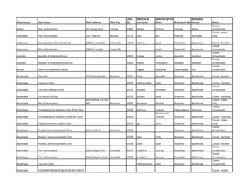
PEDS EMERGENCIES ARE COMMON INTHE CLINIC“Studies have shown that emergencies are common in primary care practices that providecare to children. In 1 study, the authors surveyed 52 pediatric offices and found that thesepractices saw a median of 24 emergencies per year. Most of the offices (82%) reportedthat they encountered, on average, at least 1 emergency per month. In another study,62% of pediatricians and family physicians in an urban setting who were asked aboutemergencies in their offices reported that they assessed more than 1 patient each week intheir offices who required hospitalization or urgent stabilization.Despite these findings, which suggest that a significant number of children present toprimary care offices with urgent or emergent problems, some health care professionalsdiscount the need for preparation because “emergencies are not very common” orbecause they feel they can rely on rapid response from emergency medical services(EMS) or proximity to a hospital. Some PPCPs have interpreted risk-managementguidelines to mean that having emergency equipment and medications on site willincrease their liability in emergency situations; however, lack of preparation may be a truecause of increased liability.” AAP
PEDS EMERGENCIES ARE COMMON INTHE CLINIC1.2.3.4.5.Children are often taken to the clinic at the time of an emergencyThe most common types of emergencies include respiratoryemergencies, seizures, infections in young infants, and dehydration.PPCPs may be required to provide urgent or emergent care in theiroffices for children with these conditions, at least until the arrival of EMS.The consequences of being unprepared are serious; therefore,appropriate stabilization of pediatric emergencies and timely transfer toan appropriate facility for definitive care are important responsibilities ofevery PPCP.We are responsible for stabilizing the patient until EMS comes after wecall 9-1-1.The office staff, not just the PPCPs have to be prepared andknowledgeable because the first person to assess patients who arrive inthe office may be the least clinically experienced employee: the secretaryor receptionist.
What are the response timesfor EMS to get here after wecall 9-1-1?
OUR CLINIC’SPREPARDNESS
RECOMMENDATIONS FROM THE AAP & AAFPü Perform a self-assessment of office readiness.ü Develop an plan for emergency response in the office, which includes: Staffcommunication, roles, and responsibilities at the time of an emergency during timesof high and low staffing; protocol to access EMS; and maintaining readinessthrough practice (mock codes).ü Maintain recommended emergency equipment and medicationsü Develop a plan to provide education and continuing medical education for all staff.ü Practice mock codes in the office on a regular basis (quarterly or biannually).ü Educate families about what to do in an emergency. E.g. 1) Encourage first aid andCPR training for parents and caregivers, etc.ü Partner with EMS and hospital-based emergency providers to ensure optimalemergency care and emergency/disaster readiness for children.
Effective Resuscitation Team Preparation
SYSTEMATICAPPROACH
PATHWAYS TO PEDIATRIC CARDIAC ARREST
PALS SYSTEMATIC APPROACH
EVALUATE-IDENTIFY-INTERVENE SEQUENCEContinue the sequence until the child is stable. Use it before and after each intervention. E.g. If you give O2, reevaluatethe child. Is he breathing a little easier? Are color and mental status improving? If you give an IV fluid bolus to tx hypovolemicshock, reevaluate. Have the HR and perfusion improved? Is another bolus needed? Use it whenever the pt’s condition changes.
EVALUATE: PRIMARY ASSESSMENT (ABCDE)ü Airwayü Assess to det. if it is patent. Is it clear, obstructed but maintainable, or obstructed & notmaintainable?ü C-A-B if patient is in cardiac arrest!ü Breathingü Assess rate, effort, chest expansion & air movement, lung & air way sounds, and O2 satsby pulse oximetry.ü Respiratory Rate: Tachypnea, bradypnea, apneaü Respiratory Effort: Retractions (Subcostal Substernal Intercostal Supraclavicular Suprasternal, Sternal), Accessory muscles, Nasal flaring, Head bobbing, Seasawrespirationsü Lung and airway sounds: Stridor, Grunting, Gurgling, Wheezing, Crackles
EVALUATE: PRIMARY ASSESSMENT (ABCDE)ü Breathing (Caution when using pulse ox)ü Interpret pulse ox in conjunction with your clinical assessment &signs like RR, effort, level of consciousness, not in isolation. A pt.may be in distress and still have normal pulse ox, esp. if gettingO2. If HR from pulse ox is different from the HR on ECG monitor,don’t trust the O2 sat from that pulse ox machine.ü Pulse ox measures saturation, not delivery. A severely anemic ptmay have 100% pulse ox.ü Pulse ox won’t be accurate in severe shock and cardiac arrest.ü If you suspect Met-hemoglobin or Carboxyhemoglobin, get ABGbecause pulse ox will be wrong.
EVALUATE: PRIMARY ASSESSMENT (ABCDE)ü Circulationü Assessment: HR & rhythm, pulses, Capillary refill, skin color & temperature, BP, UOP,level of consciousnessü Disabilityü Assessment: Quick evaluation of neurologic function [use: AVPU (Alert, responsive toVoice, responsive to Pain, Unresponsive), GCS, pupil response to light (PERRLA?)]ü Exposureü Undress the pt, one part at a time and examine the pt’s body. Keep patient coveredand only expose the part being examined.
EVALUATE: SECONDARY ASSESSMENTSecondary Assessment Focused History Focused Physical Examü Focused History – e.g. SAMPLE.ü SAMPLE Signs & symptoms; Allergies, Meds, PMH, Last Meal, Events leading tothe current illness/injury.ü You don’t have to use SAMPLE. May use the usual hx part of admit H&Ps.ü Focused Physical Exam –ü Examine primary area of concern of illness or injury (e.g. respiratory assessment withrespiratory distress) as well as do a brief head-to-toe evaluation.
EVALUATE: DIAGNOSTIC TESTSü Labsü ABG, VBG, CBC (Hgb.), Lactateü Imagingü CXR, CT, Echocardiogramü Monitoring, PFTs, etc.ü Peak Expiratory Flow Rateü Central venous oxygen satsü CVP Monitoringü Invasive arterial pressure monitoring
IDENTIFYTry to identify the type and severity of the child’s problem.TypeSeverityRespiratory-Obstruc?on, upper airway-Obstruc?on, lower airway-Lung ?ssue disease-Disordered control ofbreathing-Respiratory distress-Respiratory failureCirculatory-Hypovolemic shock-Distribu?ve shock-Cardiogenic shock-Obstruc?ve shock-Compensated shock-Hypotensive shockCardiopulmonary FailureCardiac Arrest
INTERVENESome common interventions you can make are to:ü Position the child to maintain a patent airwayü Activate emergency response (call 911)ü Start CPRü Get the Code cart and monitorü Place the child on a cardiac monitor and pulse oxü Give O2ü Support ventilationü Start meds and fluids (e.g. neb tx, IV/IO fluid bolus)
Is there a Life-Threatening Problem?Signs of a life-threatening condi?on include the following:AirwayComplete or severe airway obstruc6onBreathingApnea, significant increased work of breathing, bradypneaCircula?onAbsence of palpable pulses, poor perfusion, hypotension, bradycardia.DisabilityUnresponsiveness, decreased level of consciousnessExposureSignificant hypothermia, significant bleeding, petechiae, or purpura consistent with sep?cshock.
RESPIRATORYEMERGENCIES
Fundamental Issues Associated WithRespiratory Problemsü Respiratory problems Impairment of either oxygenation, ventilation, orboth.ü The function of the Resp. System is gas exchange (O2 in & CO2 out).ü O2 sat % of Hgb. that becomes bound to O2. Note, a small % of O2 isdissolved in blood and O2 sats don’t show that.ü Children have a higher metabolic rate, so the O2 demands per Kg/bodyweight is high. Infants consume 6 to 8ml/kg/min of O2 vs. 3 to 4ml/kg/minin adults. As such, hypoxemia and tissue hypoxia can develop morerapidly in a child than in an adult if apnea or poor ventilation occurs.ü Resp. Problems can cause: Hypoxemia, Hypercarbia, or both.
Fundamental Issues Associated WithRespiratory Problemsü Hypoxemiaü Is low PaO2 that is associated with a low O2 sat assessed by pulse ox (SpO2)ü Hypoxemia SpO2 94% in a child who is breathing room air.ü Hypoxemia indicates inadequate oxygenation.ü Hypoxemia is NOT tissue hypoxia.ü Tissue hypoxia O2 delivery is not enough to meet tissue O2 needs.ü Hypoxemic pt can compensate and avoid tissue hypoxia by increasing CO. Also byincreasing O2-carrying capacity (O2 conc.).ü You can have normal O2 sats & still have tissue hypoxia e.g. in shock or severeanemia.
Where is the respiratorycenter located?
Respiratory centerü Brainstem (Pons & Medulla)ü Pneumotaxic area controls the depth ofinspiration & prevents over-distensionof lungs. It does so by inhibiting theApneustic center.ü Apneustic center stimulates theMedulla leading to apneustic breathing(prolonged end-expiratory phases)ü Medulla controls both the inspiration &expiration. Controls the rhythm ofbreathing.
What does the body do whenthere is tissue hypoxia?
Signs of Tissue Hypoxiaü Tachycardia (early sign)ü Tachypneaü Nasal flaring, retractionsü Agitation, anxiety, irritabilityü Pallorü Cyanosis (late sign)ü Decreased level of consciousness (late sign)ü Bradypnea, apnea (late sign)ü Bradycardia (late sign)
What is the most commoncause of hypoxemia?
V/Q mismatch is the mostcommon cause of hypoxemiain both pediatric. & adultpopulations b/c the causesare very common
CAUSES OF HYPOXEMIACAUSESDISEASE / PROCESSMECHANISMTREATMENTLow atmosphericPo2High al?tude (decreasedbarometric pressure)Decreased PaO2Supplemental O2AlveolarHypoven?la?onCNS infec?on, Trauma?cbrain injury, Drug overdose,Neuromuscular weakness,Apneaé PaCO2 (hypercarbia) displaces alveolar O2,resul?ng in ê alveolar and arterial O2 tension (lowPaO2 or hypoxemiaRestore normal ven?la?on,supplementary O2.Diffusion defectPulmonary edema,Inters?al PNA, Alveolarproteinosis.Impaired movement of O2 & CO2 b/n the alveolusand blood results in ê PaO2, and if severe, é PaCO2Supplementary O2 with CPAPor ven?la?on with anadvanced airway and PEEP.PNA, Atelectasis, ARDS,Asthma, Bronchioli?s,Foreign Body, PE, COPD.Mismatch of Ven?la?on and perfusion: Blood floodthrough areas of the lung that are inadequatelyPEEP to increase mean airwayven?lated results in incomplete oxygena?on of thepressure; supplementary O2;blood returning to the le] side of the heart. Thevent ilatory support.result is a decreased arterial O2 satura?on and PaO2,and to a lesser extent, increased PaO2Cyano?c congenital heartdisease, Extracardiac(anatomical vascular shunt).Same causes listed for V/Qmistmatch.Shun?ng of deoxygenated blood from the right ofthe heart to the le] (or from the pulmonary arteryinto the aorta) results in a low PaO2. Effects similarto right to le] shunt in the lungs.Ven?la?on /Perfusion (V/Q)Mismatch(Most common causeof hypoxemia)Right-to-le]shuntCorrec?on of defect(supplementary O2 alone isinsufficient)
What are signs respiratoryeffort?
Physiology of Respiratory DiseaseImportant factors associated with increased work of breathing include:ü Increased airway resistance (upper and lower)ü Decreased lung compliance (Distensibility)ü Use of accessory muscles of respirationü Disordered CNS control of breathing.
Identify Resp. Problems by SeverityRespiratory DistressRespiratory FailureA clinical state characterized by abnormal respiratory rate oreffort.A clinical state of inadequate oxygenation, ventilation, or both.Clinical Signs of Resp. distress typically include some or all ofthe following:Suspect probably respiratory failure if some of the followingare present:-Increased RR (Tachypnea)-Marked tachypnea (early)-Increased resp. effort (e.g. nasal flaring, retrac?ons)-Bradypnea (late)-Inadequate resp. effort (e.g. hypoven?la?on, bradypnea)-Increased, decreased, or no resp. effort.-Abnormal airway sounds (e.g. stridor, wheezing, grun?ng)-Poor to absent air movement.-Increased HR (Tachycardia)-Tachychardia (early)-Pale, cool skin-Bradycardia (late)-Changes in level of consciousness-Cyanosis-Stupor, Coma (lateResp. distress is classified as mild to severe based on severity ofthe above signs.Resp. failure can result from upper or lower airway obstruc?on,lung ?ssue disease, and disordered control of breathing (e.g. apneaor shallow, slow respira?ons). When resp. effort is inadequate,resp. failure can occur without the typical signs of resp. distress)
Signs of Respiratory ProblemsClinical SignsUpper Airwayobstruc6onA PatencyBAir MovementCIncreased-Stridor (typicallyinspiratory)-Barking Cough-Hoarseness-Wheezing (typicallyexpiratory)-Prolonged Expiratoryphase)-Cough-Grun?ng-Crackles-Decreased breathsoundsDecreasedNormalVariableTachycardia (early), Bradycardia (late)SkinPallor, cool skin (early), Cyanosis (late)TemperatureDisordered Controlof BreathingVariableHeart RateD Level ofConsciousnessELung Tissue DiseaseAirway open and maintainable / not maintainableResp. Rate / EffortBreath SoundsLower AirwayObstruc6onAnxiety, agita?on (early)Lethargy, unresponsiveness (late)Variable
Identification of Resp. Problems by SeverityRespiratory Distress !ARespiratory FailureOpen and maintainable è Not MaintainableTachypnea è Bradypnea to apneaBWork of breathing (nasal flaring/retrac?ons)Increased effortè Decreased effort è ApneaGood air movement è Poor to absent air movementCTachycardia è BradycardiaPallor è CyanosisDAnxiety, agita?on è Lethargy to unresponsiveEVariable temperature
What is the major cause ofcardiac arrest in children?
Respiratory problems
Is it always possible todifferentiate betweenrespiratory distress andfailure on the basis of clinicalexam alone?
No. In children, thedeterioration in resp. functionmay progress rapidly. Plusyou can have failure withoutdistress.
What are the two mainfunctions of the lungs?
Oxygenation & Ventilation
RESPIRATORY EMERGENCIESType of Resp. ProblemCondi6onsObstruc?on, Upper Airway-Severe Croup-Epigloc?s-Foreign Body Aspira?on-Allergic Emergencies (Anaphylaxis)Obstruc?on, Lower Airway-Acute Asthma-Bronchioli?sLung Tissue DiseaseDisordered Control of Breathing-Infec?ous PNA-Chemical PNA-Aspira?on PNA-Cardiogenic Pulmonary Edema-Non-cardiogenic pulmonary edema (ARDS)*DKA may present with respiratory features-Increased ICP-Poisoning or drug overdose-Neuromuscular disease
INITIAL MGT. OF ALL RESP. EMERGENCIESFirst, ask: Is this pa.ent in cardiac arrest? If Yes, begin C-A-B. If not, do A-B-C. The following ABCs apply to all theresp. emergencies. Addi?onal measures will be given for each specific condi?on.EvaluateInterven6ons (as indicated)Airway-Support an open airway (allow the child to assume posi?on of comfort) or if necessary, open airway with:--Head ?lt-chin li]--Jaw thrust without head ?lt if cervical spine injury suspected. If jaw thrust doesn’t work, use the head ?lt-chin li] orjaw thrust with gentle head extension.-Clear airway if indicated (e.g. suc?on nose and mouth, remove visualized foreign body).-Consider an OPA or NPA to improve air way patency (in pa?ents with obstruc?on). * OPA only if the child is deeplyunconscious with no gag reflex. Use NPA for conscious pa?ents who s?ll have a gag reflex. Don’t use NPA if child hasincreased risk of bleeding.-Minimize agita?on (which o]en worsens upper airway obstruc?on)(Oxygena?on& Ven?la?on)-Monitor O2 sats with pulse ox.-Provide O2 (humidified if available). Use a high-concentra?on delivery device such as a non-rebreathing mask fortreatment of severe respiratory distress or possible respiratory failure.-Give inhaled meds (e.g. albuterol, ipratropium, epinephrine) as needed.-Use bag-mask device and supplementary oxygen to assist ven?la?on if needed (e.g. if no spontaneous respira?ons).Bag-mask ven?la?on with cricoid pressure may be used indefinitely if ven?la?ng effec?vely (look at chest rise).-Prepare to intubate if indicated.Circula?on-Monitor heart rate, rhythm, and BP-Establish vascular access (for fluid therapy and media?ons) as needed.Breathing
CROUPManagement based on severity – Mild, Moderate, Severe, Impending Resp. FailureSeverity of CroupMildModerate toSevereImpending Resp.FailureInterven6onMinimal disturbance, cool mist, hydra?on, an?pyre?cs, and consider steroids (Dexamethasone).-1) Give humidified O2; Keep NPO; The efficacy of mist therapy is not established.-2) Nebulized racemic epinephrine. A]er giving, observe for a minimum of 2 to 4 hours, owing to poten?alfor rebound obstruc?on. Hospitalize if more than one nebuliza?on required.-3) Dexamethasone, 0.3 to 0.6mg/kg IV, IM, or Po once. Effect lasts 2 to 3 days. Alterna?vely, nebulizedbudesonide (2mg) may be used, though linle data exist to support its use, and some studies find it inferiorto dexamethasone.-4) Heliox for severe disease. A helium-oxygen mixture may decrease resistance to turbulent gas flowthrough a narrowed airway.c) If a child fails to respond as expected to therapy, consider other e?ologies (e.g. retropharyngeal abscess,bacterial trachei?s, subglocc stenosis, epigloc?s, foreign body). Obtain airway radiography, CT, andevalua?on by otolaryngology or anesthesiology.-Give high conc. of O2; use non-rebreathing mask if available.-Assist ven?la?on (i.e. bag-mask ven?la?on) if necessary (e.g. persistent, severe hypoxemia [ 90% O2 Sats]despite O2 administra?on, inadequate ven?la?on, or changes in level of consciousness.-Give dexamethasone IM/IV.-Intubate if indicated. Prepare for surgical airway if needed.
CROUPManagement based on severity – Mild, Moderate, Severe, Impending Resp. FailureSeverity of CroupMildModerate toSevereImpending Resp.FailureInterven6onMinimal disturbance, cool mist, hydra?on, an?pyre?cs, and consider steroids (Dexamethasone).-1) Give humidified O2; Keep NPO; The efficacy of mist therapy is not established.-2) Nebulized racemic epinephrine. A]er giving, observe for a minimum of 2 to 4 hours, owing to poten?alfor rebound obstruc?on. Hospitalize if more than one nebuliza?on required.-3) Dexamethasone, 0.3 to 0.6mg/kg IV, IM, or Po once. Effect lasts 2 to 3 days. Alterna?vely, nebulizedbudesonide (2mg) may be used, though linle data exist to support its use, and some studies find it inferiorto dexamethasone.-4) Heliox for severe disease. A helium-oxygen mixture may decrease resistance to turbulent gas flowthrough a narrowed airway.c) If a child fails to respond as expected to therapy, consider other e?ologies (e.g. retropharyngeal abscess,bacterial trachei?s, subglocc stenosis, epigloc?s, foreign body). Obtain airway radiography, CT, andevalua?on by otolaryngology or anesthesiology.-Give high conc. of O2; use non-rebreathing mask if available.-Assist ven?la?on (i.e. bag-mask ven?la?on) if necessary (e.g. persistent, severe hypoxemia [ 90% O2 Sats]despite O2 administra?on, inadequate ven?la?on, or changes in level of consciousness.-Give dexamethasone IM/IV.-Intubate if indicated. Prepare for surgical airway if needed.
Why are infants and smallchildren especially prone toupper airway obstruction?
ü Large tongue in proportion to oropharyngeal.cavityü Prominent Occiput – can easily cause flexion ofthe neck obstructing upper airway.ü Smaller airway – the smaller the airway, theeasier it is to obstruct.
EPIGLOTTITISü Most often affects children between 2 and 7 years, but may occur at any age. This is a true emergency involvingcellulitis and edema of the epiglottis, aryepiglottic folds, and hypopharynx.ü -a) Patient is usually febrile, anxious, and toxic appearing, with sore throat, drooling, respiratory distress, stridor,tachypnea, and tripod positioning (sitting forward supported by both arms, with neck extended and chin thrustout). Any agitation of the child may cause complete obstruction, so avoid invasive procedures/evaluation untilairway is secured.ü -b) Unobtrusively give oxygen (blow-by). Nothing by mouth, monitor with pulse ox, allow the parent to holdpatient.ü -c) Summon epiglottitis team (most senior pediatrician, anesthesiologist, intensive care physician, andotolaryngologist in the hospital).ü -d) Management options:ü–1) If unstable (unresponsive, cyanotic, bradycardic) emergently intubate.ü –2) If stable with high suspicion take the patient to the operating room for laryngoscopy and intubation under generalanesthesia.ü –3) If stable with moderate or low suspicion obtain lateral neck radiographs to confirm.ü -e) After airway is secure, obtain cultures of blood and epiglottic surface. Begin antibiotics to cover Haemophilusinfluenza type B, Streptococcus pneumoniae, group A streptococci, Staphylococcus aureus.ü -f) Epiglottitis may be caused by thermal injury, caustic ingestion, or foreign body.
FOREIGN BODY ASPIRATIONü Mostly seen in 6 months to 3 year olds. It frequently involves hot dogs, candy, peanuts, grapes, or balloons. Mostevents are unwitnessed, so suspect this in children with sudden-onset choking, stridor, or wheezing.ü a) If partial airway obstruction is suspected – i.e. the Pt. is stable (can make sounds, cough forcefully, is welloxygenated), do not intervene. Call for help and allow the child to clear the obstruction by coughing. Removal of theFBAO by bronchoscopy or laryngoscopy should be attempted in a controlled environment.ü b) If complete airway obstruction is suspected – i.e. the pt. makes no sounds, can’t speak, unable to cough, moves airpoorly / unable to breathe adequately, or is cyanotic:ü 1) 1 year: Place infant over arm or rest on lap. Give five back blows/slaps between the scapulae. If unsuccessful, turn infant overand give five chest thrusts (not abdominal thrusts).ü 2) 1 year: Perform five abdominal thrusts (Heimlich maneuver) from behind a sitting or standing child.ü 3) After back, chest, and/or abdominal thrusts, open mouth using tongue-jaw lift and remove foreign body if visualized. Do notattempt blind finger sweeps. Magill forceps may be used to retrieve objects in the posterior pharynx. Ventilate if unconscious, andrepeat sequence as needed.ü 4) If there is complete airway obstruction and the patient cannot be ventilated by bag-valve mask or ETT, consider percutaneous(needle) cricothyrotomy.ü If the infant or child becomes unresponsive, start CPR beginning with chest compressions (even if pulse is palpable),until additional expertise is available. This may help to dislodge the foreign body. Before you give breaths, look intothe mouth. Remove the foreign body if you see it.
BRONCHIOLITISRisk Factors for developingBronchioli?s include: Male gender,Lack of breast-feeding,Those living in crowdedcondi?ons,Maternal smoking,Preterm birth, andChronic lung disease.
BRONCHIOLITISü ABCsü Oral or nasal suctioning as neededü Consider viral studies (RSV, influenza A & B), ABG, CXR, CBC, CMPü Supplemental O2 to keep O2 sat above 94%ü Trial of Albuterol Nebs 2.5mg/0.5ml – No longer recommended!ü Nebulized Hypertonic saline 3% solution. Give 4ml q2h for 3 doses followed by 4ml q4h for 5doses, followed by 4ml q6h till discharge.ü Saline drops in the nose and nasal suctioning PRN.ü Bronchiolitis is self-limiting and requires only supportive care. *Watch for bacterialsuperimposition.ü Most common cause is RSV. RSV season is from October to late January, latest Aprilü Usually affects children younger than 2 years, with a peak in infants aged 3-6 months.
ASTHMA, mod to severeü Results from a triad of inflammation, bronchospasm, and increasedsecretions:ü Check RR, resp. effort, oxygen sat, peak expiratory flow, HR, alertness, color.ü Initial Management:a) Oxygen: Give humidified oxygen to keep sats 95%. Use non-rebreather Mask ifneeded.b) Albuterol (beta-agonists): Give MDI or Nebulized solution. If wheezing and aeration arenot alleviated, continues albuterol administration may be needed.c) Ipratropium bromide by nebs. May use duonebs.d) Steroids: Methylprednisolone, 2 mg/kg IV/IM bolus, then 2mg/kg/day IV or IM, dividedevery 6 hours; or prednisone 2mg/kg PO every 24 hours; requires 3 or 4 hours to takeeffect.e) Diagnostic tests. Get CXR, ABG, etc. as indicated. NOTE: A normalizing PCO2 on ABGis often a sign of impending respiratory failure.
ASTHMA, Impending Resp. FailureIf air movement is still poor despite maximizing above therapy:ü Epinephrine: 0.01mg/kg (0.01ml/kg) of 1:1000 SQ or IM (Maximum dose 0.5mg). -a) Epinephrinehas bronchodilator, vasopressor, and inotropic effects. -b) It is short-acting ( 15 min) and shouldbe used as temporizing rather than definitive therapy.ü Magnesium Sulfate: 25 to 75mg/kg/dose IV or IM (maximum 2g) infused over 20 minutes. – a)Smooth muscle relaxant; relieves bronchospasm. -b) Many clinicians advise giving a salinebolus prior to administration because hypotension may result. -c) Contraindicated if the patientalready has significant hypotension or renal insufficiency.ü Terbutaline: 0.01 mg/kg SQ (maximum dose 0.4mg) every 15 minutes up to three doses. -a)Systemic beta-2 agonist limited by cardiac intolerance. -b) Monitor continuous 12-lead ECG,cardiac enzymes, UA, and electrolytes. May use terbutaline as an infusion.ü Consider bilevel positive-pressure airway pressure (noninvasive PPV), especially in alert,cooperative children.ü Consider intubating children with refractory hypoxemia (low O2 sats), worsening clinicalcondition (e.g. decreasing level of consciousness, irregular breathing), or both.
RESOURCES FORMGT. OF RESP.EMERGENCIES
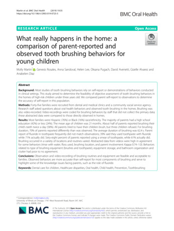
Self-Infla6ng Ven6la6onBag, with Oxygenreservoir (A & B), andwithout (C & D).
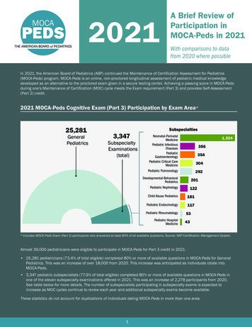
Correct Posi6oning of the child 2years of age for ven6la6on andEndotracheal intuba6on.A & D: Incorrect posi6ons of the neck.B: A folded sheet or towel placedunder the occiput aligns thePharyngeal (P) and Tracheal (T) axes.C: Extension of the atlanto-occipitaljoint results in the alignment of theoral (O), pharyngeal, and trachealaxes.Note that proper posi6oning placesthe external ear canal anterior to theshoulder.
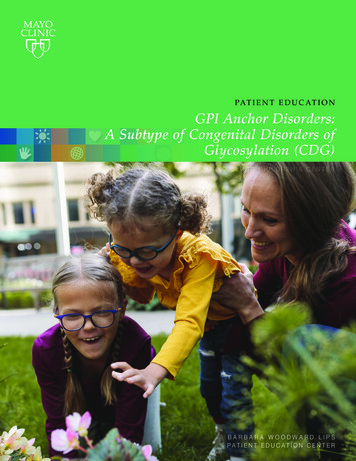
One-handed E-C clampface-mask applica6ontechnique. Three fingersof one hand liZ the jaw(they form the “E” whilethe thumb and indexfinger hold the mask tothe face (making a “C”
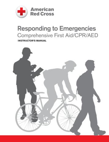
Two-person bag-maskven6la6on technique ismore effec6ve than the1-person technique.
ALLERGICEMERGENCIES(ANAPHYLAXIS)
What is the most commonorgan involved in anaphylaxis?
ANAPHYLAXIS - Definitionü 1. A rapid-onset IgE-mediated release of histamine and other mediators from mastcells and basophils leading to a systemic allergic reaction involving multiple organsystems, including two or more of the following:ü a) Cutaneous/mucosal (flushing, urticaria, pruritis, angioedema); seen in 90%.ü b) Respiratory (laryngeal edema, bronchospasm, dyspnea, wheezing, stridor,hypoxemia); seen in 70%ü c) GI ( N/V/D, crampy abdominal pain); seen in about 40-50%ü d) Circulatory (tachycardia, hypotension, syncope); seen in about 30-40%ü 2. Initial reaction may be delayed for several hours AND symptoms may recur up to72 hours after initial recovery. Patients should therefore be observed for a minimumof 6 to 24 hours for late-phase symptoms.
ANAPHYLAXIS - TreatmentStop exposure to precipitating antigen /allergenEpinephrine mainstay of therapy. While performing ABCs, immediately give IM epinephrine, 0.01mg/kg(0.01ml/kg) of 1:1000 SQ or IM (maximum dose 0.5mg). Repeat every 5 minutes as needed. The site of choiceis the lateral aspect of the thigh, owing to its vascularity.Airway - Establish airwayBreathing – Give Oxygen and PPV as needed.Treat hypotension: Obtain IV access, Trendelenburg position with head 30 degrees below feet, give IV fluidboluses (20ml/kg) followed by pressors (epinephrine infusion) as needed.Anti-histamines: Histamine-1 receptor antagonists such as diphenhydramine, 1 to 2mg/kg via IM, IV, or oral (PO)route (maximum dose, 50mg). Also, consider a histamine-2 receptor antagonist (e.g. ranitidine).Corticosteroids help prevent the late phase of the allergic response. Administer methylprednisolone in a 2-mg/kgIV bolus, followed by 2mg/kg/day IV or IM, divided every 6 hours, or prednisone 2mg/kg PO once daily.Albuterol 2.5mg for 30kg, 5mg for 30mg, for bronchospasm(wheezing). Repeat q15mins prn. MDI or Nebs.Racemic epinephrine 0.5ml of 2.25% solution inhaled for signs of upper airway obstruction.Patient should be discharged with an Epi-pen ( 30kg), Epi-pen junior ( 30kg), or comparable injectableepinephrine product with specific instructions on appropriate use, as well as anaphylaxis action plan.
SHOCK
SHOCKü Shock is a critical condition that results from inadequate tissuedelivery of oxygen and nutrients to meet tissue metabolic demands.ü Shock is often, but not always, characterized by inadequateperipheral and end-organ perfusion.ü The definition of shock doesn’t depend on BP measurement. Shockcan occur with normal, increased, or decreased BP.ü All type of shock can result in impairment of vital organs like thebrain (AMS) and kidneys (low UOP)ü In children, most shock is characterized by low CO. However,sepsis and anaphylactic shock is high CO shock.
Shock can result from:MechanismSHOCKType of ShockCommon CausesInadequate blood volume oroxygen-carrying capacityHypovolemic shock-GI losses: Diarrhea; Vomi?ng(Hypovolemia is the most -Inadequate fluid intakecommon cause of shock in -Hemorrhage (internal & external)childr
"Studies have shown that emergencies are common in primary care practices that provide care to children. In 1 study, the authors surveyed 52 pediatric offices and found that these practices saw a median of 24 emergencies per year. Most of the offices (82%) reported that they encountered, on average, at least 1 emergency per month. In another .